0107
ODF-Fingerprinting reconstruction of corticospinal tracts for preoperative planning of brain tumor resection1Bernard and Irene Schwartz Center for Biomedical Imaging, Department of Radiology, New York University Grossman School of Medicine, New York, NY, United States, 2Center for Advanced Imaging Innovation and Research (CAI2R), Department of Radiology, New York University Grossman School of Medicine, New York, NY, United States, 3Herricks High School, New Hyde Park, NY, United States, 4Department of Neurosurgery, Perlmutter Cancer Center, Neuroscience Institute, Kimmel Center for Stem Cell Biology, NYU Langone Health, New York, NY, United States, 5Vilcek Institute of Graduate Biomedical Sciences, New York University Grossman School of Medicine, New York, NY, United States
Synopsis
Keywords: Tractography, Tractography & Fibre Modelling, preoperative planning, ODF-fingerprinting, pyramidal tract, corticospinal tract, brain tumor, BOLD activation, task fMRI
Motivation: Tractography enables preoperative visualization of major neural pathways altered or displaced by a brain tumor; however, it often fails to reconstruct the cortical terminations of corticospinal tracts due to the complex bending and branching formations of fibers.
Goal(s): We aim to improve tracking of corticospinal tracts in their most challenging regions of hand and face projections to the motor cortex.
Approach: We refine reconstruction of fibers inside corticospinal tracts by incorporating ODF-Fingerprinting into the tracking pipeline.
Results: With ODF-Fingerprinting, we increased the overlap between the reconstructed corticospinal tracts and the cortical regions activated during task-based functional MRI involving hand and face movement.
Impact: Our improved reconstruction can help decrease the incidence of postoperative deficits by identifying the structural neural connections that need to be spared during tumor resection.
Introduction
Many gliomas occur in the frontoparietal subcortical areas where they disturb the neural connections involved in the language or motor functions [1]. Diffusion MRI tractography is the sole in-vivo imaging modality to visualize white matter fibers altered or infiltrated by a growing tumor [2]. However, complex neural structures with bending or branching fibers, like the lateral projections of corticospinal tracts, remain challenging for tracking algorithms. Therefore, the accuracy of visualizations used in tumor resection planning is often compromised in the critical regions of hand and face projections to the motor cortex.In this work, we incorporated Orientation Distribution Function Fingerprinting (ODF-FP) [3,4] into the fiber tractography pipeline to improve reconstruction of corticospinal tracts from preoperative Diffusion-Weighted Images (DWIs) of brain tumor patients. ODF-FP is a dictionary-based reconstruction method that refines identification of narrow-angled fiber structures [4]. To verify our approach, we quantified the overlap between the cortical terminations of the reconstructed fascicles and the Blood Oxygenation Level Dependent (BOLD) maps obtained from task-based functional MRI (fMRI).
Our results showed that ODF-FP can improve tracking of the lateral projections of corticospinal tracts altered by a tumor. This can provide more accurate preoperative visualizations of white matter tracts located in proximity to the tumor. Our improved tractography outcomes can aid identification of the brain areas that need to be spared during tumor resection and thus potentially decrease the risk of postoperative deficits.
Methods
Data:We considered preoperative MRI of 8 patients (7 males / 1 female, 46±12 years old) diagnosed with glioma located in the proximity to the corticospinal tract. The DWIs were acquired at $$$2.0\times2.0\times2.0~$$$mm resolution, $$$TE/TR=92/5900~$$$ms, $$$b=300,1100,2500,5000~$$$s/mm2, 60 encoding directions each, interleaved with 8$$$~b=0$$$ images. During fMRI acquisition at $$$3.0\times3.0\times3.0~$$$mm ($$$TR=1$$$s) or $$$2.3\times2.3\times2.3~$$$mm ($$$TR=2$$$s), all 8 patients performed the finger tapping task (with the hand contralateral to the tumor) and 6 of them also performed the lip puckering task.
Processing:
Our DWI postprocessing in MRtrix3 [5] included denoising, Gibbs ringing removal, correction of B1 field inhomogeneity and eddy currents. We applied Generalized Q-sampling Imaging (GQI) [6] reconstruction in DSI Studio [7] to obtain ODFs. We then executed ODF-FP with a dictionary of 1,000,000 items having $$$0≤N≤3$$$ crossing fibers per voxel and the remaining parameters drawn randomly with a uniform distribution (ODF-FP prior). Next, we ran one iteration of the stepwise stochastic learning of ODF-dictionary [8] followed by another execution of ODF-FP (referred to as ODF-FP posterior). Simultaneously, we preprocessed the task-based fMRI using fMRIprep [9] and then completed the first-level analysis in fMRI Expert Analysis Tool (FEAT) [10] with Z-threshold=3.1, masked at the motor cortex.
Evaluation:
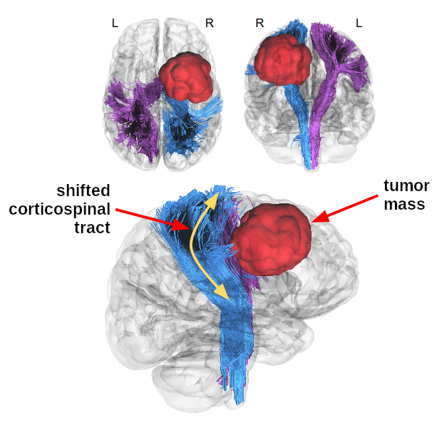
We reconstructed the corticospinal tracts (Figure 1) using deterministic tractography [11] in DSI Studio by tracking from the brainstem to the motor cortex. Next, we quantified the overlap between the cortical terminations of the tracts and the BOLD activation regions with the following measures:
(a) True Positive Rate $$TPR=\frac{|tractography∩BOLD|}{|BOLD|}\in[0,1];\qquad\mbox{(higher is better)}$$
(b) Dice coefficient $$DC=\frac{2\cdot|tractography∩BOLD|}{|tractography|+|BOLD|}\in[0,1];\qquad\mbox{(higher is better)}$$
(c) 95th percentile of the Hausdorff distance (HD95) between $$$tractography$$$ and $$$BOLD$$$ (lower is better).
Results
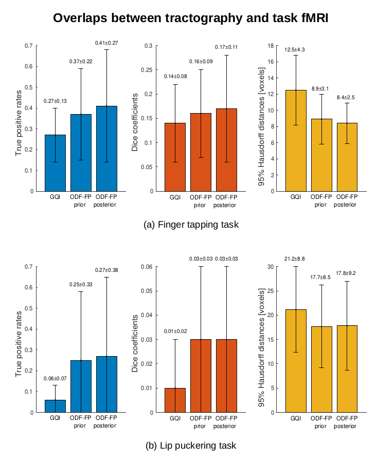
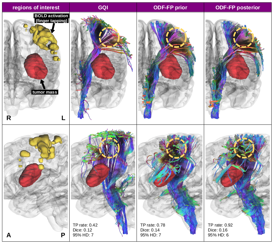
The corticospinal tracts reconstructed with ODF-FP posterior reached higher TPR (0.41±0.27) and DC (0.17±0.11) and lower HD95 (8.4±2.5) than GQI and ODF-FP prior in the finger tapping task (Figure 2a). Our dictionary-based method helped generate more streamlines representing the lateral projections of the corticospinal tracts despite deformation by a tumor mass, as shown in a representative example (Figure 3).Similarly, both ODF-FP approaches outperformed GQI in the lip puckering task (Figure 2b), although with relatively lower scores because of the smaller BOLD activation regions.
Discussion
Preoperative visualization of corticospinal tracts is challenging due to the bending and branching of fibers projecting to the motor cortex. The deformation of the fascicles by the tumor mass adds another confounding factor for tractography. Our study showed that ODF-FP in both its variants – prior and posterior – can alleviate these difficulties. The observed improvement in overlaps between the reconstructed cortical terminations and the BOLD activation during the motor-related tasks confirmed the accuracy of streamlines generated with ODF-FP.The low values of DC (0.03 in lip puckering or 0.17 in finger tapping) should not be mistaken for poor overlap. Indeed, we tracked the entire shapes of corticospinal tracts, whereas our BOLD activation regions only represented the subregions attributed to hand or lips. We thus compared relative changes in DC rather than absolute values.
Considering that 40% of brain tumor patients experience functional deficits after the surgery [12], future work should further improve identification of peritumoral white matter fibers that need to be spared during tumor resection.
Acknowledgements
This project was supported in part by the National Institutes of Health (NIH, R01 EB028774 and R01 NS082436) under the rubric of the Center for Advanced Imaging Innovation and Research (CAI2R, https://www.cai2r.net), a NIBIBBiomedical Technology Resource Center (NIH P41 EB017183).References
[1] Larjavaara et al. (2007). Incidence of gliomas by anatomic location. Neuro-oncology, 9(3), 319-325.
[2] Bucci et al. (2013). Quantifying diffusion MRI tractography of the corticospinal tract in brain tumors with deterministic and probabilistic methods. NeuroImage: Clinical, 3, 361-368.
[3] Baete et al. (2019). Fingerprinting Orientation Distribution Functions in diffusion MRI detects smaller crossing angles. Neuroimage, 198, 231-241.
[4] Filipiak et al. (2022). Performance of orientation distribution function‐fingerprinting with a biophysical multicompartment diffusion model. Magnetic Resonance in Medicine, 88(1), 418-435.
[5] Tournier et al. (2019). MRtrix3: A fast, flexible and open software framework for medical image processing and visualisation. NeuroImage, 202, 116137.
[6] Yeh et al. (2010). Generalized q-sampling imaging. IEEE transactions on medical imaging, 29(9), 1626-1635.
[7] Yeh, F. (2017). Diffusion MRI reconstruction in DSI Studio. Advanced Biomedical MRI Lab, National Taiwan University Hospital. Available online at: http://dsi-studio.labsolver.org
[8] Filipiak et al. (2022). Stepwise Stochastic Dictionary Adaptation Improves Microstructure Reconstruction with Orientation Distribution Function Fingerprinting. Computational Diffusion MRI, 89-100.
[9] Esteban et al. (2020). Analysis of task-based functional MRI data preprocessed with fMRIPrep. Nature protocols, 15(7), 2186-2202.
[10] Woolrich et al. (2004). Multilevel linear modelling for FMRI group analysis using Bayesian inference. Neuroimage, 21(4), 1732-1747.
[11] Yeh et al. (2013). Deterministic diffusion fiber tracking improved by quantitative anisotropy. PloS one, 8(11), e80713.
[12] Zetterling et al. (2020). Time course of neurological deficits after surgery for primary brain tumours. Acta neurochirurgica, 162, 3005-3018.
Figures