0092
Use of 4D Flow MRI to Indirectly Predict Cardiac Adverse Events Through Hemodynamic Alterations Induced by Exercise1Department of Radiology, The Affiliated Hospital of Southwest Medical University, Lu Zhou, China, 2MR Research Collaboration, Siemens Healthineers, Chengdu, China
Synopsis
Keywords: Flow, Cardiovascular
Motivation: Cardiovascular-related deaths are increasing in athletes, which necessitates a deeper understanding of hemodynamics.
Goal(s): Our goal was to use 4D flow MRI to assess athletes' cardiac and aortic hemodynamics and their links to myocardial fibrosis and cardiac remodeling risks.
Approach: Cardiac MRI was performed on 213 athletes and 32 matched controls. Hemodynamic parameters were measured and analyzed against myocardial fibrosis and cardiac remodeling risks.
Results: Athletes exhibited increased wall shear stress and energy loss. Hemodynamics differed markedly between groups. Our prediction model reliably displayed the potential of 4D flow in assessing cardiac risks.
Impact: Exercise can elevate aortic wall shear stress and energy loss. Four-dimensional flow cardiac MRI may allow predicting myocardial fibrosis or cardiac remodeling risks in athletes, thus informing clinicians of adverse event associations and guiding follow-up adjustments.
Introduction
Athlete health is a concern worldwide. Increasing numbers of sudden deaths due to cardiovascular adverse events are being reported in athletes1,2. Sports increase the cardiovascular load, which is reflected by hemodynamic parameters3. Four-dimensional (4D) flow magnetic resonance imaging (MRI) can be used to analyze complex hemodynamic models in vivo by quantifying blood flow parameters and deriving characteristics4. This study was conducted to evaluate the hemodynamic changes in the heart and aorta in athletes to predict the risk of myocardial fibrosis (MF) and/or cardiac remodeling (CR).Methods
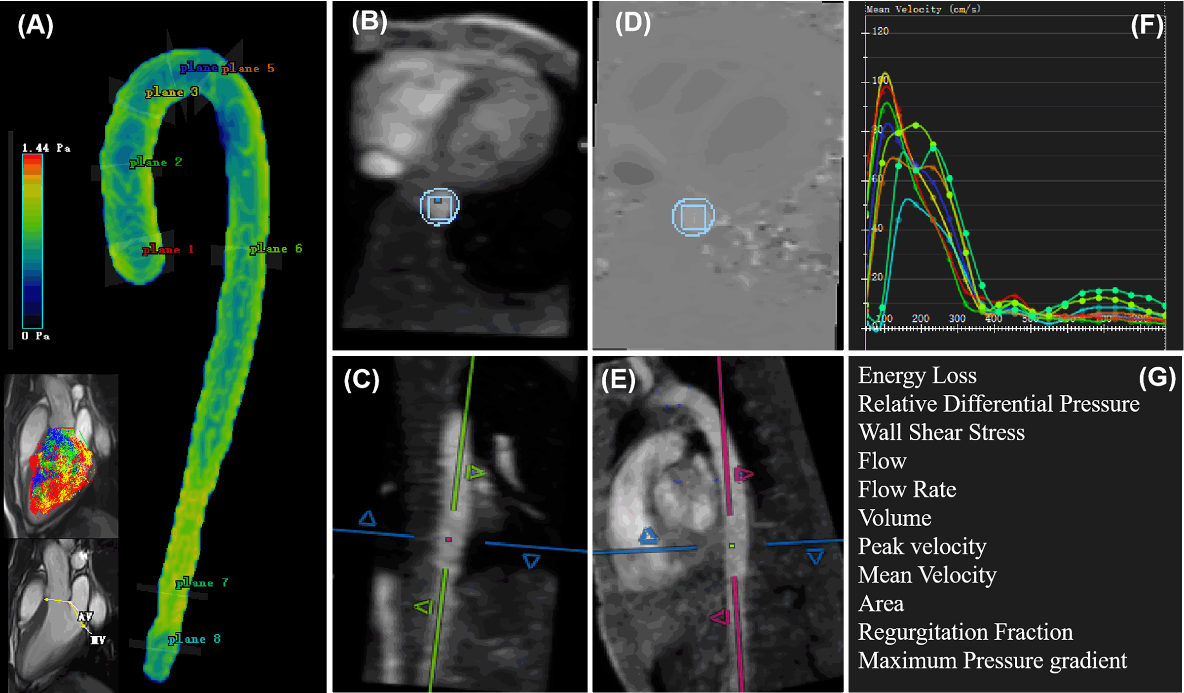
Our institutional ethics committee approved this study. We prospectively enrolled 213 athletes and 32 matched sedentary healthy controls for cardiac magnetic resonance (CMR) examinations on a clinical 3T system (MAGNETOM Prisma, Siemens Healthcare, Erlangen, Germany) equipped with an 18-channel body coil. 4D flow was performed with the following scanning parameters: echo time = 2.88 ms, echo spacing = 5.7 ms, repetition time = 45.36 ms, spatial resolution = 1.8 × 1.8 × 3.5 mm3, and velocity encoding = 150 cm/s in all three velocity-encoding directions. Routine cardiac cine and late gadolinium enhancement sequences were measured for reference. As showing in Figure 1 the cardiac function and 4D flow parameters for the left ventricle (LV) and aorta (planes 1–8) were measured using cvi42 (v. 5.12.4; Circle Cardiovascular Imaging, Calgary, Canada). Through this cardiac morphology and function analysis, athletes were categorized into groups either exhibiting or lacking MF and/or CR. The least absolute shrinkage and selection operator (LASSO) penalized regression was used to construct the clinical prediction model for MF and/or CR. Subsequently, identified scores were calculated by summing selected features weighted by their coefficients for each participant. Continuous variables were compared between athletes with MF and/or CR (positive group) and athletes lacking both MF and CR (negative group) using t-tests or Mann-Whitney U tests. P<0.05 was considered statistically significant.Results
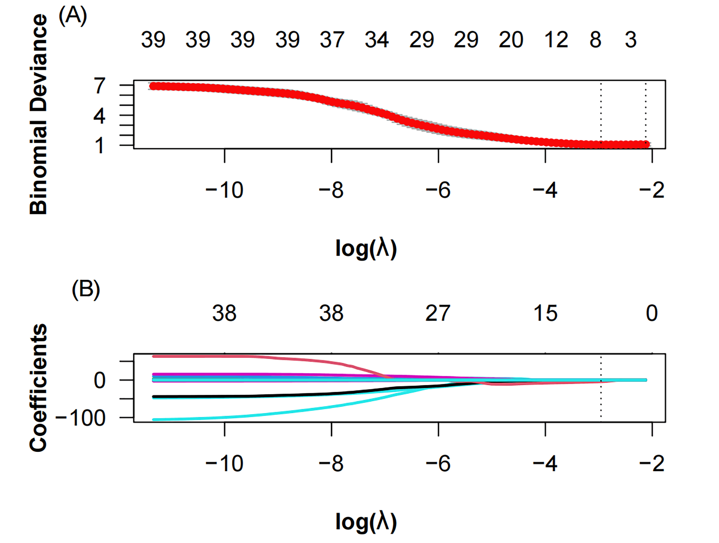
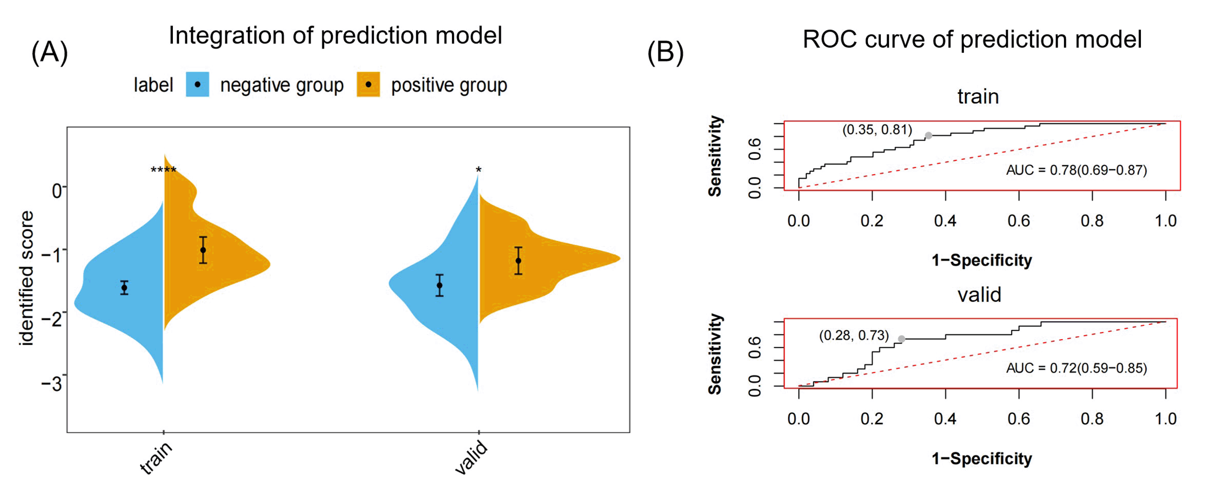
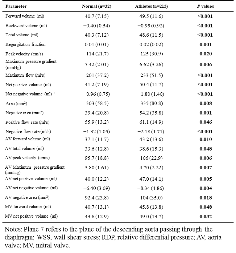
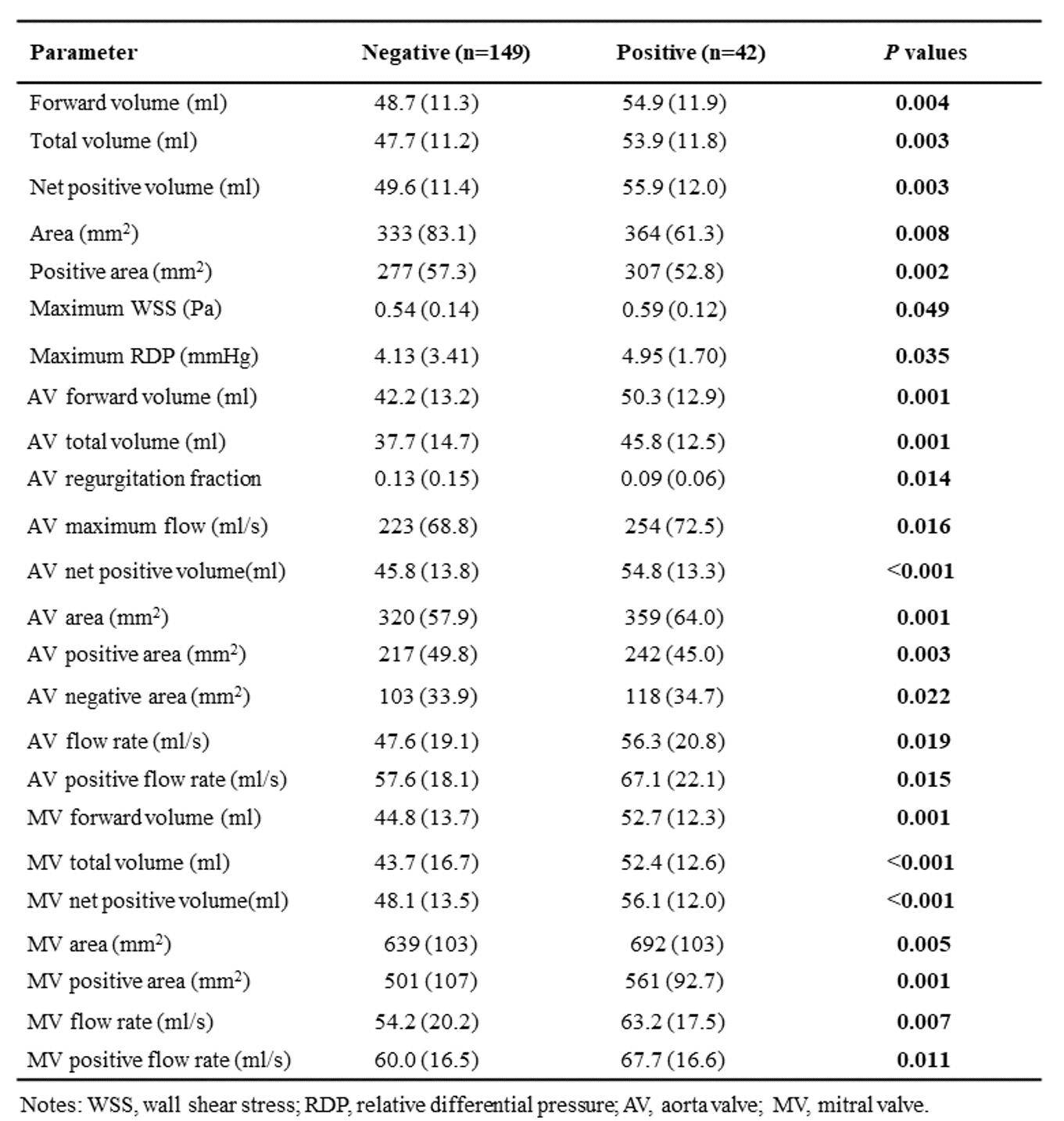
Among the athletes, 35 men had CR, and nine exhibited MF in the LV. Table 1 delineates the differences in 4D flow parameters between athletes and controls, specifically focusing on aortic plane 7 and LV measurements. Athletes exhibited significantly heightened wall shear stress (WSS) and energy loss. However, athletes had significantly lower general aortic parameters and LV peak velocity than did the controls (P<0.05). Forty-two athletes were categorized into the positive group with MF and/or CR (n=42); 149 were categorized into the negative group without these conditions (Table 2). The positive group exhibited higher forward volume, total volume, net positive volume, area, WSS, and relative differential pressure. Leveraging clinical significance and statistical relevance from redundancy analysis and LASSO regression (Figure 2), we formulated a clinical prediction model on eight important indicators, listed by descending importance: average WSS, maximum WSS, positive areas of the mitral valve (MV) and of plane 7, net positive flows of the MV and of plane 7, body mass index, and body weight. Figure 3 shows the performance of our predictive model and its capability to discriminate between the positive and negative athlete groups. Figure 3A shows a graph of the predicted scores for both the training and validation datasets. The negative and positive groups are easily distinguished. The areas under the curve (AUCs) were 0.78 for the training set and 0.72 for the validation set.Discussion and Conclusion
The AUCs of our model corroborate its robust diagnostic efficiency. Our results revealed that excessive exercise can escalate the aortic WSS and energy loss, potentially causing endothelial damage and aortic eddy currents, culminating in detrimental cardiac effects. We constructed a highly accurate predictive model for CR and/or MF onset. These insights affirm the efficacy of 4D flow CMR in evaluating hemodynamic shifts and forecasting potential cardiac risks in athletes, demonstrating the suitability of 4D flow CMR as an innovative athletic monitoring tool, especially for tracking aortic WSS alterations at the diaphragmatic passage. While these results are promising, further research with larger sample sizes and longitudinal tracking is required to validate these findings and determine optimal predictive and intervention strategies.Acknowledgements
None.References
1. Emery MS, Kovacs RJ. Sudden cardiac death in athletes. JACC Heart Fail. 2018;6:30–40.
2. Landry CH, Allan KS, Connelly KA, et al. Sudden cardiac arrest during participation in competitive sports. N Engl J Med. 2017;377:1943–1953.
3. Liu H-L, Chen X-Y, Li J-R, et al. Efficacy and safety of pulmonary arterial hypertension-specific therapy in pulmonary arterial hypertension: a meta-analysis of randomized controlled trials. Chest. 2016;150:353–366.
4. Eriksson J, Bolger AF, Ebbers T, et al. Assessment of left ventricular hemodynamic forces in healthy subjects and patients with dilated cardiomyopathy using 4D flow MRI. Physiol Rep. 2016;4:e12685.
Figures