0091
Impact of flow encoding strategies on velocity and turbulence quantification in 4D flow MRI1University and ETH Zurich, Zurich, Switzerland
Synopsis
Keywords: Synthetic MR, Velocity & Flow, Simulation
Motivation: Blood flow turbulence and velocity quantification with 4D flow MRI is sensitive to imaging parameters such as Venc and undersampling factor, but their impact has not been clearly quantified yet.
Goal(s): Use synthetic 4D flow MRI data to determine the impact of encoding directions and strengths for turbulence and velocity quantification.
Approach: Personalized synthetic 4D flow MRI data are simulated for a given set of encoding velocity strengths and directions assuming a fixed scan budget of 15 minutes.
Results: Turbulent kinetic energy shows large variations depending on the encoding strategy, while velocity magnitudes are marginally affected by the choice.
Impact: Based on the simulations, a 7-point dual- approach represents an efficient approach for accurate velocity and turbulence quantification. However, the low-Venc needs to be tuned to be sensitive to the ranges of expected intra-voxel standard deviations.
Introduction
The assessment of aortic flow velocity and turbulence with 4D flow MRI has the potential to enhance the diagnostic accuracy of aortic stenosis1. Patients with aortic stenosis typically present large velocity ranges, rendering single-Venc acquisitions suboptimal. Various multi-Venc approaches have been proposed, to improve the precision of velocity measurements2-5 and to increase the sensitivity to turbulence6-8. Since additional velocity encoding points would result in longer scans, advanced acceleration techniques need to be employed. Turbulence quantification under 10 minutes scans was shown to be possible9 and highly undersampled scans have been shown to accurately measure velocity10,11. However, a standard protocol for clinical turbulent 4D flow MRI acquisitions does not yet exist.In this study we use a personalized synthetic 4D flow MRI dataset to investigate the impact of multiple encoding strategies on the quantification of velocity and turbulence assuming a clinically reasonable fixed scan budget of 15 minutes. The synthetic data consists of simulated foreground aortic flow data embedded into a 4D flow MRI background, resulting in realistic MR images with known flow and turbulence ground truth.
Methods
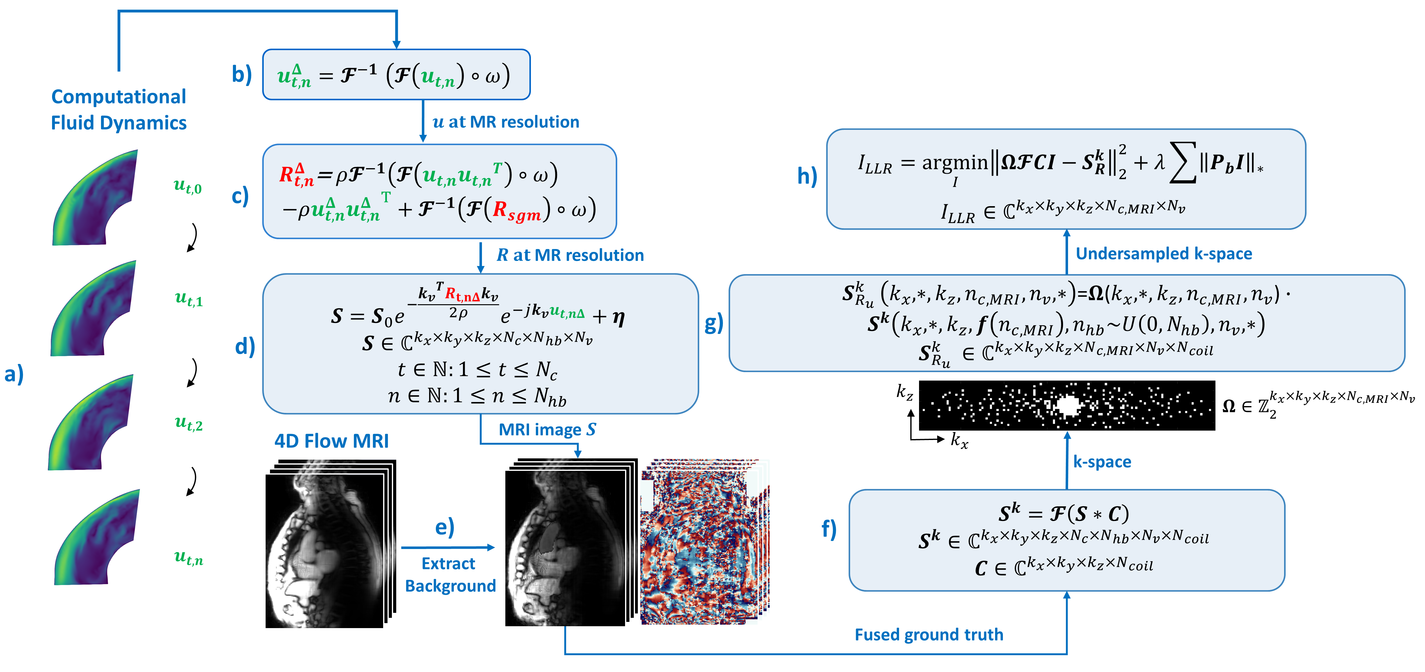
Spatiotemporal segmentation of a patient’s aorta and inflow, flow simulations and synthesis of 4D flow MRI data (Figure 1a, 1b, 1c and 1d) were performed as described previosuly12. Synthetic 4D flow MRI foreground was embedded into the end-expiratory bin of the same patient’s 5D flow MRI background data, serving as ground truth (Figure 1e). This process was repeated for every CFD simulated cardiac phase and cardiac cycle, resulting in a set of 2400 fully sampled 3D k-space grids. Prospective undersampling was achieved using pseudo-spiral Cartesian k-space filling masks10 (Figure 1g), where each k-space point in the trajectory was sampled from the pool of k-space grids previously computed. This allowed to include the effect of scan-to-scan variability. Undersampled k-space acquisitions were reconstructed (Figure 1h) using a locally low-rank approach13 (LLR) as implemented in the BART toolbox14. All reconstruction parameters were optimized with a grid search to ensure optimal reconstructions (including different parameters for velocity and turbulence). Bayesian unfolding was performed for all multi-Venc acquisitions as described previously12. Error metrics were computed using the structural similarity index measure (SSIM) with respect to a reference obtained using noiseless cycle averaged flow quantities. The impact of scan-to-scan variance on turbulent kinetic energy (TKE) and velocity magnitude were measured using the turbulence-to-noise ratio7 (TNR) and velocity-to-noise ratio (VNR), respectively. Various encoding schemes including 4, 7, 13 and 19-point velocity encoding were studied. In order to have identical scan budgets, the undersampling factor R was adapted for each scan.Results
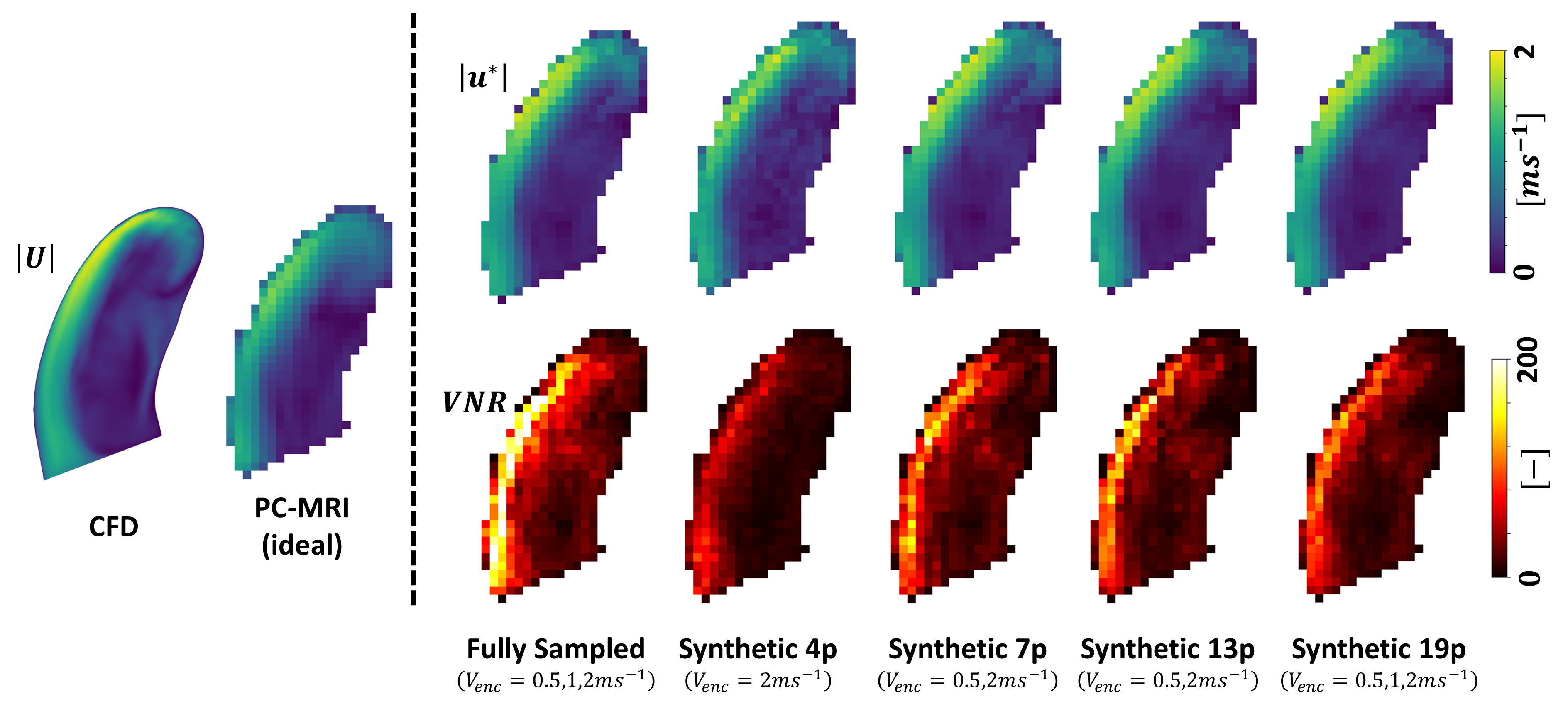
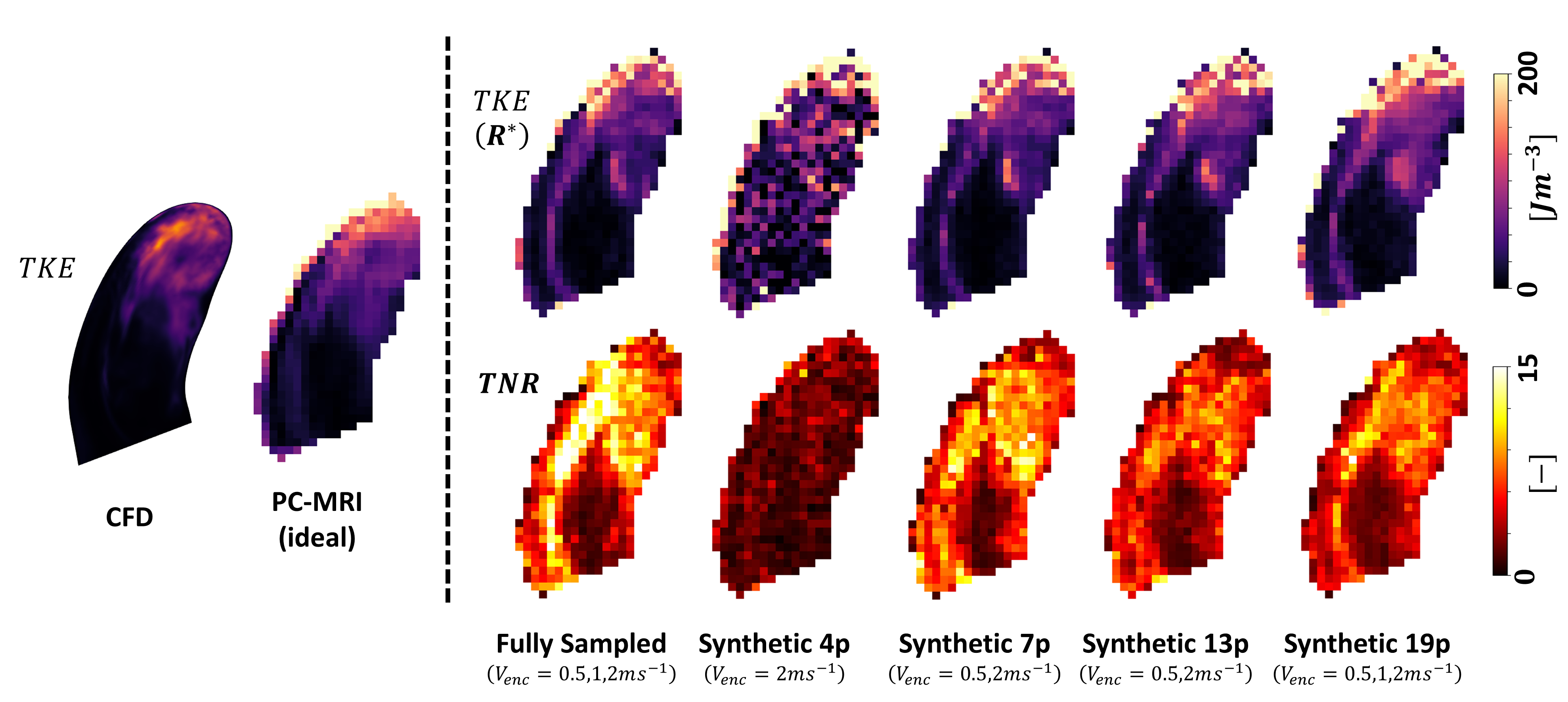
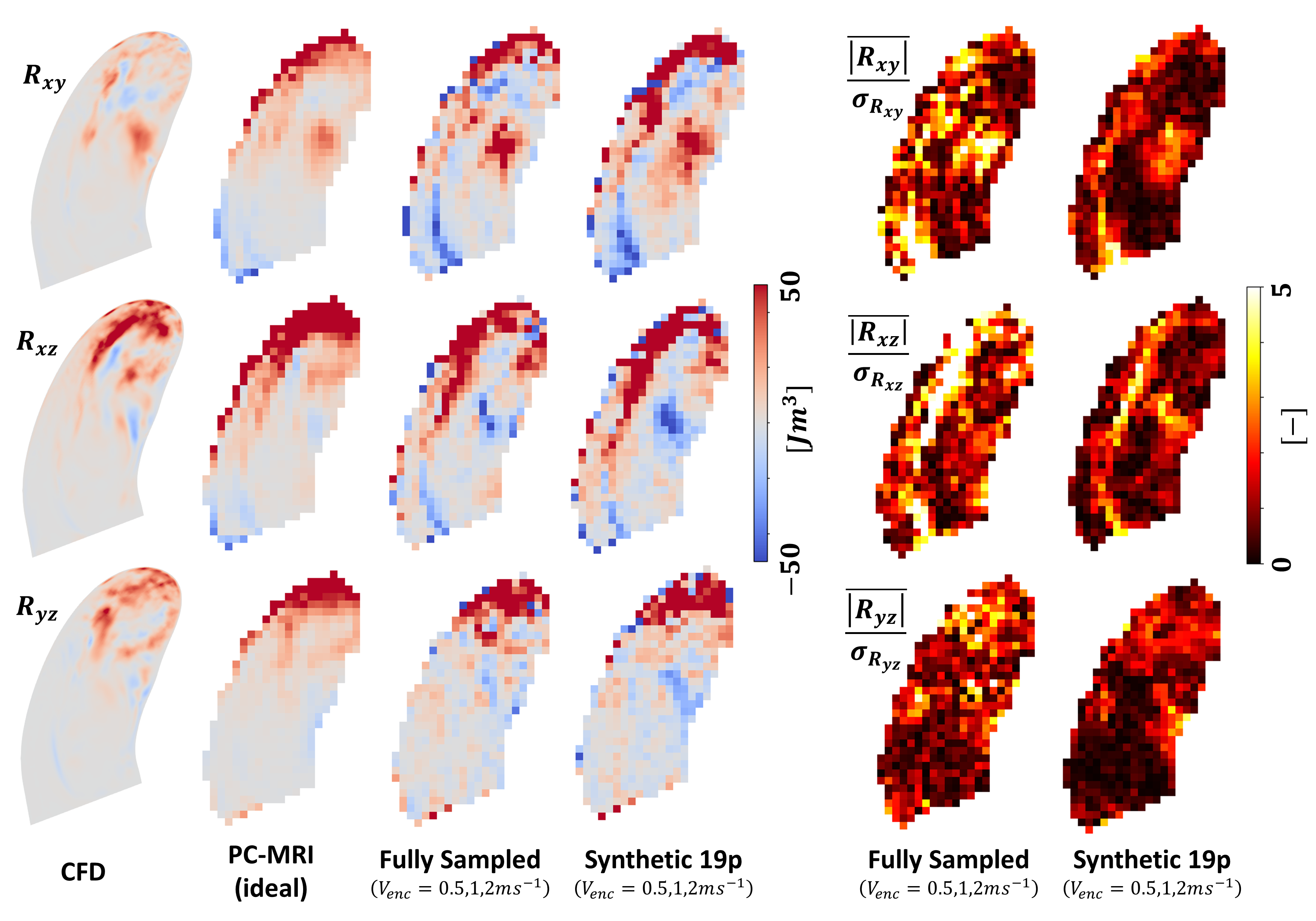
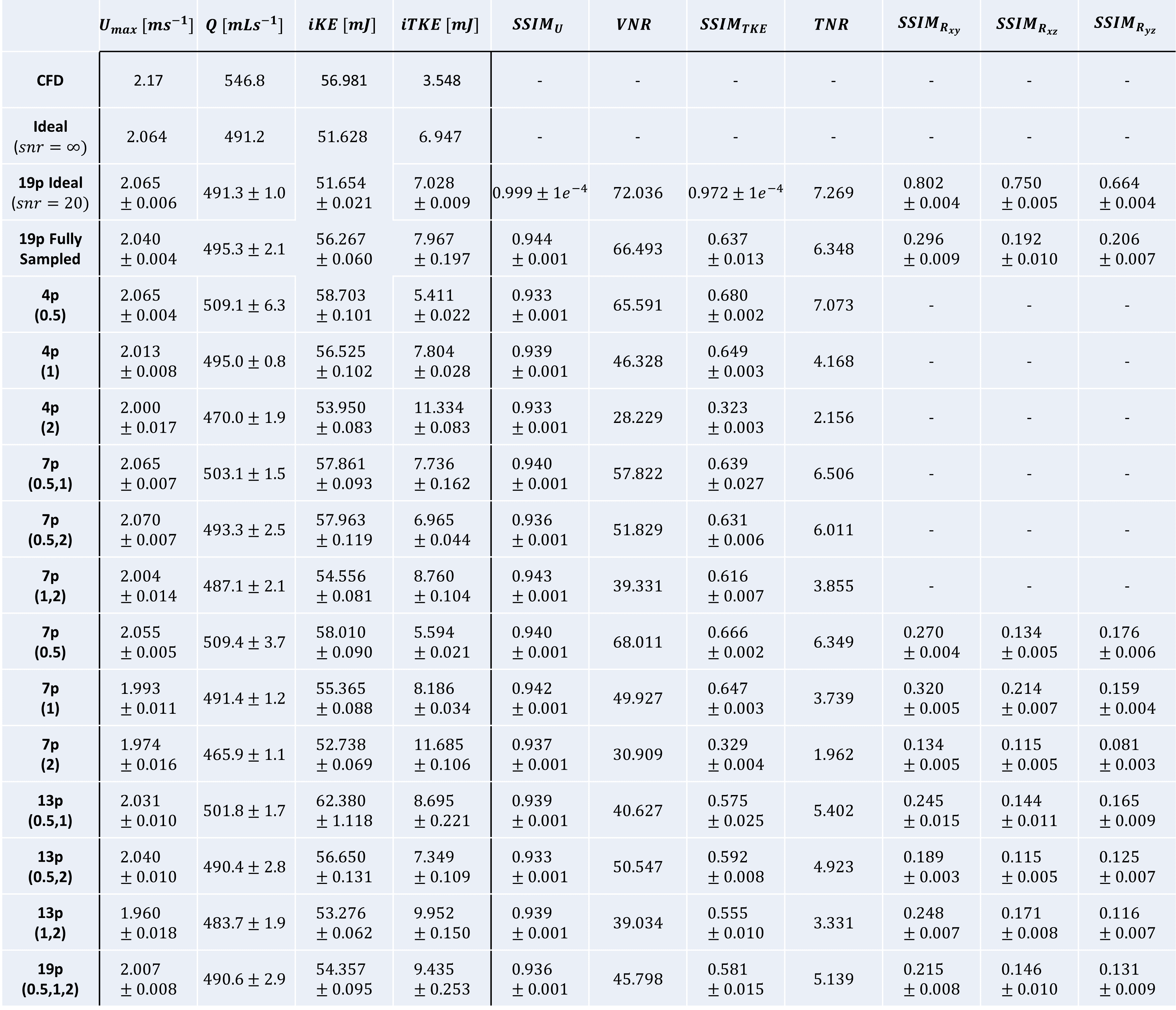
Reconstructed velocity maps and TKE maps are presented in Figures 2 and 3. The velocity-to-noise ratio (VNR) is an order of magnitude larger than TNR in the regions of interest. TNR of non-diagonal RST terms is around threefold lower than TKE (Figure 4). In Figure 5 we summarize a selection of flow metrics quantified using various encoding strategies. While peak velocity was consistently underestimated with respect to CFD due to the limited spatiotemporal MR resolution it was limited to 10% across all encoding strategies. Velocity SSIM was >0.933 for all encoding strategies, while TKE SSIM values varied from 0.323 to 0.680 and all SSIM values of non-diagonal RST terms were <0.320.Discussion
We simulated 15 minutes scans and re-scans with 14 different encoding strategies, resulting in a total of 280 scans or 110 hours of scanning (including fully sampled scans), which allowed us to better understand the impact of encoding strategy on flow quantification.In our model, velocity quantification is marginally affected by the encoding strategy. However, in this work, all data was ideally unwrapped using the CFD data. In practice, unwrapping algorithms would need to be applied to recover correct velocity fields when Venc is lower than the velocities in the aorta. When compared to a 4p low-Venc encoding, a 7p dual-Venc determines a loss of 20% in VNR but with no significant impact on velocity SSIM and peak velocity and with no need of unwrapping the measurements. Similarly, TKE SSIM drops by 7% and TNR by 8%, when comparing a 7p to a 4p encoding. TNR on non-diagonal terms of the RST does not seem to justify the additional encoding directions. Our results suggest that the optimal encoding consists in a 7p dual-Venc with a low-Venc between 0.5 and 1ms-1, for high VNR (>50) and sensitivity to the expected range of turbulence in stenotic patients, and a high-Venc larger than the expected peak velocity to allow for correct Bayesian unfolding of the low-Venc.
Acknowledgements
No acknowledgement found.References
1. Garcia, Julio, Alex J. Barker, and Michael Markl. "The role of imaging of flow patterns by 4D flow MRI in aortic stenosis." JACC: Cardiovascular Imaging 12.2 (2019): 252-266.
2. Ha, Hojin, et al. "Multi‐VENC acquisition of four‐dimensional phase‐contrast MRI to improve precision of velocity field measurement." Magnetic resonance in medicine 75.5 (2016): 1909-1919.
3. Callaghan, Fraser M., et al. "Use of multi‐velocity encoding 4D flow MRI to improve quantification of flow patterns in the aorta." Journal of Magnetic Resonance Imaging 43.2 (2016): 352-363.
4. Schnell, Susanne, et al. "Accelerated dual‐venc 4D flow MRI for neurovascular applications." Journal of Magnetic Resonance Imaging 46.1 (2017): 102-114.
5. Kroeger, Jan Robert, et al. "Velocity quantification in 44 healthy volunteers using accelerated multi-VENC 4D flow CMR." European Journal of Radiology 137 (2021): 109570.
6. Binter, Christian, et al. "Bayesian multipoint velocity encoding for concurrent flow and turbulence mapping." Magnetic resonance in medicine 69.5 (2013): 1337-1345.
7. Ha, Hojin, et al. "In vitro experiments on ICOSA6 4D flow MRI measurement for the quantification of velocity and turbulence parameters." Magnetic Resonance Imaging 72 (2020): 49-60.
8. Walheim, Jonas, Hannes Dillinger, and Sebastian Kozerke. "Multipoint 5D flow cardiovascular magnetic resonance-accelerated cardiac-and respiratory-motion resolved mapping of mean and turbulent velocities." Journal of Cardiovascular Magnetic Resonance 21.1 (2019): 1-13.
9. Walheim, Jonas, et al. "5D flow tensor MRI to efficiently map Reynolds stresses of aortic blood flow in-vivo." Scientific reports 9.1 (2019): 18794.
10. Peper, Eva S., et al. "Highly accelerated 4D flow cardiovascular magnetic resonance using a pseudo-spiral Cartesian acquisition and compressed sensing reconstruction for carotid flow and wall shear stress." Journal of Cardiovascular Magnetic Resonance 22.1 (2020): 1-15.
11. Liu, Jing, et al. "Highly accelerated aortic 4D flow MR imaging with variable-density random undersampling." Magnetic resonance imaging 32.8 (2014): 1012-1020.
12. Dirix, Pietro, et al. "Synthesis of patient-specific multipoint 4D flow MRI data of turbulent aortic flow downstream of stenotic valves." Scientific Reports 12.1 (2022): 16004.
13. Zhang, Tao, John M. Pauly, and Ives R. Levesque. "Accelerating parameter mapping with a locally low rank constraint." Magnetic resonance in medicine 73.2 (2015): 655-661.
14. Uecker, Martin, et al. "Berkeley advanced reconstruction toolbox." Proc. Intl. Soc. Mag. Reson. Med. Vol. 23. No. 2486. 2015.
Figures