0086
Predicting Ischemic Stroke Prognosis based on Lesion Structural and Functional Disconnections1Department of Medical Imaging Technology, Yanjing Medical College, Capital Medical University, Beijing, China
Synopsis
Keywords: Data Processing, Stroke
Motivation: Assessing stroke patients' prognosis is challenging due to complex neurophysiological mechanisms involved, with only lesion location accessible from DWI sequence.
Goal(s): This study aims to use patients' lesion information, alongside its structural and functional disconnections, to predict their recovery.
Approach: We designed a retrospective study using lesion information at admission along with its strctural and funcitonal disconnetion, combined with machine learning to predict the prognosis of 148 stroke patients six months post-stroke.
Results: Our results suggested that the structural and functional disruptions of the lesion could explain and predict National Institutes of Health Stroke Scale score and prognosis of stroke.
Impact: The results not only help us understand the neurophysiological mechanisms underpinning stroke prognosis from the perspective of brain structural and functional connections, but also reveal potential targets for intervention treatments aimed at stroke recovery.
PURPOSE
Ischemic stroke is a leading global cause of both mortality and disability,1 and it is challenging and important to provide the precise prognostic information for intervention strategies and therapeutic targets. The prognosis of a stroke has traditionally depended on clinical assessments, such as the National Institutes of Health Stroke Scale (NIHSS) score, which inadequately capture the intricate neurophysiological processes involved in stroke recovery. While the voxel-based lesion-symptom mapping (VLSM) based on diffusion-weighted imaging (DWI) sequences can provide symptom-related lesion locations, it is limited by local information and cannot reflect associated distant neurobiological abnormalities.2 Recent researches have revealed that the structural and functional disconnections based on lesions, are important factors correlated with stroke outcome.3 Consequently, we hypothesize that the connection disruptions are imaging biomarkers for stroke prognosis, and may provide effective approach for therapeutic planning.METHOD
Data Collection: This study was a retrospective analysis of 148 patients diagnosed with acute ischemic stroke at Liangxiang Hospital (Beijing, China) from October 2020 to May 2022, approved by the ethics committee of Liangxiang Hospital (approval number 2016126). MRI scans were performed within 3 days of stroke onset, and the modified Rankin Scale (mRS) score after six months post-stroke, was used as a prognostic judgment index, with good prognosis defined as mRS scores of 0-2 (N= 83), and poor prognosis defined as mRS scores of 3-5 (N = 65). The demographic and MR image acquisition were detailed in our previous study.4 Three-dimensional T1w, DTI and resting-state functional MRI (R-fMRI) images from 50 healthy controls (HCs) were also included for building structural and functional connection.Lesion Processing: Segmentation of the lesion was performed by manually outlining a mask of the lesion on DWI using MRIcron software. The lesions were then coregistered to T1w images and normalized to MNI standard space using SPM. To ensure the consistency of the analysis, all lesions were all flipped to the left hemisphere.
Structural Disconnection (SD): Fifty Whole-brain tractographies in MNI standard space were constructed from DTI images of HCs using the DSI Studio. For each lesion, tracking was performed on the whole-brain tractographies and the results were overlapped and threshed at 60% to get the SD map. Analysis was done using DSI Studio and modified code from BCBtoolbox.
Functional Disconnection (FD): R-fMRI images of HCs were preprocessed, and then seed-to-whole-brain functional connectivity analysis was performed for each lesion. A threshold of 15% was selected for functional connectivity strength following previous research.5 The overlapped results from 50 people were threshed at 60% to generate the FD map.3 Analysis was done with SPM and modified MATLAB code.
Statistical Analysis: We tested for the associations of lesion, SD and FD map with prognosis using multivariate lesion symptom mapping approach (SVR-LSM), results with p<0.05 at voxel-level and p<0.05 at cluster-level were considered as significance, using age, gender and lesion volume as covariates.
Model Building: SD and FD score of each lesion were calculated according to previous study 6, and then used to predict the good and poor prognosis through the application of 5 SVM models, with synthetic minority oversampling technique and five-fold cross-validation.
RESULTS AND CONCLUSION
Lesion information of each patient was represented as a lesion map, SD map and FD map in the MNI standard space. Figure 1 represented the averaged lesion (middle), SD (left) and FD (right) from all subjects.Based on the above results, we used the SVR-LSM method to test relationship between NIHSS and lesion map (Figure 2A), SD map (Figure 3A) and FD map (Figure 4A), and compared the differences between the two groups with good and poor prognosis on the three maps (Figure 2-4B). In good prognostic group, regions of SD and FD had a large overlap with the lesions. However, patients with poor prognosis exhibited damage in distal regions such as frontal and occipital lobes.
Finally, we used the statistically significant regions as ROIs to extract and calculate the lesion scores, SD scores and FD scores separately for each subject. Then, based on these scores, five SVM model were built and achieved outstanding performance (best accuracy=0.80), especially when compared with just using lesion volume (accuracy=0.58). Detailed results were shown in Figure 5.
In conclusion, our findings suggest that the prognosis of stroke is closely related to lesion structural and functional damage, and different prognostic outcomes showed significant differences on lesion, SD and FD maps. Moreover, by integrating lesion distribution maps with lesion damage map information, a high-performance prediction model can be achieved.
Acknowledgements
We want to thank all patients who participated in the study. We thank Beijing CerebroTech, Inc., for assistance with data processing.References
1. Katan M, Luft A. Global burden of stroke. Thieme Medical Publishers; 2018:208-211.
2. Lo R, Gitelman D, Levy R, Hulvershorn J, Parrish T. Identification of critical areas for motor function recovery in chronic stroke subjects using voxel-based lesion symptom mapping. Neuroimage. 2010;49(1):9-18.
3. Klingbeil J, Brandt M-L, Stockert A, et al. Associations of lesion location, structural disconnection, and functional diaschisis with depressive symptoms post stroke. Frontiers in Neurology. 2023;14:1144228.
4. Yu H, Wang Z, Sun Y, et al. Prognosis of ischemic stroke predicted by machine learning based on multi-modal MRI radiomics. Frontiers in Psychiatry. 2022;13:1105496.
5. Dosenbach NU, Fair DA, Miezin FM, et al. Distinct brain networks for adaptive and stable task control in humans. Proceedings of the National Academy of Sciences. 2007;104(26):11073-11078.
6. Ding L, Liu H, Jing J, et al. Lesion network mapping for neurological deficit in acute ischemic stroke. Annals of Neurology. 2023;94(3):572-584.
Figures
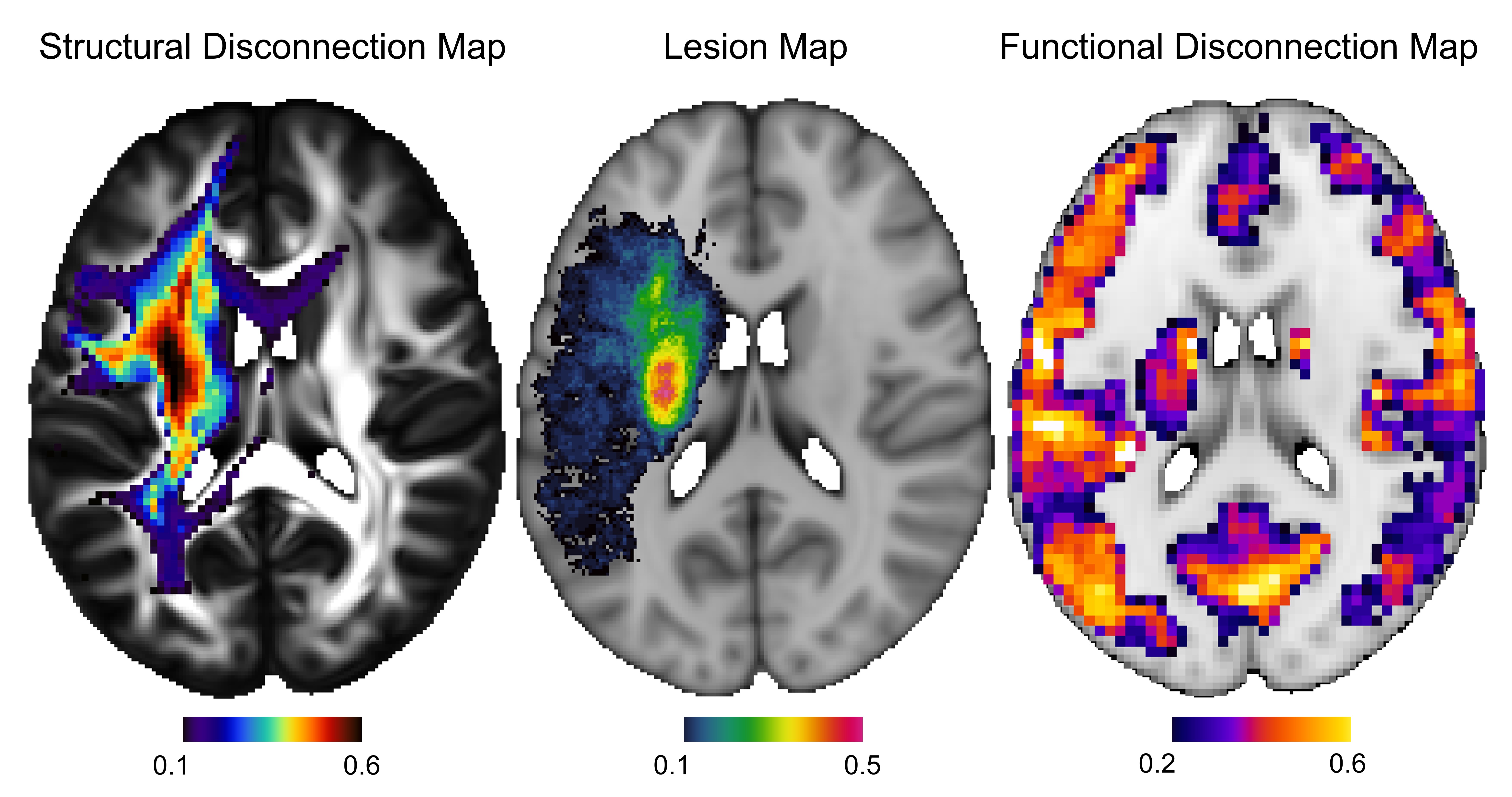
Figure 1. The averaged lesion map (middle), structural disconnections map (left) and functional disconnections map (right) for all subjects.
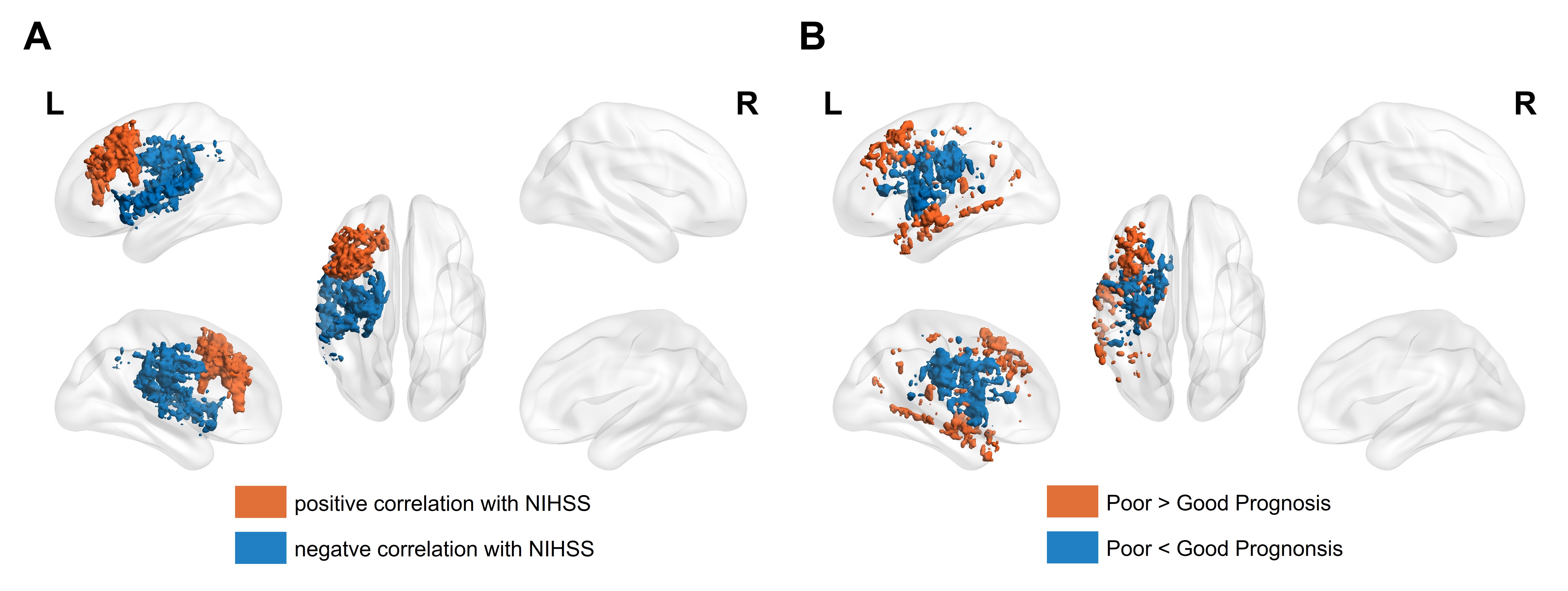
Figure 2. Significant results for lesion map. A, correlation with NIHSS; B, differences between the two groups with good and poor prognosis.
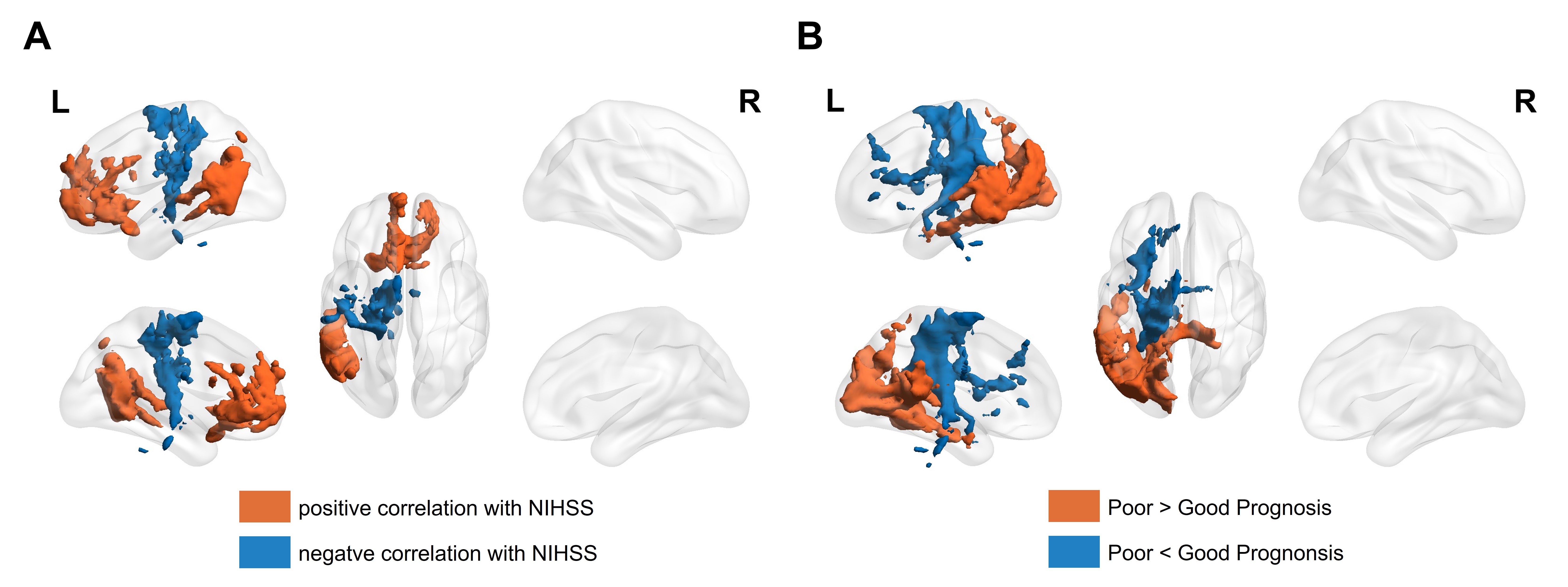
Figure 3. Significant results for structural disconnection map. A, correlation with NIHSS; B, differences between the two groups with good and poor prognosis.
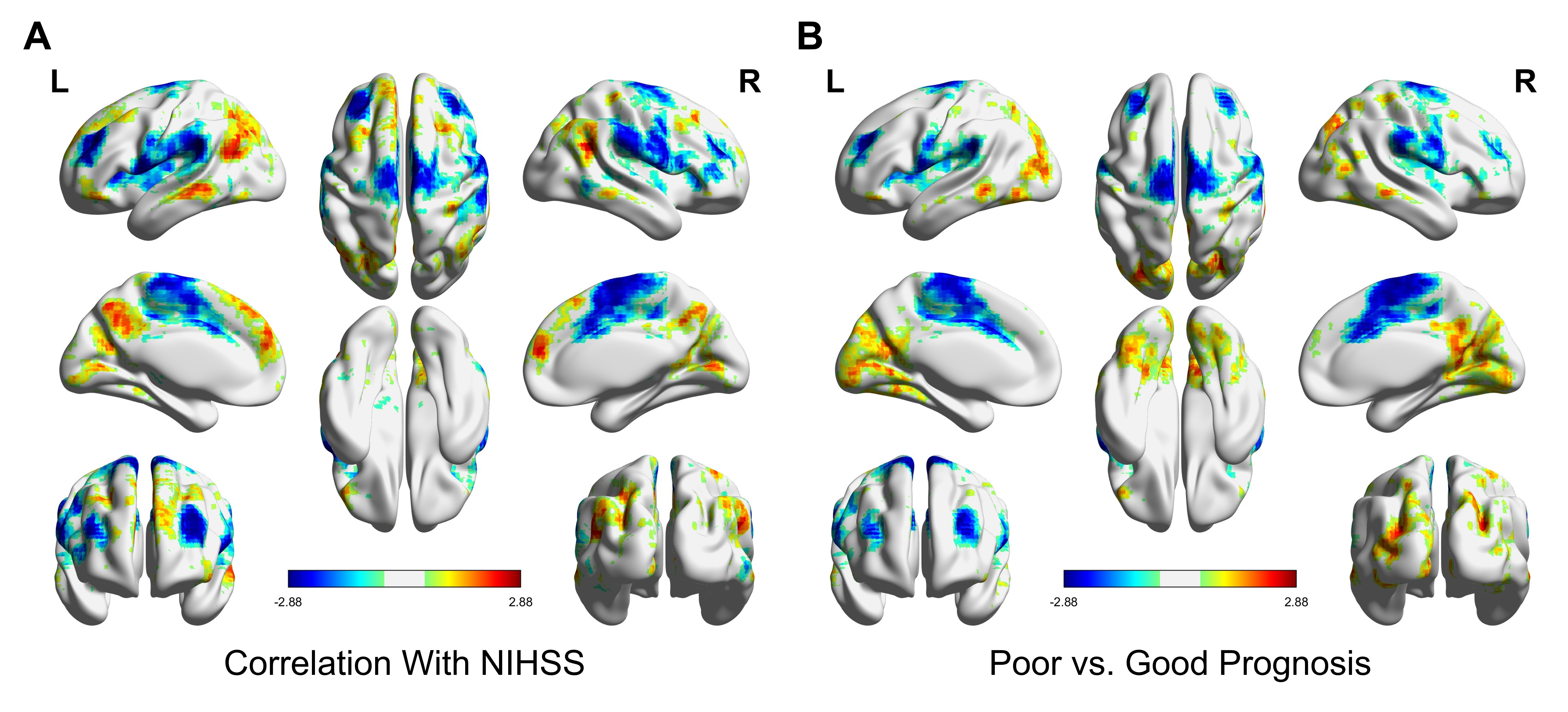
Figure 4. Significant results for functional disconnection map. A, correlation with NIHSS; B, differences between the two groups with good and poor prognosis.
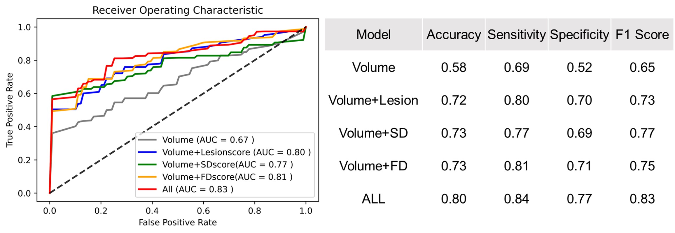
Figure 5. Results of good and poor prognosis prediction using lesion information. With lesion, structural and functional disconnection scores, the model significantly outperformed than with only volume.