0079
Prognosis prediction based on penumbra and infarct core radiomics features in patients with acute ischemic stroke1Department of Radiology, The First Affiliated Hospital of Jinan University, Guangzhou, Guangdong, China, 2Institute of Research and Clinical Innovations, Neusoft Medical Systems Co., Ltd, Shanghai, China, 3Institute of Research and Clinical Innovations, Neusoft Medical Systems Co., Ltd, Guangzhou, China, 4Institute of Research and Clinical Innovations, Neusoft Medical Systems Co., Ltd, Beijing, China, 5Neusoft Medical Systems Co. Ltd,, Shenyang, China
Synopsis
Keywords: Stroke, Radiomics
Motivation: The identification and assessment of the penumbra are crucial for making the right treatment decisions and improving clinical outcomes in acute ischemic stroke (AIS) patients.
Goal(s): To develop a radiomics model based ASL and DWI to predict outcomes of AIS patients with clinical factors.
Approach: Radiomics features were extracted from penumbra and infarct core in 151 patients with clinical parameters. Five-fold cross-validation was performed on 70% data sets, and the model performance was evaluated by an independent test cohort.
Results: The joint model with 4 radiomics features from infarct core and NHISS score yielded highest AUC of 0.802.
Impact: The combined model incorporating clinical factors and radiomics features based on infarct core and penumbra has achieved satisfactory performance in predicting the outcomes of AIS patients, which provides a non-invasive approach to optimize individualized treatment for AIS patients.
Introduction
Acute ischemic stroke (AIS) is the leading cause of death and adult disability globally1. Penumbra refers to the hypoperfused ischemic tissue surrounding the center of infarction lesions2. The aim of AIS treatment is to salvage the penumbra and to achieve the reperfusion of ischemic brain tissue. The identification and evaluation of penumbra is crucial for AIS patients to make correct treatment decision to improve clinical outcomes. Arterial spin labeling (ASL) is a non-invasive MRI sequence without either radiation exposure or intravenous administration of contrast agent3, providing a powerful tool to predict clinical outcomes for AIS patients4. In recent years, artificial intelligence has been demonstrated to have the potential to guide early stroke treatment. Radiomics is an emerging methodology that quantifies high-dimensional features from radiological images and has been applied to predict clinical outcomes5. There were few studies exploring the prediction of clinical outcomes based on penumbral radiomics features derived from ASL. To this end, we aimed to develop and validate a prediction model, in which the collective predictive performance for clinical factors, penumbra and infarct core radiomics features were evaluated; this may provide valuable evidence to develop personalized therapy during AIS management.Methods
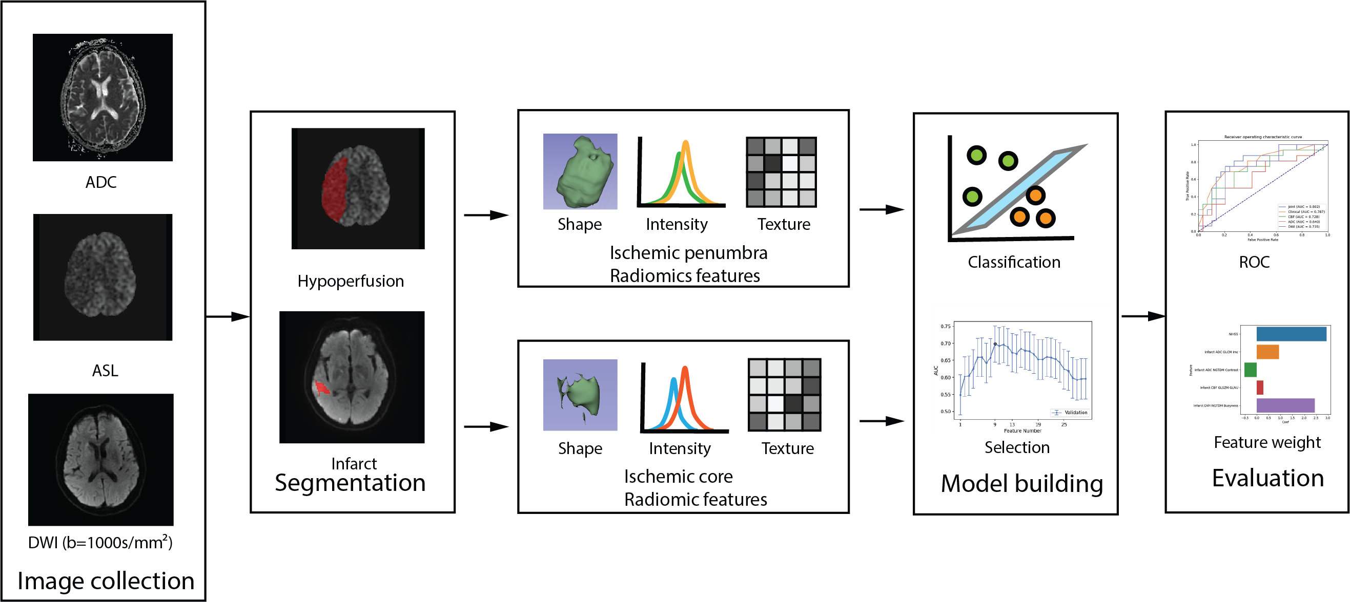
Data acquisition: We retrospectively analyzed data from 151 AIS patients (training cohort, n=105; test cohort, n=46) who underwent MRI examinations include diffusion-weighted Imaging (DWI) and ASL. The CBF maps were generated from ASL images. According to the modified Rankin Scale (mRS) at 90-day after hospital discharge, patients were divided into good (mRS ≤ 2) and poor (mRS > 2) prognosis. Penumbra and infarct segmentations: The ischemic core and hypoperfusion areas were segmented semi-automatically on the apparent diffusion coefficient (ADC) and CBF maps by applying appropriate thresholds, which were then manually corrected and segmented by a neuroradiologist (6 years of experience) who was blinded to the patients’ clinical details. Subsequently, a senior neuroradiologist with a decade of experience in neuroimaging verified the segmentation. When there was a conflict on the result, discussion was held until a final consensus was achieved. Radiomics features were extracted from DWI, ADC, and CBF images, in which a total of 214 radiomics features were extracted from ischemic core and penumbra in each single image. Machine learning models construction: Logistic regression algorithm and support vector machine (SVM) were used to build the clinical (using clinical parameters only), radiomics (using radiomics features only), and joint models (using a combination of clinical and radiomics features). The radiomics model were trained with five-fold cross validation for evaluating the performance of the prognosis model. All operations were automatically conducted on FeAture Explorer (Figure 1)6 . The Delong test was used for comparisons between the area under the curve (AUC), while the nomogram, decision curve analysis (DCA) was conducted to evaluate the clinical utility.Results
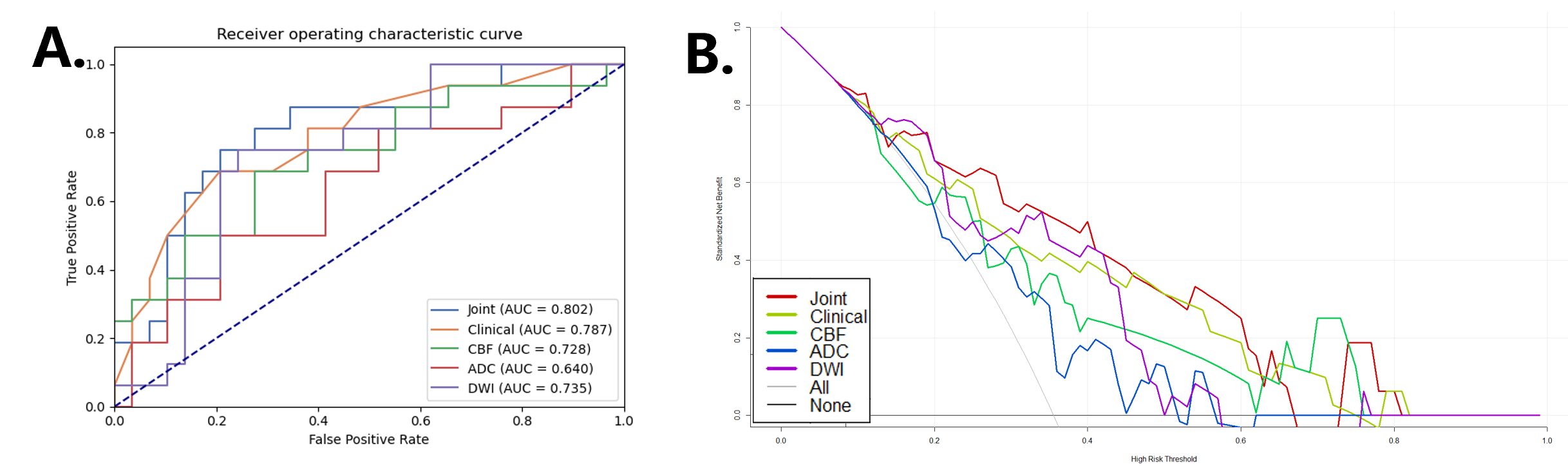
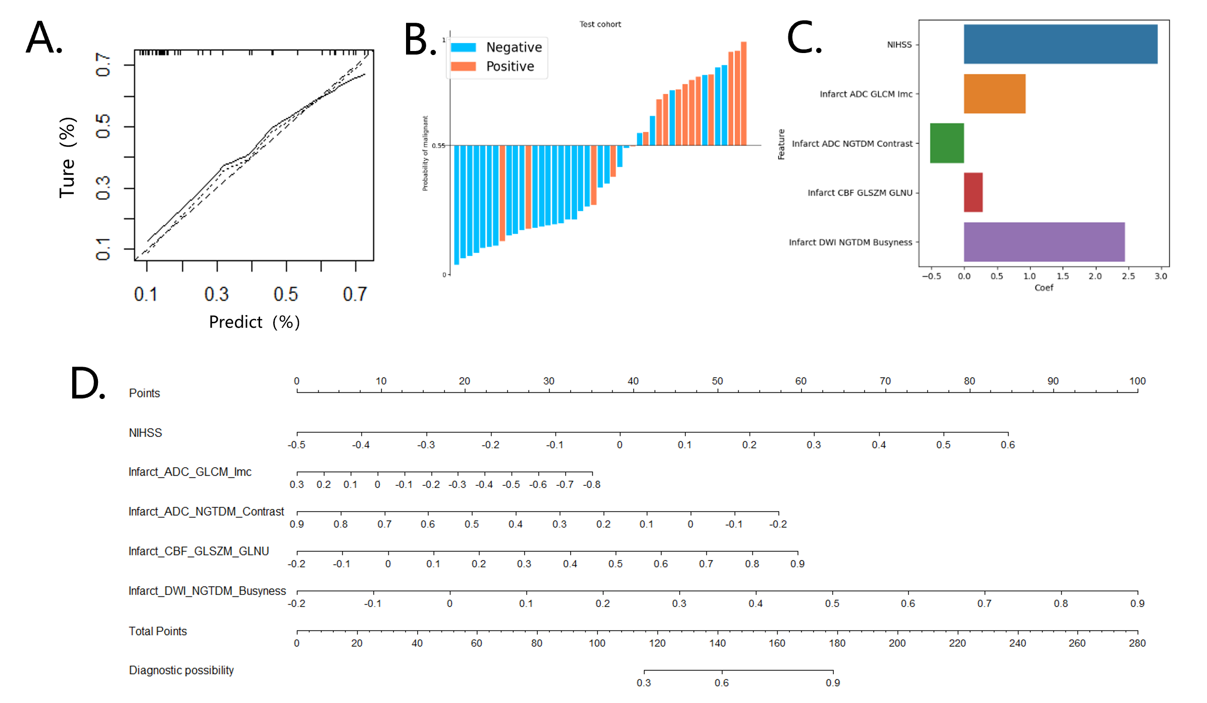
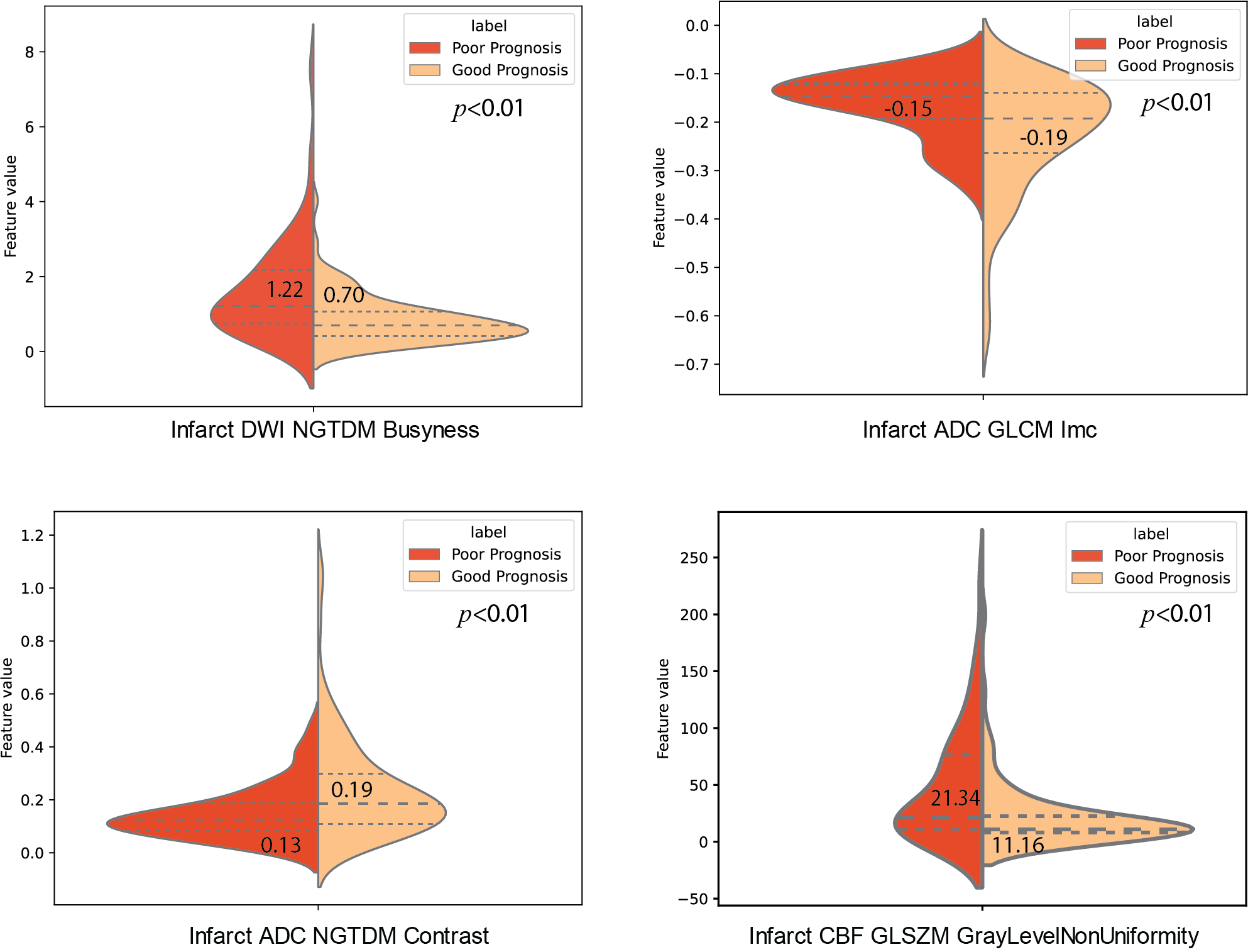
DWI model achieved highest AUC of 0.735(0.584-0.886), and the joint model integrating the fusion radiomics signature and clinical variables showed the best performance with AUC of 0.802 (0.663-0.940) in the test cohort, and the DCA indicated that the joint model added more net benefit (Figure 2 and Table 1). Calibration curve and water fall curve demonstrated the excellent performance of the joint model in the test cohort (Figures 3A & 3B). The most relevant radiomics parameters were texture features from the infarct, whereas the NIHSS score showed the most positive contribution in the joint model (Figure 3C). Furthermore, clinical validity of the proposed model was demonstrated with a nomogram (Figures 3D). Distribution of texture features also showed a significant difference between good and poor prognosis patients (Figure 4).Discussion
Previous studies have demonstrated that infarcted tissue and penumbra estimated on radiological imaging may contain additional prognostic information that could improve outcome prediction for AIS7-9, which is consistent with our findings. In this study, we developed and validated a prediction model incorporating clinical factors and radiomics features extracted from DWI and ASL images of the infarct core and penumbra, which achieved satisfactory prediction of AIS outcomes with an AUC of 0.802. The nomogram provided multi-dimensional information and achieved higher predictive accuracy than a previous study10.Conclusion
It has been demonstrated that the proposed joint model integrating clinical factors and radiomics features extracted from penumbra and infarct core performed well in predicting the outcomes of AIS patients. This could provide a noninvasive and reliable approach to assist clinicians in optimizing individualized treatment plans at the early stage of onset for patients with AIS, which may significantly improve the ultimate outcome of stroke patients.Acknowledgements
We would like to acknowledge the equal contributions of Xiaoling Wu and Jing Zhang to this work. Both authors contributed equally to the experimental design, data analysis, and manuscript preparation.References
1. Campbell BCV, Khatri P. Stroke. Lancet 2020;396(10244):129-142.
2. Ginsberg MD. Adventures in the pathophysiology of brain ischemia: penumbra, gene expression, neuroprotection: the 2002 Thomas Willis Lecture. Stroke. 2003;34(1):214-23.
3. Hernandez-Garcia L, Aramendía-Vidaurreta V, Bolar DS, et al. Recent Technical Developments in ASL: A Review of the State of the Art. Magn Reson Med 2022;88(5):2021-2042.
4. Lyu J, Duan Q, Xiao S, et al. Arterial Spin Labeling-Based MRI Estimation of Penumbral Tissue in Acute Ischemic Stroke. J Magn Reson Imaging 2023;57(4):1241-1247.
5. Chen Q, Xia T, Zhang M, Xia N, Liu J, Yang Y. Radiomics in Stroke Neuroimaging: Techniques, Applications, and Challenges. Aging Dis 2021;12(1):143-154.
6. Song Y, Zhang J, Zhang Yd, Hou Y, Yan X, et al. (2020) FeAture Explorer (FAE): A tool for developing and comparing radiomics models. PLOS ONE 15(8): e0237587.
7. Tolhuisen ML, Hoving JW, Koopman MS, et al. Outcome Prediction Based on Automatically Extracted Infarct Core Image Features in Patients with Acute Ischemic Stroke. Diagnostics (Basel). 2022;12(8):1786.
8. Jiang L, Peng M, Chen H, et al. Diffusion-weighted imaging (DWI) ischemic volume is related to FLAIR hyperintensity-DWI mismatch and functional outcome after endovascular therapy. Quant Imaging Med Surg. 2020;10(2):356-367.
9. Jiang L, Zhang C, Wang S, et al. MRI Radiomics Features From Infarction and Cerebrospinal Fluid for Prediction of Cerebral Edema After Acute Ischemic Stroke. Front Aging Neurosci. 2022;14:782036.
10. Tang TY, Jiao Y, Cui Y, et al. Penumbra-based radiomics signature as prognostic biomarkers for thrombolysis of acute ischemic stroke patients: a multicenter cohort study. J Neurol. 2020;267(5):1454-1463.
Figures