0068
Reduced GABA level in ipsilateral thalamus correlates with cognitive impairment in stroke patients1Department of Radiology, Guangzhou First People's Hospital, Guangzhou, China, 2School of Medicine, South China University of Technology, Guangzhou, China, 3Philips Healthcare, Shanghai, China, 4Philips Healthcare, Guangzhou, China
Synopsis
Keywords: Stroke, Stroke, GABA, Glx
Motivation: Neurotransmitters are involved in diseases associated with cognitive impairment.
Goal(s): Investigate the changes of the main inhibitory (gamma-aminobutyric acid, GABA) and excitatory neurotransmitters (glutamate and glutamine, Glx) for stroke patients and their correlation with cognitive impairment.
Approach: GABA and Glx were measured using Meshcher-Garwood point-resolved spectroscopy (MEGA-PRESS) sequence in 20 ischemic stroke patients.
Results: GABA to total creatine ratio (GABA/Cr) were reduced in the ipsilateral thalamus compared to the contralateral thalamus, and reduced GABA/Cr in ipsilateral thalamus was strong correlated with cognitive impairment. Thalamic GABA level could serve as a potential target for the evaluation and treatment of patients with post-stroke cognitive impairment.
Impact: The levels of GABA, glutamate and glutamine (Glx) in thalamus can be noninvasively quantified using MRS based on MEGA-PRESS technique in ischemic stroke patients. Reduced GABA level in ipsilateral thalamus was associated with cognitive impairment in ischemic patients.
Introduction
Neurotransmitter alterations play a critical role in the development of cognitive impairment. 1 In this study, we aimed to utilize magnetic resonance spectroscopy (MRS) imaging to noninvasive assessment the changes of gamma-aminobutyric acid (GABA) and glutamate/glutamine (Glx) levels in the bilateral thalamus of ischemic stroke patients. Additionally, we aimed to explore the associations between neurotransmitter levels and cognitive scores, thereby enhancing our understanding of the pathophysiological mechanisms underlying post-stroke cognitive impairment.Methods
A total of 20 ischemic stroke patients with infarcts located in one cerebral hemisphere were enrolled in the study. GABA and Glx levels in bilateral thalamus were measured by MEGA-PRESS on a 3.0T scanner (Elition, Philips Healthcare, Best, the Netherlands) with a 32-channel head coil. Detailed imaging parameters were: Voxel Size: 30×30×30 cm3; Echo time/Repetition time: 68/2000 ms; Spectral samples: 1024; Spectral Bandwidth: 2000 Hz; Edit on frequency: 1.89 ppm; Edit off frequency: 7.46 ppm; Signal average for each dynamic: 16; Dynamic: 9. Before MR scanning, all participants underwent cognitive assessments using the Mini-Mental State Examination (MMSE) and Montreal Cognitive Assessment (MoCA). The GABA to total creatine ratio (GABA/Cr), the Glx to total creatine ratio (Glx/Cr) in bilateral thalamus were calculated by Gannet toolkit and compared, their relationship with cognitive scores was analyzed as well.Results
Figure 1 demonstrates the voxel location and spectral fitting from a representative stroke patient. GABA/Cr were significant reduced in the ipsilateral thalamus compared to the contralateral thalamus in ischemic stroke patients (p = 0.002). GABA/Cr in ipsilateral thalamus were associated with cognitive scores of MMSE (R = 0.641, p = 0.002) and MoCA (R = 0.733, p < 0.001). However, no significant alterations were observed in Glx/Cr levels (p = 0.219) and they were not correlated with cognitive scores (MMSE: R = -0.002, p = 0.994; MoCA: R = 0.019, p = 0.937) (Figure 2 and 3).Discussion
In patients with ischemic stroke, we observed a decrease in GABA levels in the ipsilateral thalamus compared to the contralateral thalamus, while Glx levels did not show significant differences between the bilateral thalamus. Furthermore, we found a correlation between GABA levels in the ipsilateral thalamus and cognitive impairment, whereas no such correlation was observed for Glx.Given that the thalamus serves as an information processing relay station and is rich in neurotransmitters, it plays a crucial role in cognitive function regulation. 2 There may be the following reasons for the decreased GABA in the ipsilateral thalamus after stroke: 1) ischemia-related damage or dysfunction of GABAergic neurons in the ipsilateral corticothalamic circuit may contribute to this reduction; 2) the alterations in glial cells due to damage or proliferation could affect the release, uptake, and metabolism of GABA; 3) neuroinflammation and oxidative stress due to ischemia may lead to a reduced neurotransmitters in the ipsilateral thalamus. 3,4 Furthermore, the positive correlation between GABA and cognitive function in the ipsilateral thalamus implies that the changes of GABAergic neurotransmission in thalamus are closely associated with cognitive impairment after stroke. Thalamus and GABA level could serve as potential targets for the assessment of post-stroke cognitive impairment.
However, our study did not find any difference in Glx levels between the bilateral thalamus, and there was no association between ipsilateral Glx levels and cognitive function. It's important to note that due to methodological limitations, the glutamate and glutamine are mixed as a single component (Glx) for analysis, so opposite changes in the two metabolites may result in unnoticeable changes of Glx. 5 Previous study experienced the similar challenges. 6 More advanced technology is needed to solve this mystery.
Conclusion
Our study demonstrates that ischemic stroke can lead to decreased GABA in the ipsilateral thalamus, and the decreased GABA level is associated with cognitive impairment. These findings suggest that thalamic GABA levels have the potential to serve as a potential metabolic marker for the study of the pathophysiological mechanism of cognitive impairment after stroke.Acknowledgements
No acknowledgement found.References
1. Isaacson JS, Scanziani M. How inhibition shapes cortical activity. NEURON 2011;72(2):231-243.2.Pinault D. Dysfunctional thalamus-related networks in schizophrenia. Schizophr Bull 2011;37(2):238-243.
3. Kleerekooper I, Herbert MK, Kuiperij HB, et al. CSF levels of glutamine synthetase and GFAP to explore astrocytic damage in seronegative NMOSD. J Neurol Neurosurg Psychiatry 2020;91(6):605-611.
4. Radak D, Resanovic I, Isenovic ER. Link between oxidative stress and acute brain ischemia. ANGIOLOGY 2014;65(8):667-676.
5. Sanaei NF, Anton A, Michou E, Jung J, Parkes LM, Williams SR. Quantification of GABA, glutamate and glutamine in a single measurement at 3 T using GABA-edited MEGA-PRESS. NMR BIOMED 2018;31(1).
6. Yang Y, Rui Q, Han S, et al. Reduced GABA levels in the medial prefrontal cortex are associated with cognitive impairment in patients with NMOSD. Mult Scler Relat Disord 2022;58:103496.
Figures
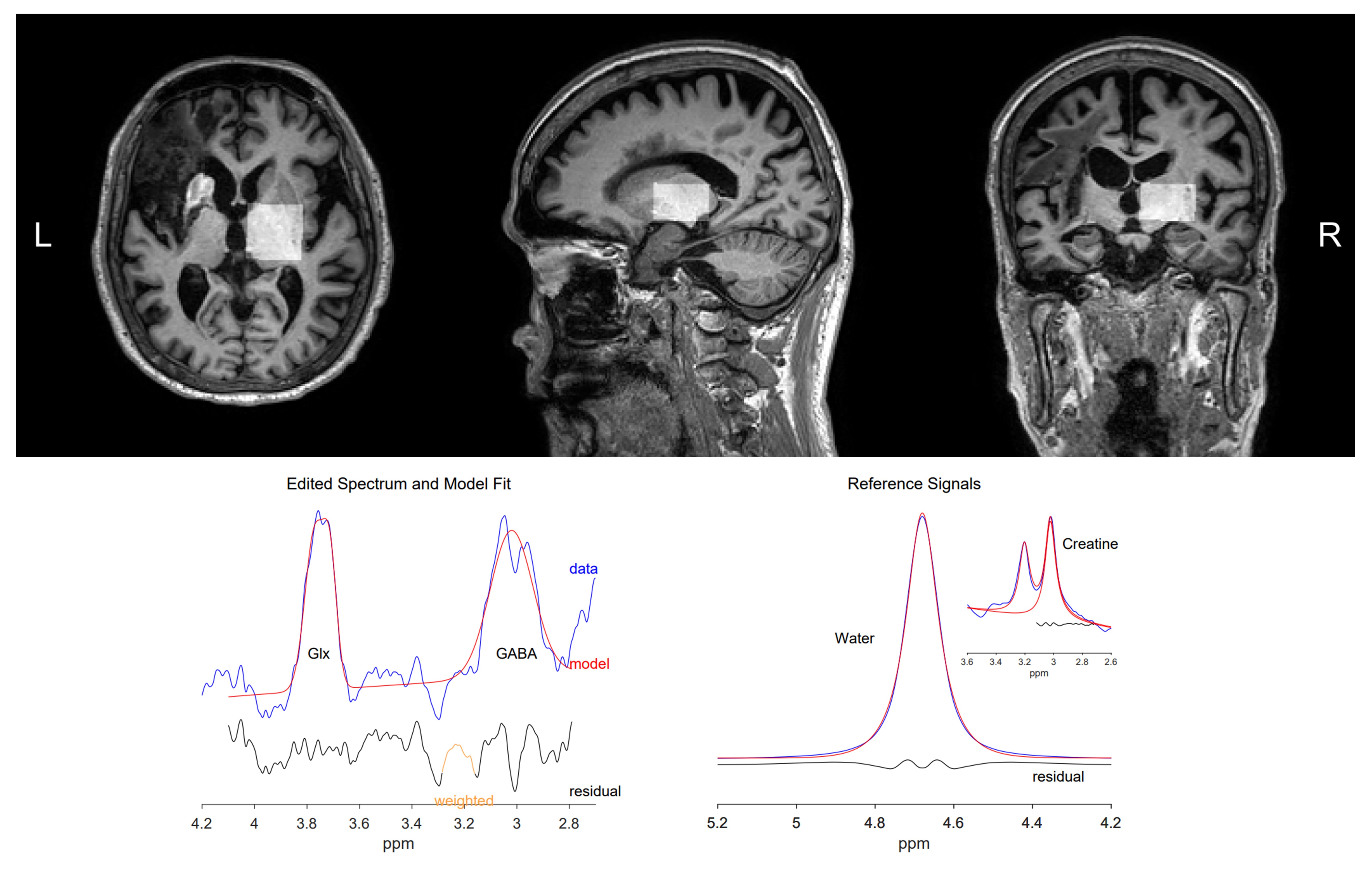
Figure 1. The voxel location and spectral fitting using Gannet toolkit from a representative stroke patient.
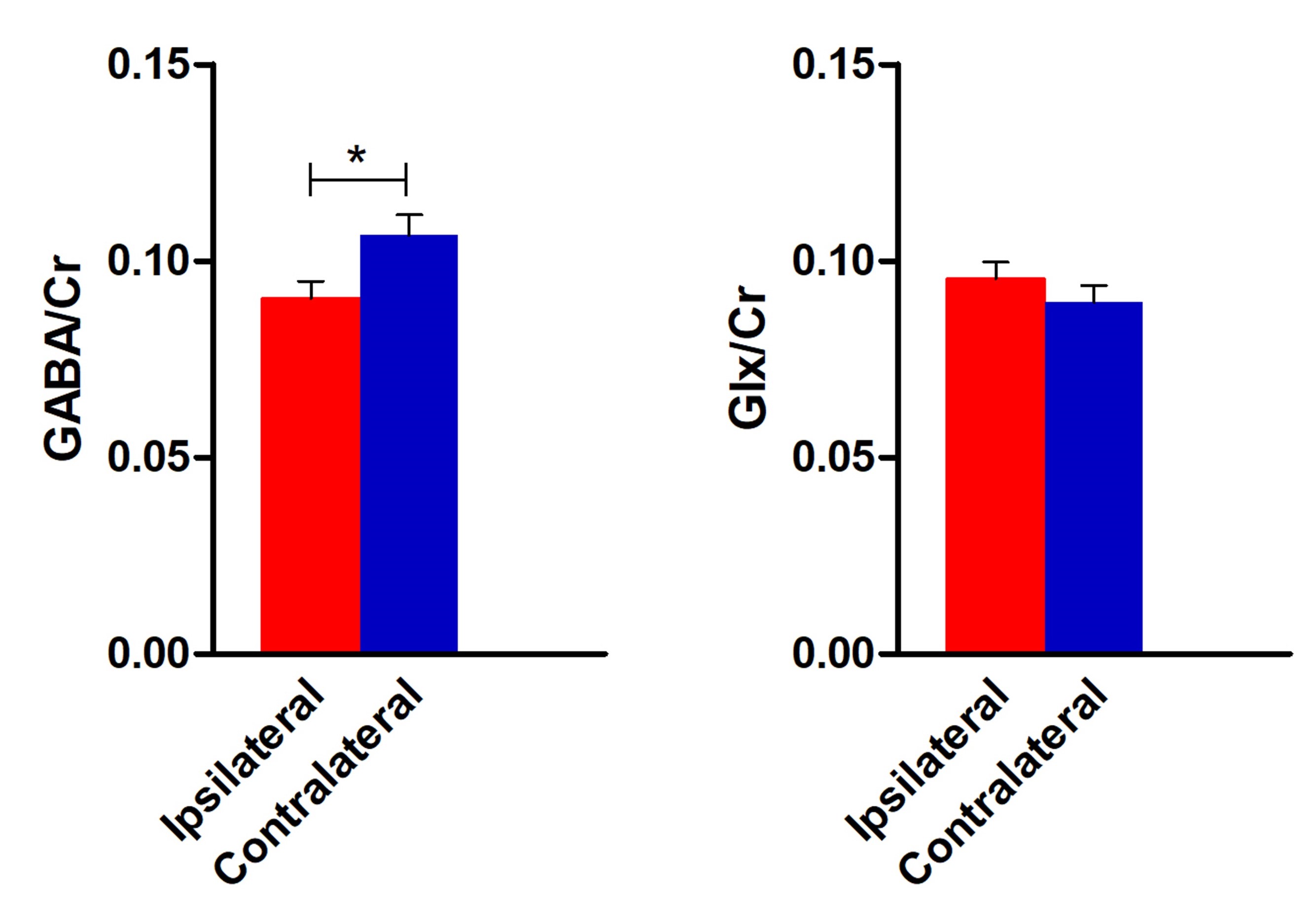
Figure 2. Comparison of GABA and Glx levels between ipsilateral and contralateral thalamus.
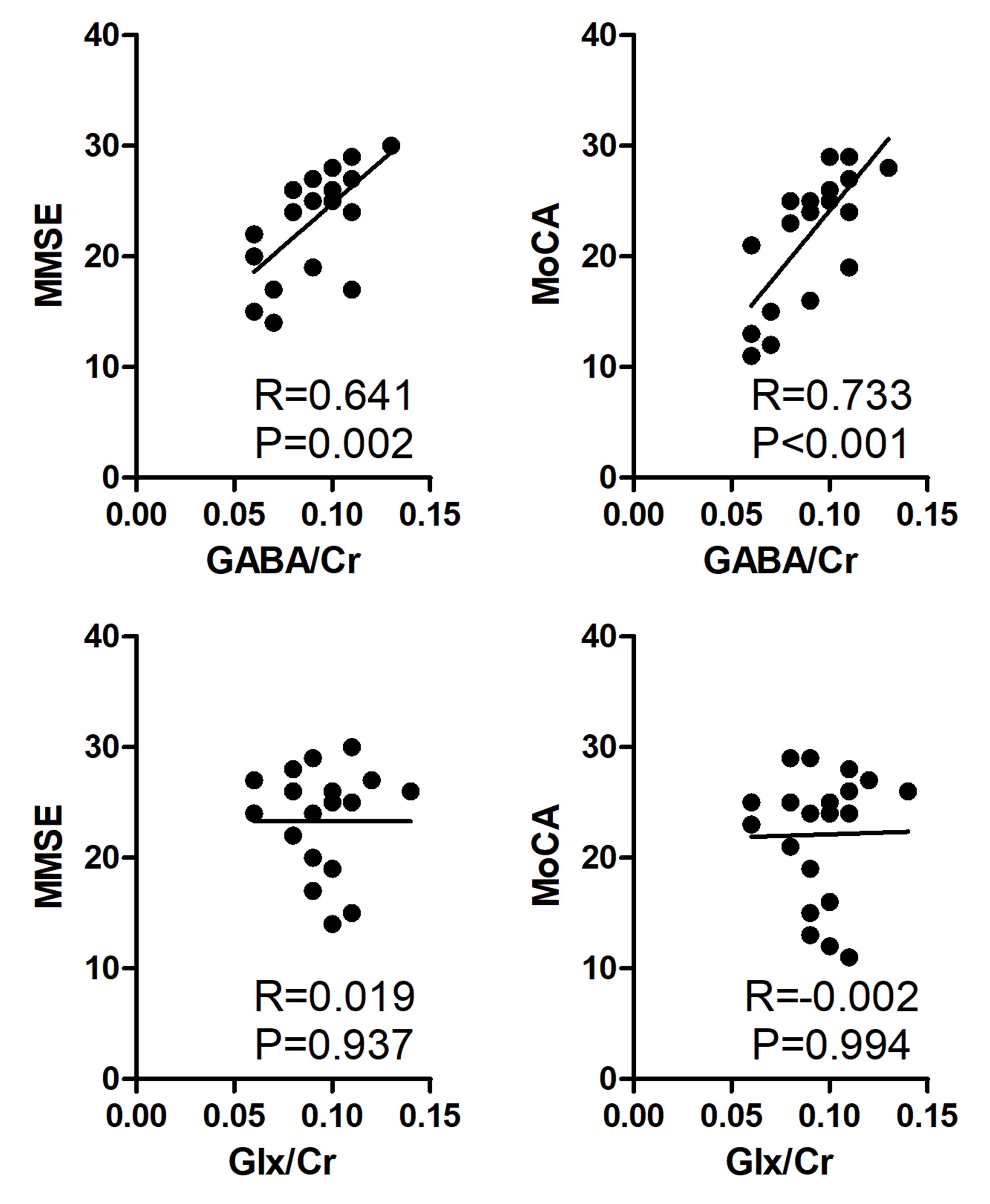
Figure 3. Correlation between cognitive scores and GABA and Glx levels.