0066
High resolution free-breathing respiratory-resolved volumetric lung imaging at 0.55T using stack-of-spiral out-in bSSFP1Electrical and Computer Engineering, University of Southern California, Los Angeles, CA, United States, 2Biomedical Engineering, University of Southern California, Los Angeles, CA, United States
Synopsis
Keywords: Lung, Lung, Low-Field MRI, Data Sampling and Reconstruction, Non-Cartesian Trajectory
Motivation: High resolution free-breathing structural lung imaging at 0.55T has been demonstrated using bSSFP half-radial dual-echo imaging with constrained reconstruction within ~10-min scan time.
Goal(s): To develop faster high-resolution free-breathing lung imaging using spiral sampling.
Approach: We employ a stack-of-spirals out-in trajectory with constrained reconstruction along with pilot-tone based respiratory navigation.
Results: Structural lung imaging is demonstrated with 2mm isotropic resolution, 5-7 respiratory states, and 5-7 min scan time. 3D ventilation maps are demonstrated, showing -3.1% ~ 70.2% lung capacity during normal and deep breathing.
Impact: Free-breathing SoSOi can provide simultaneous structural and functional lung imaging at 0.55T within a 5-min scan, with improved sampling efficiency and lower undersampling factor compared to bSTAR. This has implications for the evaluation of lung function and chronic lung diseases.
INTRODUCTION
Lung MRI can provide structural and functional information for the screening, diagnosis, and longitudinal assessment of lung diseases such as cancer, emphysema, and chronic obstructive pulmonary disease. Lung MRI is also technically demanding. At conventional field strengths (≥1.5T), the major challenge is very short T2* (<1ms), due to the susceptibility difference between air (alveoli) and tissue (parenchyma/blood), resulting in low intrinsic SNR1.High-performance 0.55T systems provide improved field homogeneity and prolonged T2*2, resulting in significantly higher lung parenchyma signal3,4. Recently, submillimeter bSTAR at 0.55T has been developed to achieve a free-breathing scan with more than 10min5,6. A 3D stack-of-spirals out-in (SoSOi) bSSFP7 has been proposed for breath-hold lung imaging with improved sampling efficiency but relatively coarse spatial resolution. Here, we develop a free-breathing SoSOi bSSFP sequence for high resolution respiratory-resolved lung volumes that simultaneously provide structural and functional information, requiring only 5 min.
METHODS
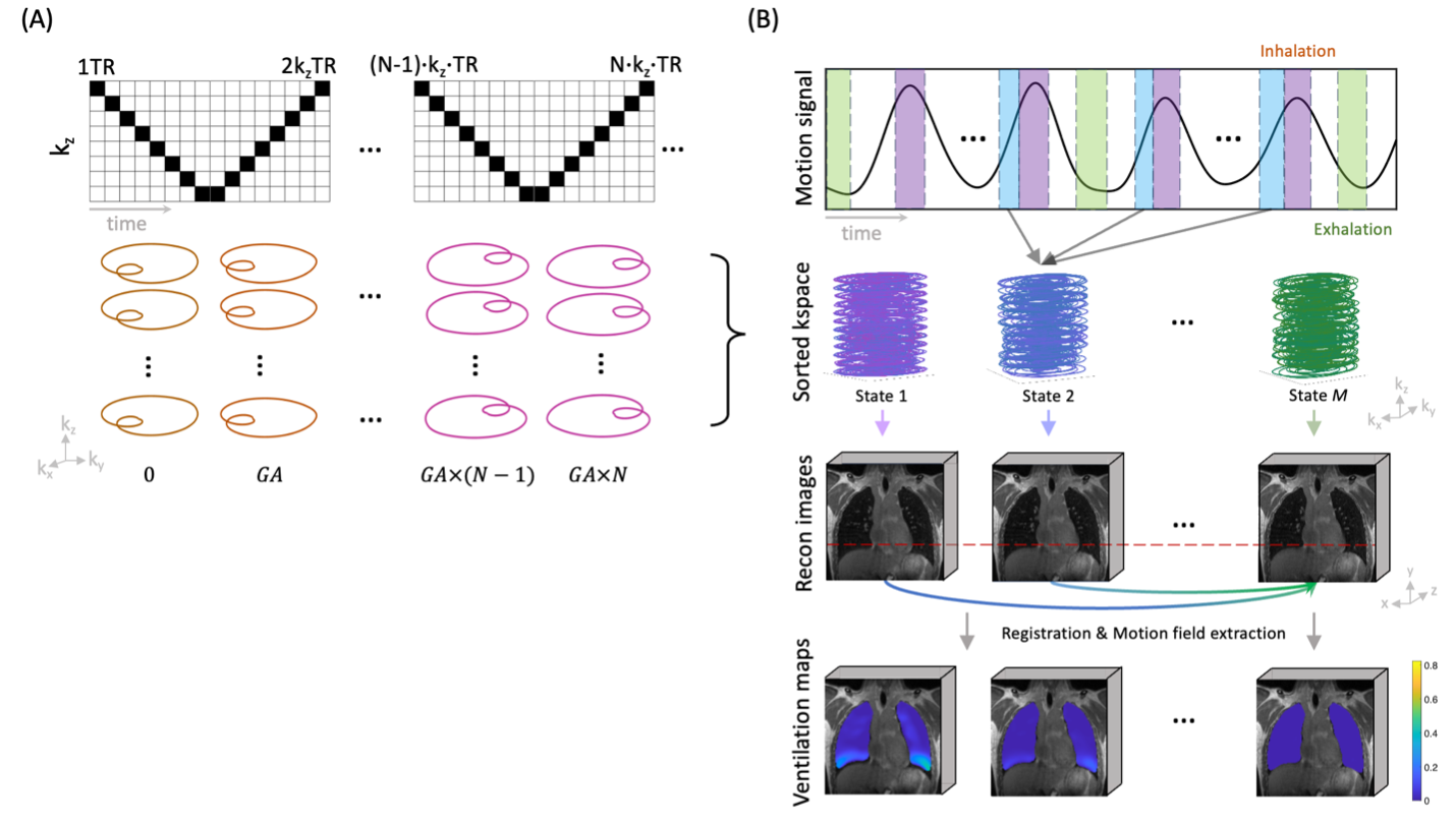
Pulse sequence: Figure 1 illustrates the proposed free-breathing SoSOi pipeline. Imaging was performed in the supine position during free-breathing with a coronal prescription. A non-selective hard pulse (100 µs, FA = 25°) was used and partition encoding is placed in the anterior-posterior direction. A spiral out-in readout creates a dual echo. A “ping pong” kz order was used while a fixed kx-ky spiral staring angle. This lasts 120 TRs and the takeoff angle was increased by a tiny golden angle8 (GA = 23.6281°) for the next pass. Imaging parameters were: FOV=384×384×240mm3, 2mm isotropic resolution, and TE1/TE2/TR = 0.52/2.3/2.9 ms. The proposed sequence was implemented with the open-source Pulseq framework9.Respiratory signal extraction: Pilot Tone (PT) 10 was used to estimate respiratory phases. A sinusoid signal was generated with a general-purpose signal generator (AFG3252, Tektronix) and transmitted through a dipole antenna. A respiratory navigator was extracted using the pipeline11. Residual artifacts in k-space due to the increased PT amplitude was removed with EDITER 12,13. The PT respiratory signal was then processed with a bandpass filter (0.1 to 0.5Hz) and used for data binning.
Image reconstruction: All respiratory phases were simultaneously reconstructed using a constrained reconstruction with a circular finite difference along the respiratory dimension and spatial total variation. Regularization parameters were manually tuned. Image reconstructions were performed with GIRF-predicted trajectories14 using the BART toolbox15.
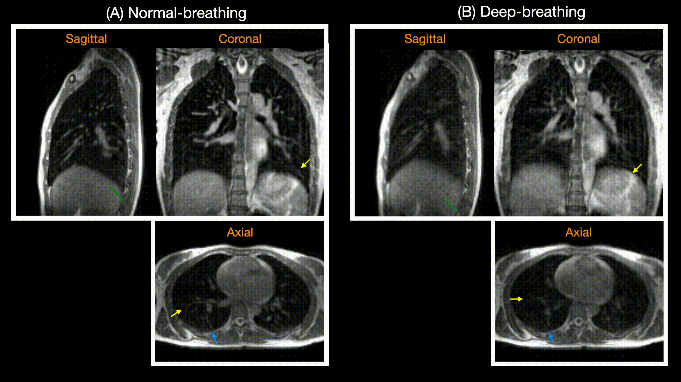
In-vivo experiments: Experiments were performed using a whole body 0.55T system (prototype MAGNETOM Aera, Siemens Healthineers, Erlangen, Germany) equipped with high-performance shielded gradients (45 mT/m amplitude, 200 T/m/s slew rate). Data were collected using a 6-channel body array (anterior) and 6 elements from a table-integrated 18-channel spine array (posterior). 1 healthy volunteer (28F) was scanned. The volunteer was asked to perform normal breathing (4:59min) and deep breathing (7:13min) during the continuous acquisition.
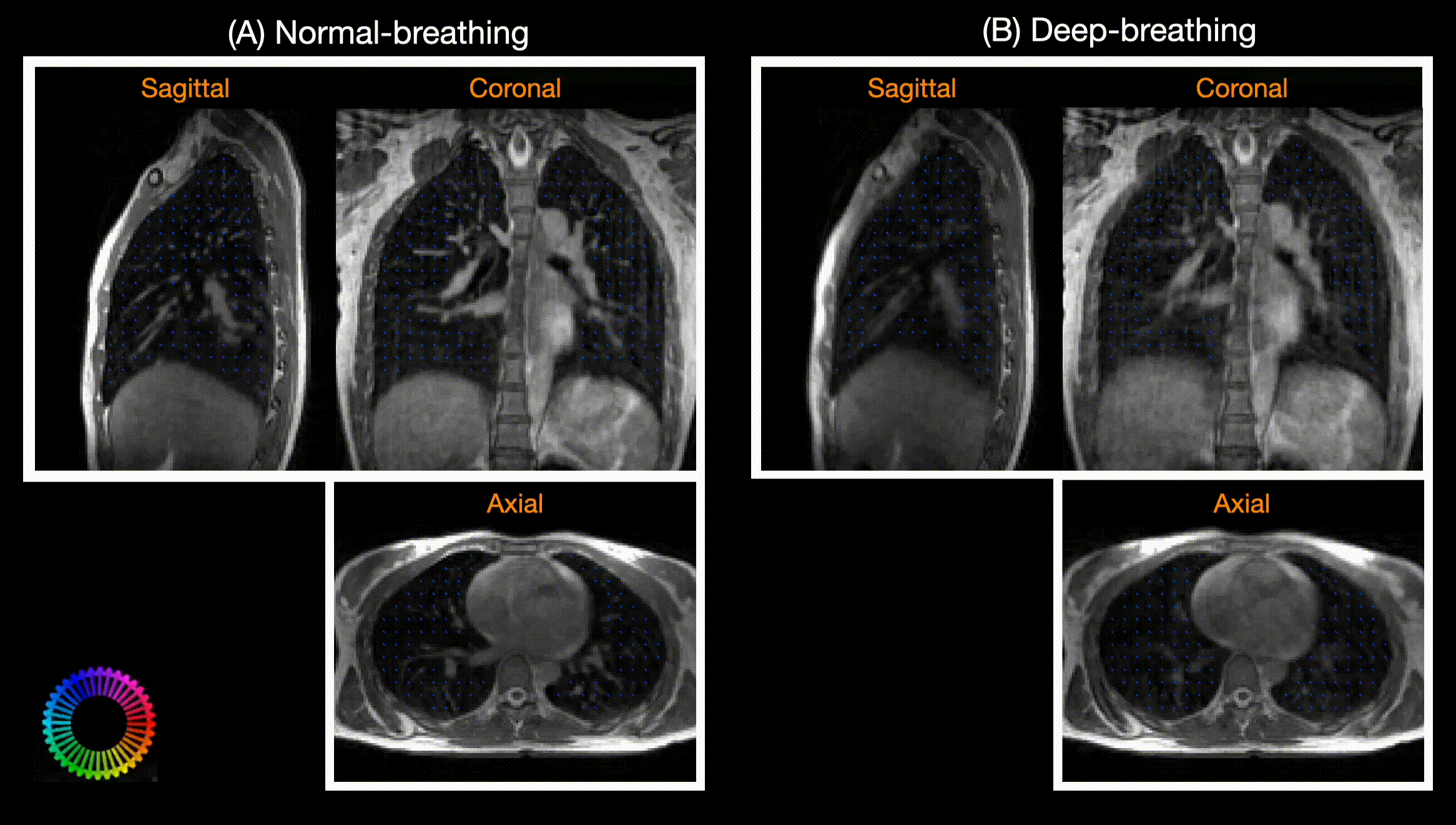
Ventilation analysis: 3D segmentation and registration were performed using an in-house semi-automatic segmentation method and the ANTS toolbox16, respectively. Deformation maps were calculated using the Jacobian determinant of the estimated deformation fields from the registration step17,18.
RESULTS
Figure 2 shows representative respiratory phase-resolved images during normal and deep breathing acquired with 4:59min and 7:13min scans, respectively. Vessel structures and diaphragm movements are clearly visualized in both breathing patterns.Figure 3 shows the corresponding 3D ventilation maps for normal and deep breathing, showing -3.1% ~ 26.7% and 0% ~ 70.2% total lung capacity, respectively. In both breathing patterns, the inferior lung experiences higher volume change in sagittal and coronal views, which we infer as proportional to ventilation and gas exchange. In deep breathing, the motion fields in axial plane show expansion and contraction during inhale and exhale respectively, and reflects the hysteresis of respiration.
DISCUSSION
This work demonstrates an efficient lung imaging that simultaneously provides structural and functional information within a 5-7 min free-breathing scan, utilizing efficient 3D sampling and a widely accessible processing pipeline. Due to its high scan efficiency, the proposed method can be augmented with magnetization preparation to create tissue contrasts that would be beneficial for lung cancer screening and nodule characterization.Future work includes jointly optimizing kz-t sampling and the constrained reconstruction. Aliasing artifacts from both arms were observed and could be eliminated with arms-up positions. We anticipate that additional fine tuning of regularization parameters improves the vessel delineation and sharpness of the liver diaphragm during deep breathing.
CONCLUSION
We demonstrate an efficient free-breathing technique for 3D high resolution lung imaging at 0.55T with SoSOi bSSFP acquisition. Isotropic 2mm 3D lung imaging with 5 respiratory phases providing sufficient parenchyma signals and detailed vessel structures, along with ventilation maps, can be achieved within a 5min scan.Acknowledgements
We acknowledge grant support from the National Science Foundation (#1828736) and researchsupport from Siemens Healthineers. We thank Ye Tian for helpful discussions.References
1. Yu J, Xue Y, Song HK. Comparison of lung T2* during free-breathing at 1.5 T and 3.0 T with ultrashort echo time imaging. Magn Reson Med. 2011;66(1):248-254. doi:10.1002/mrm.228292.
2. Li B, Lee NG, Cui SX, Nayak KS. Lung parenchyma transverse relaxation rates at 0.55 T. Magn Reson Med. 2023;89(4):1522-1530. doi:10.1002/mrm.295413.
3. Campbell-Washburn AE, Malayeri AA, Jones EC, et al. T2-weighted lung imaging using a 0.55-t mri system. Radiol Cardiothorac Imaging. 2021;3(3). doi:10.1148/ryct.20212006114.
4. Campbell-Washburn AE, Ramasawmy R, Restivo MC, et al. Opportunities in interventional and diagnostic imaging by using high-performance low-field-strength MRI. Radiology. 2019;293(2):384-393. doi:10.1148/radiol.20191904525.
5. Bauman G, Lee NG, Tian Y, Bieri O, Nayak KS. Submillimeter lung MRI at 0.55 T using balanced steady-state free precession with half-radial dual-echo readout (bSTAR). Magn Reson Med. 2023. doi:10.1002/mrm.297576.
6. Lee NG, Bauman G, Bieri O, Nayak KS. Reproducibility of the bSTAR sequence and open-source implementation. In: Proc. Intl. Soc. Mag. Reson. Med. 31. Vol 84. John Wiley and Sons Inc; 2023:0397. doi:10.1002/mrm.281197.
7. Tian Y, Lee NG, Zhao Z, Nayak KS. Rapid 3D lung imaging with bSSFP stack-of-spiral out-in (SoSoi) sampling at 0.55T. In: Proceedings of the 31st Annual Meeting of ISMRM. ; 2023:1409.8.
8. Wundrak S, Paul J, Ulrici J, et al. Golden ratio sparse MRI using tiny golden angles. Magn Reson Med. 2016;75(6):2372-2378. doi:10.1002/mrm.258319.
9. Layton KJ, Kroboth S, Jia F, et al. Pulseq: A rapid and hardware-independent pulse sequence prototyping framework. Magn Reson Med. 2017;77(4):1544-1552. doi:10.1002/mrm.2623510.
10. Vahle T, Bacher M, Rigie D, et al. Respiratory Motion Detection and Correction for MR Using the Pilot Tone: Applications for MR and Simultaneous PET/MR Examinations. Invest Radiol. 2020;55(3):153-159. doi:10.1097/RLI.000000000000061912.
11. Falcão MBL, Di Sopra L, Ma L, et al. Pilot tone navigation for respiratory and cardiac motion-resolved free-running 5D flow MRI. Magn Reson Med. 2022;87(2):718-732. doi:10.1002/mrm.2902313.
12. Srinivas SA, Cauley SF, Stockmann JP, et al. External Dynamic InTerference Estimation and Removal (EDITER) for low field MRI. Magn Reson Med. 2022;87(2):614-628. doi:10.1002/mrm.2899214.
13. Tasdelen B, Yagiz Ecrin, Ramasawmy Rajiv, et al. Assessment and mitigation of EMI from in-room equipment in the setting of interventional 0.55T MRI. In: Proceedings of the 31st Annual Meeting of ISMRM. ; 2023:4428.
14. Duyn JH, Yang Y, Frank JA, Van Der Veen JW. Simple Correction Method for k-Space Trajectory Deviations in MRI The new method measures the actual k-space trajectories. Journal of magnetic resonance. 1998;132:150-153. doi:10.1006/jmre.1998.139611.
15. Uecker M, Ong F, Tamir JI, et al. Berkeley Advanced Reconstruction Toolbox. doi:10.5281/zenodo.1249516.
16. Avants BB, Tustison NJ, Song G, Cook PA, Klein A, Gee JC. A reproducible evaluation of ANTs similarity metric performance in brain image registration. Neuroimage. 2011;54(3):2033-2044. doi:10.1016/j.neuroimage.2010.09.02517.
17. Castillo R, Castillo E, Martinez J, Guerrero T. Ventilation from four-dimensional computed tomography: Density versus Jacobian methods. Phys Med Biol. 2010;55(16):4661-4685. doi:10.1088/0031-9155/55/16/00418.
18. Tan F, Zhu X, Chan M, et al. Motion-compensated low-rank reconstruction for simultaneous structural and functional UTE lung MRI. Magn Reson Med. 2023;90(3):1101-1113. doi:10.1002/mrm.29703
Figures