0055
An optimised framework for relating microstructural MRI to multi-stain histology metrics in the mouse brain1Wellcome Centre for Integrative Neuroimaging, FMRIB, Nuffield Department of Clinical Neurosciences, University of Oxford, Oxford, United Kingdom, 2SJTU-Ruijin-UIH Institute for Medical Imaging Technology, Ruijin Hospital, Shanghai Jiao Tong University School of Medicine, Shanghai, China, 3Athinoula A. Martinos Centre for Biomedical Imaging, Harvard University, Cambridge, MA, United States, 4British Heart Foundation Centre of Research Excellence, King's College London, London, UK, London, United Kingdom, 5Mouse Imaging Centre, Hospital for Sick Children, Toronto, ON, Canada
Synopsis
Keywords: Biology, Models, Methods, Microstructure, Validation
Motivation: Voxel-wise MRI-histology comparisons routinely rely on manual segmentations of ROIs and subjective quantitative histological metrics, an error-prone and labour-intensive process.
Goal(s): We developed an automated framework for investigating relationships between multiple MRI metrics and immunostains.
Approach: We used MRI and histology protocols optimised for ex-vivo mouse brains. We co-registered this data and derived quantitative histological metrics. We conducted voxel-wise correlations in grey and white matter between MRI (diffusion, R2* and susceptibility) and immunohistochemistry (myelin, neurofilament and extracellular matrix proteins).
Results: Our framework successfully recapitulated known relationships for myelin and neurofilaments and, interestingly, demonstrated new relationships between MRI metrics and extracellular matrix protein stains.
Impact: Our optimised framework combines openly-shared software and MRI-histology protocols, addressing current challenges, such as obtaining high-quality histology data, MRI-to-histology registration and automatic extraction of quantitative histological metrics. This can benefit future MRI-histology studies in mouse brains prepared for ex-vivo MRI.
Introduction
Linking microstructural MRI to quantitative histology metrics can help pinpoint the biological drivers of MRI signal change. Existing techniques require considerable expertise, are laborious and, due to a lack of MRI-histology co-registration, do not fully take advantage of the complementary benefits of the two imaging modalities.To overcome this challenge, we need accurately co-registered MRI and histology data from the same brain, automatically-derived quantitative histological metrics and automated ROI delineations. These tools allow us to capture meaningful biological variability across tissue and leverage the statistical potential of spatially-resolved measurements.
We present a general framework to acquire multi-modal MRI and multi-stain histological data in the same mouse brain and relate the extracted metrics from both modalities at the MRI voxel level. We achieved this by leveraging a histology protocol we recently developed1. This protocol was designed to account for the long storage times of mouse brains prepared for ex-vivo MRI, which can lead to tears and folds in the obtained brain slices and inconsistent immunostaining quality.
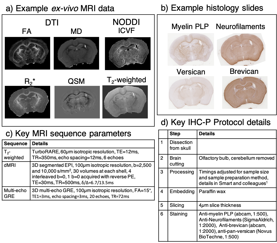
Our MRI metrics and histological stains capture a broad range of biological properties of mouse brain tissue. The ex-vivo MRI data includes T2-weighted structurals, two-shell dMRI, and multi-echo GRE MRI data. We used immunohistochemistry paraffin to acquire myelin proteolipid protein (PLP) and neurofilament (NF) stains (targetting myelin and neurites, respectively). We also stained for the extracellular matrix proteins versican and brevican, which act as remyelination2 and neurite growth3 inhibitors following neural injury.
We demonstrate the successful application of our framework, which facilitates sophisticated, automated MRI-histology comparisons with higher throughput than can be achieved with current manually-intensive protocols. To evaluate it, we conducted simple correlations between microstructural MRI metrics and all stains in grey matter (GM) and white matter (WM) separately.
Methods
Wild-type adult mouse brains were perfusion-fixed (4% PFA, Gd-based contrast agent) and scanned in skull on a Bruker 7T scanner (BioSpec 70/20 USR, receive-only cryoprobe) using an ex-vivo multi-modal MRI protocol4,5 (Figure 1c). The dMRI and multi-echo GRE data were processed using custom-built pipelines producing DTI (FA, MD), NODDI (ICVF), R2* and QSM maps6,7 (Figure 1a).The brain samples were subsequently processed, embedded and sliced (coronal sections, 4µm thickness). Neighbouring slides were stained using our optimised protocols1 (Figures 1b and d). The slides were digitised (0.25µm resolution).
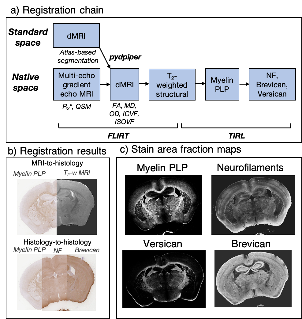
We estimated stain area fraction (SAF) maps from all stains using an automated pipeline8 and a colour matrix (used for stain separation during colour deconvolution) that was either fixed for all slides (PLP stain) or estimated for each slide (other stains) (Figure 2c). SAF is the number of stained pixels within a given patch (8x8µm2) and should be considered a semi-quantitative estimate of the immuno-targeted protein.
The registration chain (Figure 2a) for all acquired data uses FLIRT9,10 and TIRL11. The dMRI data were registered to a cohort-specific standard space12, where atlas-based segmentations13 of the major WM tracts were obtained and manually-refined. For GM, an eroded isocortex mask was obtained via non-linear registration to the Allen Mouse Brain CCFv3 atlas14.
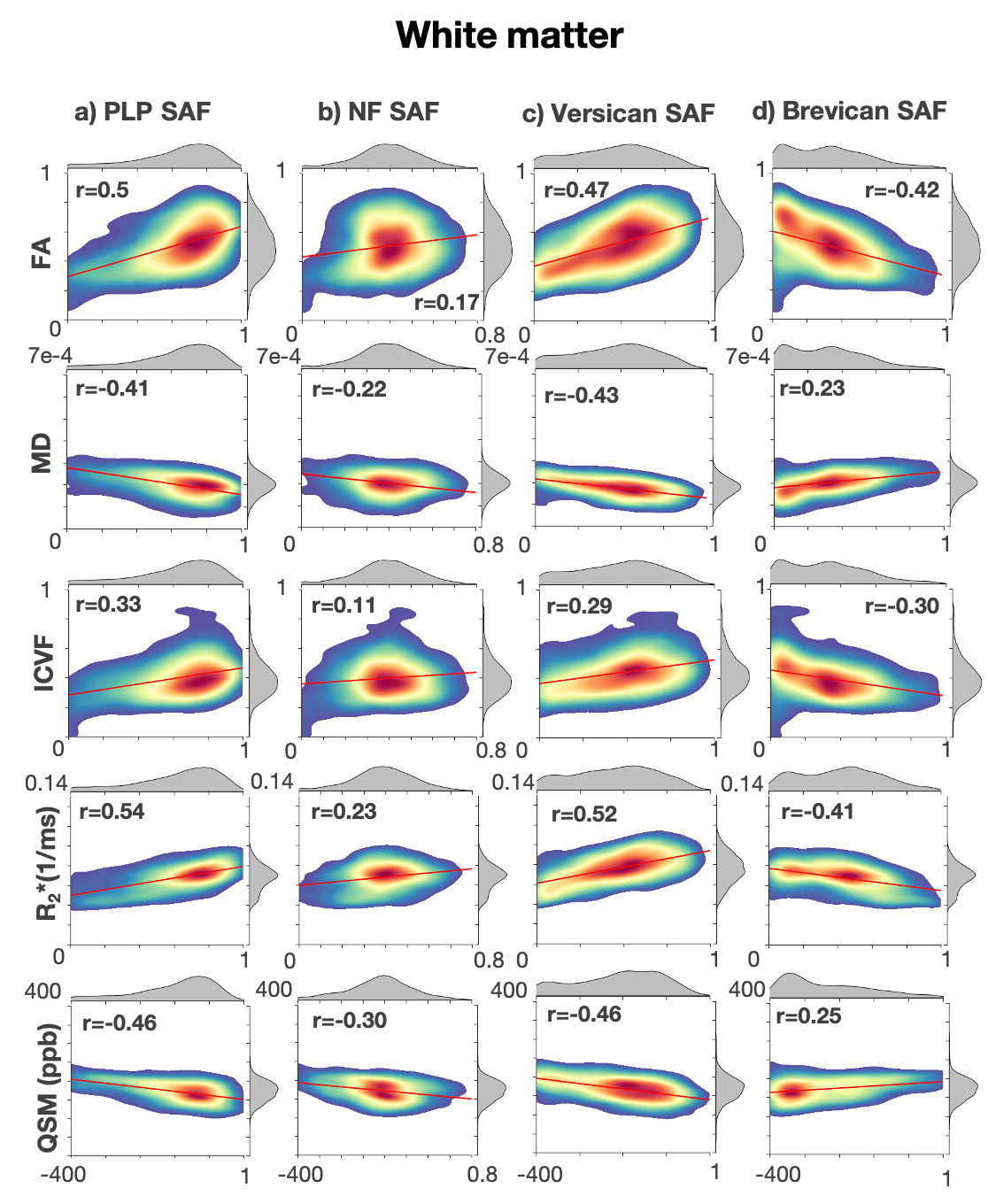
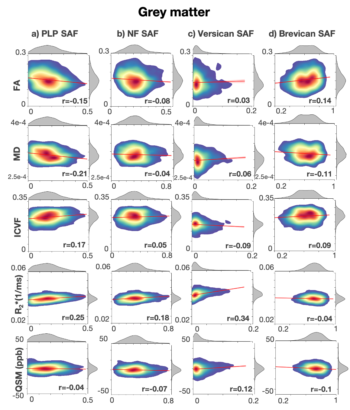
To test our acquisition and automated analysis pipeline, MRI metrics and SAF values of all stains were correlated at the MRI voxel level across the major WM tracts in the mouse brain and across GM.
Results
Our stained slides demonstrated good quality (Figure 1b), minimal artifacts, and registered successfully to MRI (Figure 2b).We observed strong correlations of PLP with all five MRI metrics in WM (Figure 3a) but not GM (Figure 4a). Similar trends are observed for NF, although the correlation strength is reduced (Figure 3b and Figure 4b). Interestingly, we observed strong correlations between versican and the five MRI metrics in WM, with the same trends as PLP (Figure 3c). Brevican showed opposite trends to PLP in WM (Figure 3d). The versican and brevican stains showed no strong trends in GM (Figure 4c, d).
Discussion
The correlations between PLP and NF metrics in WM agree with those reported in literature8,15–20. We report novel correlations between the MRI metrics of interest and the extracellular matrix proteins, versican and brevican. It is known that versican and brevican inhibit myelin and neurites; this might explain the negative correlations for brevican, but not versican. Our correlations suggest that versican and brevican might have more complex, differential roles, especially in WM.Conclusion
In this work, we present a framework combining an end-to-end set of protocols, tools and pipelines to reliably relate multi-modal MRI and multi-stain histology metrics by minimising artifacts and automating workflows. Our MRI-histology correlations ascertain that the framework is functioning as intended. All our protocols and tools are openly-available online.Acknowledgements
Aurea B. Martins-Bach and Karla L. Miller contributed equally to this work. This work was supported by the Wellcome Trust (grant 202788/Z/16/Z), the Engineering and Physical Sciences Research Council and Medical Research Council (grant EP/L016052/1). The Wellcome Centre for Integrative Neuroimaging is supported by core funding from the Wellcome Trust (203139/Z/16/Z).References
1. Smart, A. et al. An optimised tissue processing and paraffin embedding protocol for mouse brains following ex-vivo MRI. in STAR Protocols (in press) (2023).2. Harlow, D. E. & Macklin, W. B. Inhibitors of myelination: ECM changes, CSPGs and PTPs. Exp Neurol 251, 39–46 (2014).
3. Schmalfeldt, M., Bandtlow, C. E., Dours-Zimmermann, M. T., Winterhalter, K. H. & Zimmermann, D. R. Brain derived versican V2 is a potent inhibitor of axonal growth. J Cell Sci 113, 807–816 (2000).
4. Tisca, C. et al. Vcan mutation induces sex-specific changes in white matter microstructure in mice. in Proc. Intl. Soc. Mag. Reson. Med. 29 1226 (2021).
5. Tisca, C. et al. White matter microstructure changes in a Bcan knockout mouse model. in Proc. Intl. Soc. Mag. Reson. Med. 31 (2022).
6. Tisca, C. et al. A diffusion-weighted MRI post-processing pipeline for ex vivo rodent brains to extract DTI, DKI and NODDI metrics. https://doi.org/https://doi.org/10.5281/zenodo.8129321 (2023).
7. Tisca, C. et al. R2*- and quantitative susceptibility mapping (QSM) post-processing pipelines for ex vivo rodent brains. https://doi.org/https://doi.org/10.5281/zenodo.8130910 (2023).
8. Kor, D. Z. L. et al. An automated pipeline for extracting histological stain area fraction for voxelwise quantitative MRI-histology comparisons. Neuroimage 264, 119726 (2022).
9. Jenkinson, M. Improved Optimization for the Robust and Accurate Linear Registration and Motion Correction of Brain Images. Neuroimage 17, 825–841 (2002).
10. Jenkinson, M. & Smith, S. A global optimisation method for robust affine registration of brain images. Med Image Anal 5, 143–156 (2001).
11. Huszar, I. N. et al. Tensor image registration library: Deformable registration of stand‐alone histology images to whole‐brain post‐mortem MRI data. Neuroimage 265, 119792 (2023).
12. Friedel, M., van Eede, M. C., Pipitone, J., Mallar Chakravarty, M. & Lerch, J. P. Pydpiper: A flexible toolkit for constructing novel registration pipelines. Front Neuroinform (2014) doi:10.3389/fninf.2014.00067.
13. Chakravarty, M. M. et al. Performing label-fusion-based segmentation using multiple automatically generated templates. Hum Brain Mapp (2013) doi:10.1002/hbm.22092.
14. Wang, Q. et al. The Allen Mouse Brain Common Coordinate Framework: A 3D Reference Atlas. Cell 181, 936-953.e20 (2020).
15. Chandran, P. et al. Magnetic resonance imaging and histological evidence for the blockade of cuprizone-induced demyelination in C57BL/6 mice. Neuroscience 202, 446–453 (2012).
16. Chang, E. H. et al. The role of myelination in measures of white matter integrity: Combination of diffusion tensor imaging and two-photon microscopy of CLARITY intact brains. Neuroimage 147, 253–261 (2017).
17. Hametner, S. et al. The influence of brain iron and myelin on magnetic susceptibility and effective transverse relaxation - A biochemical and histological validation study. Neuroimage 179, 117–133 (2018).
18. Howard, A. F. et al. Joint modelling of diffusion MRI and microscopy. Neuroimage (2019) doi:10.1016/j.neuroimage.2019.116014.
19. Wang, C. et al. Methods for quantitative susceptibility and R2* mapping in whole post-mortem brains at 7T applied to amyotrophic lateral sclerosis. Neuroimage 222, 117216 (2020).
20. Lazari, A. & Lipp, I. Can MRI measure myelin? Systematic review, qualitative assessment, and meta-analysis of studies validating microstructural imaging with myelin histology. Neuroimage 230, (2021).
Figures