0054
Post-Mortem Changes of Anisotropic Mechanical Properties in the Porcine Brain Assessed by MR Elastography1Washington University in St. Louis, St. Louis, MO, United States, 2University of Delaware, Newark, DE, United States, 3Dartmouth College, Hanover, NH, United States
Synopsis
Keywords: Elastography, Elastography
Motivation: Anisotropic mechanical properties of brain tissue define the mechanobiology of injury and disease, but most measurements of direction-dependent properties have been performed post-mortem.
Goal(s): To characterize the post-mortem changes
Approach: We use magnetic resonance elastography and diffusion tensor imaging with a transversely-isotropic nonlinear inversion algorithm to estimate anisotropic mechanical properties of minipig brain, both in vivo and at specific times after death.
Results: White matter is stiffer, more dissipative, and more anisotropic than gray matter when the minipig is alive, but except for tensile anisotropy, these differences largely disappear post-mortem. Overall, brain tissue becomes stiffer, less dissipative, and less mechanically anisotropic after death.
Impact: Our demonstration of significantly different mechanical properties in living versus post-mortem minipig brains is critical to improving computational models of TBI and correctly interpreting their predictions, which have relied on post-mortem measurements of brain material properties for several decades.
Introduction
Computational models of brain biomechanics are important tools to simulate traumatic brain injury and improve methods for injury diagnosis and treatment, which require accurate material properties of brain tissue1,2. However, most mechanical testing of brain tissue has been performed post-mortem3,4, which may not necessarily reflect in vivo mechanical behavior5. Thus, understanding how brain mechanical properties change after death is important to correctly interpret post-mortem measurements. Brian white matter (WM) is mechanically anisotropic due to the alignment of axon fibers6. The objective of this research is thus to measure anisotropic mechanical properties in the minipig brain alive and at various times post-mortem, and to characterize changes in tissue behavior.Methods
Eight female Yucatan minipigs (age 6 to 8 months, weight 27 to 46 kg) were scanned on a Siemens Prisma® 3T scanner. The anesthetized minipig was positioned supine on the scanner table with head placed in the bottom half of the Siemens Head/Neck 20 coil. Shear waves were induced at 100 Hz using a pneumatic driver (ResoundantTM) and custom actuator in three different configurations6. MRE imaging was performed with a 2D multishot spiral sequence with OSCILLATE acceleration7. Phase contrast images proportional to displacement (1.497 microns/rad) were acquired with 8 samples per period of harmonic motion; TR/TE = 4800/60 ms; FOV=180×180×72 mm3 with 1.5 mm isotropic resolution. Wave motion was isolated by subtracting the rigid-body motion from the total displacement field. T1- and T2-weighted MR images were acquired at 0.8 mm isotropic resolution. DTI was performed to estimate WM fiber directions: single-shot echo-planar imaging acquisition; 30 diffusion-weighted directions with two averages; FOV=192×192×72 mm3 with 1.5 mm isotropic resolution. After in vivo scanning, minipigs were euthanized by intravenous (IV) injection of sodium pentobarbital; extension lines were used to allow IV injection while the animal is in the scanner without inducing motion between scanning. In situ scanning was initiated 5 minutes after heartbeat cessation, and performed with three post-mortem time intervals, 60 minutes (all 8 animals), 150 minutes (4 animals), and 240 minutes (3 animals). A transversely isotropic non-linear inversion (TI-NLI) algorithm8 was used to estimate anisotropic mechanical properties using the multi-excitation displacement data from all actuator configurations and DTI-derived fiber directions. Four mechanical parameters, as well as fractional anisotropy (FA) and mean diffusivity (MD) from DTI, were analyzed using a one-way ANOVA with the time point as a categorical factor, and four mechanical parameters were analyzed using two-way ANOVA with both time point and tissue type as categorical factors.Results and Discussion
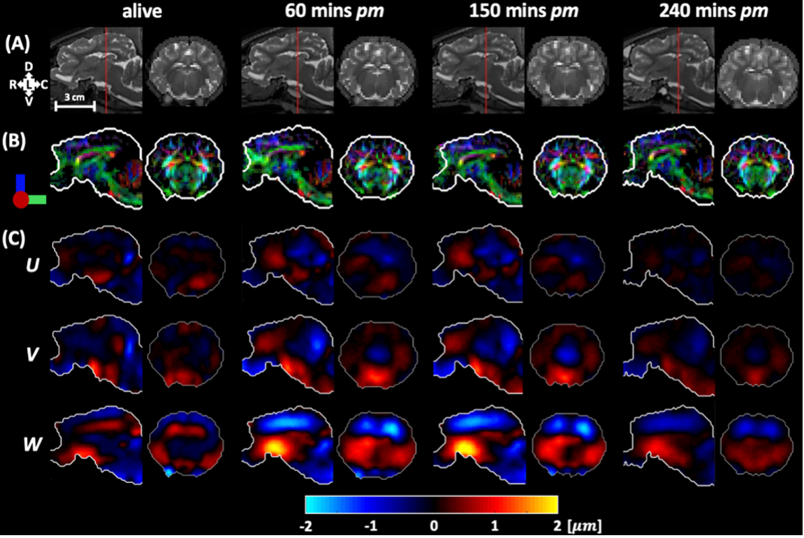
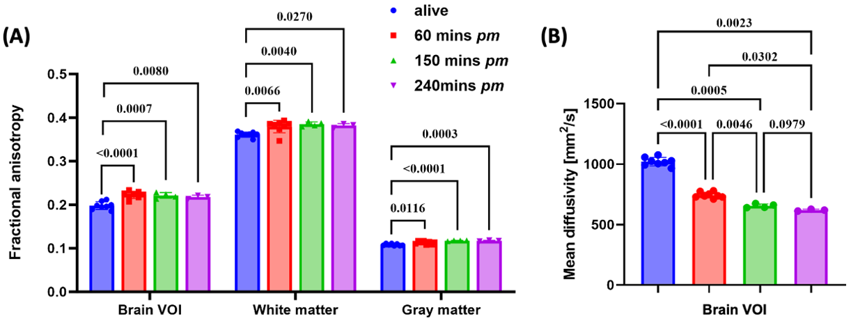
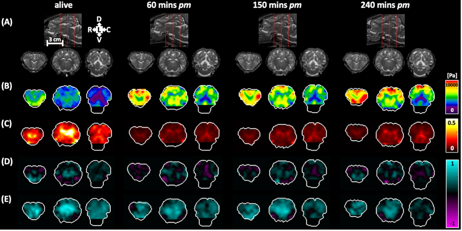
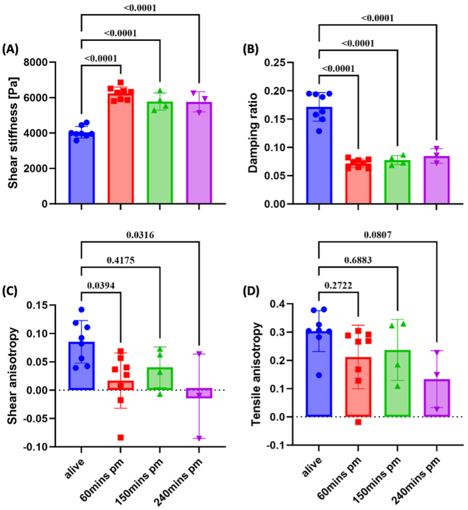
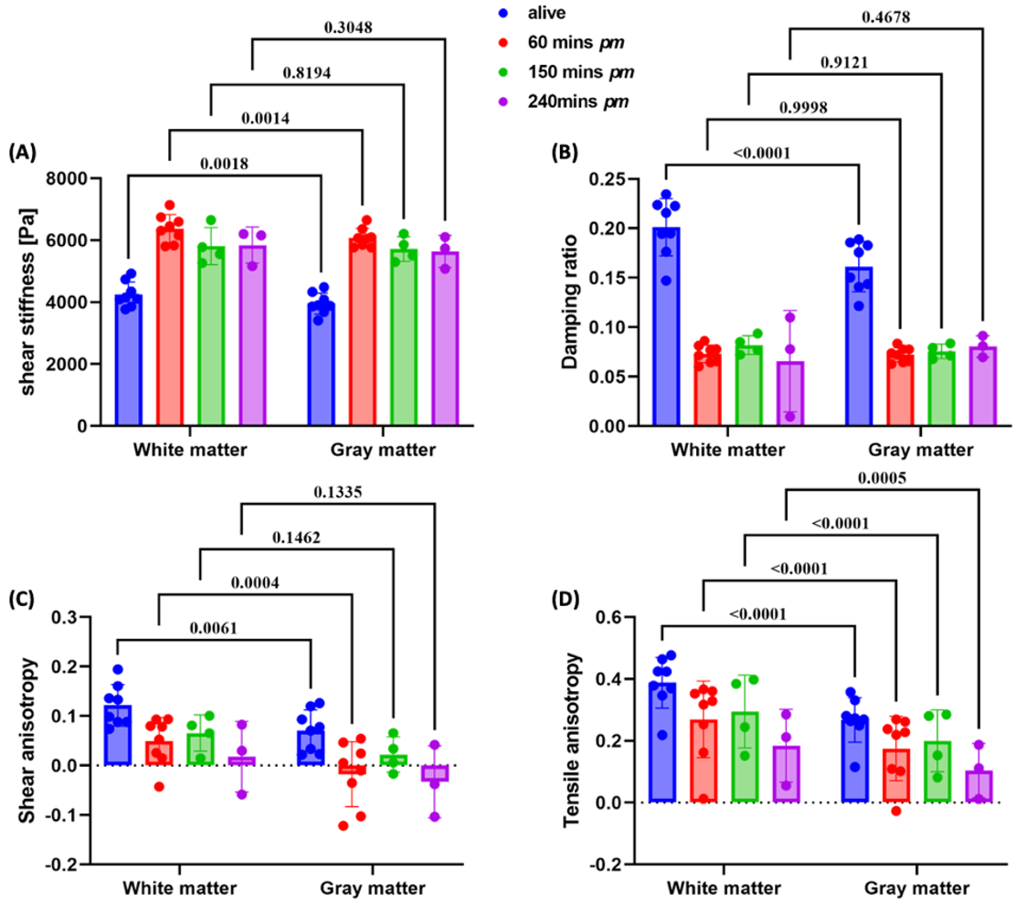
Figure 1 shows examples of anatomical structure, fiber direction, and wave displacement fields. The maximum displacement amplitude is about 2 µm. The wavelength of shear wave fields from the three post-mortem in situ MRE scans are visibly greater than those of in vivo scans, particularly in the rostral-caudal (W) component, indicating higher stiffness. Figure 2 shows the mean FA and MD values of all eight animals at different time points. FA values increase slightly after death but no statistically-significant difference was found between the three post-mortem in situ scans. MD decreases after death and appears to stabilize by 4 hours post-mortem time. Figure 3 displays representative TI-NLI-estimated material properties. Baseline shear stiffness is noticeably higher after death, which is consistent with the observed longer shear wavelength. Damping ratio, shear anisotropy, and tensile anisotropy are generally lower after death, but no clear temporal changes in these parameters were observed with increasing time post-mortem. The means and standard deviations of the parameters in brain volume of interest (VOI) are plotted versus time points in Figure 4. Shear stiffness increases and damping and shear anisotropy decrease after death, while tensile anisotropy was relatively unchanged between in vivo and post-mortem conditions. Figure 5 displays the estimated mechanical parameters in WM and GM over all four scan times.Conclusion
This study is the first to investigate post-mortem changes in the anisotropic mechanical properties of the brain. The current results reveal that WM is more anisotropic than GM, both structurally and mechanically, as well as stiffer and more dissipative. These differences generally diminish post-mortem for all parameters except tensile anisotropy. Overall, brain tissues become more anisotropic in diffusion, but stiffer, less damped, and less mechanically anisotropic after death. These differences between the anisotropic mechanical properties of living and dead brain tissue have important consequences for understanding and predicting the response of the brain in injury, neurosurgery, and development.Acknowledgements
NIH Grant R01EB027577 and ONR Grant N00014-22-1-2198.References
1. Liu Y, Lu Y, Shao Y, et al. Mechanism of the traumatic brain injury induced by blast wave using the energy assessment method. Med Eng Phys. 2022; 101:103767.
2. Panzer MB, Myers BS, Capehart BP, Bass CR. Development of a finite element model for blast brain injury and the effects of CSF cavitation. Ann Biomed Eng. 2012; 40(7): 1530-1544.
3. Forte AE, Gentleman SM, Dini D. On the characterization of the heterogeneous mechanical response of human brain tissue. Biomech Model Mechanobiol. 2017; 16(3): 907-920.
4. Chatelin S, Deck C & Willinger R. An anisotropic viscous hyperelastic constitutive law for brain material finite-element modeling. J Biorheol. 2013; 27: 26–37.
5. Gefen A, Margulies SS. Are in vivo and in situ brain tissues mechanically similar?. J Biomech. 2004; 37(9): 1339-1352.
6. Wang S, Guertler CA, Okamoto RJ, et al. Mechanical stiffness and anisotropy measured by MRE during brain development in the minipig. Neuroimage. 2023; 277: 120234.
7. McIlvain G, Cerjanic AM, Christodoulou AG, at al. OSCILLATE: A low-rank approach for accelerated magnetic resonance elastography. Magn Reson Med. 2022; 88(4): 1659-1672.
8. McGarry M, Van Houten E, Sowinski D, et al. Mapping heterogenous anisotropic tissue mechanical properties with transverse isotropic nonlinear inversion MR elastography. Med Image Anal. 2022; 78: 102432.
Figures