0053
Magnetic Resonance Elastography captures tumor invasiveness and therapy response in the invasive S24-glioma model1Neuroradiology, Heidelberg University Hospital, Heidelberg, Germany, 2Clinical Cooperation Unit Translational Radiation Oncology, Deutsches Krebsforschungszentrum, Heidelberg, Germany, 3Neurology, Heidelberg University Hospital, Heidelberg, Germany, 4Clinical Cooperation Unit Neurooncology, Deutsches Krebsforschungszentrum, Heidelberg, Germany, 5Clinical Cooperation Unit Neuroimmunology and Brain Tumor Immunology, Deutsches Krebsforschungszentrum, Heidelberg, Germany, 6School of Biomechanical Engineering and Imaging Science, King's College London, London, United Kingdom, 7INSERM UMRS1148 - Laboratory for Vascular Translational Science, University of Paris, Paris, France, 8Radiation Oncology, Heidelberg University Hospital, Heidelberg, Germany
Synopsis
Keywords: Elastography, Elastography, Cancer, Neuro
Motivation: In a neurooncological setting clinically established MRI-sequences have shortcomings with regards to tumor invasion and therapy-associated parenchyma changes.
Goal(s): Our goal was to determine if MRE and the ADC could improve detection of tumor cell invasion and radiotherapy effects.
Approach: 23 tumor-bearing mice and 9 sham injected mice underwent MRE- and MRI-scans for up to 16 weeks, a subgroup of animals underwent additional radiotherapy.
Results: MRE was sensitive to early tumor invasion and MRE and ADC captured radiotherapy effects so far not detectable with established MRI-sequences.
Impact: In a preclinical setting the ADC and especially MRE allow for a better characterization of therapeutic effects and tumor cell invasion and should thus also be evaluated in a clinical setting.
Introduction
Glioblastoma is the most common primary malignant brain tumor and is characterized by a particularly invasive growth pattern. Magnetic resonance imaging (MRI) is a key element for tumor diagnosis and therapy monitoring. Yet, clinically established MRI-sequences do not allow a precise tumor delineation, especially with regards to its “invasive front”. Furthermore, it remains difficult to differentiate between tumor progression and therapy-associated tissue changes such as radionecrosis or pseudoprogression (1). Magnetic Resonance Elastography (MRE) is an imaging technique akin to palpation evaluating and quantifying biomechanical tissue properties, in particular tissue stiffness (2). This study investigates in a preclinical invasive glioma model if MRE, alongside with other multiparametric MRI-parameters, allows for a more precise delineation of tumor cell invasion and therapy-associated tissue changes. We hypothesize that the structural organization and thereby also the biomechanical properties of the brain parenchyma are affected differently and thus amenable to MRE.Methods
100000 S24-glioma cells were orthotopically implanted into the right striatum of 23 male NMRI-nude mice (3). 13 animals underwent whole brain radiotherapy (3 x 6 Gy) 9 weeks after surgery, the other 10 animals served as untreated, tumor-bearing controls. 9 NMRI-nude mice received a 1µl PBS sham injection into the right striatum, 5 animals underwent whole brain radiotherapy in week 9 after surgery. Imaging was performed for all animals in week 4, 8 and 12 after tumor implantation on a 9.4 T small animal MRI-scanner. 5 irradiated, tumor-bearing mice received an additional scan in week 16. The imaging protocol was composed of T2-weighted sequences, Diffusion Tensor Imaging and MRE (900Hz vibration frequency). After their respectively last scan, mice were euthanized, brains were harvested and optically cleared following the iDISCO protocol (4) with turboGFP-staining for S24-cells and Propionate Iodide-staining for cell nuclei. Brains were imaged on a light sheet microscope (LSM). Images were analyzed with the open access Slicer-Software and MRE-data additionally with KIR-Software. Tumor core volumes were determined and the corpus callosum was segmented on T2w2D sequences for all measurement points. The Apparent Diffusion Coefficient (ADC) and the magnitude of the complex valued shear modulus IG*I (“stiffness”) were assessed within the tumor core and the corpus callosum.Results
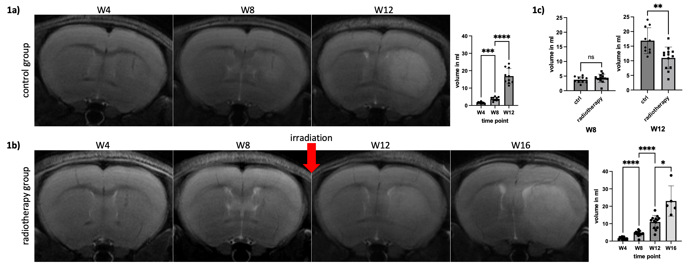
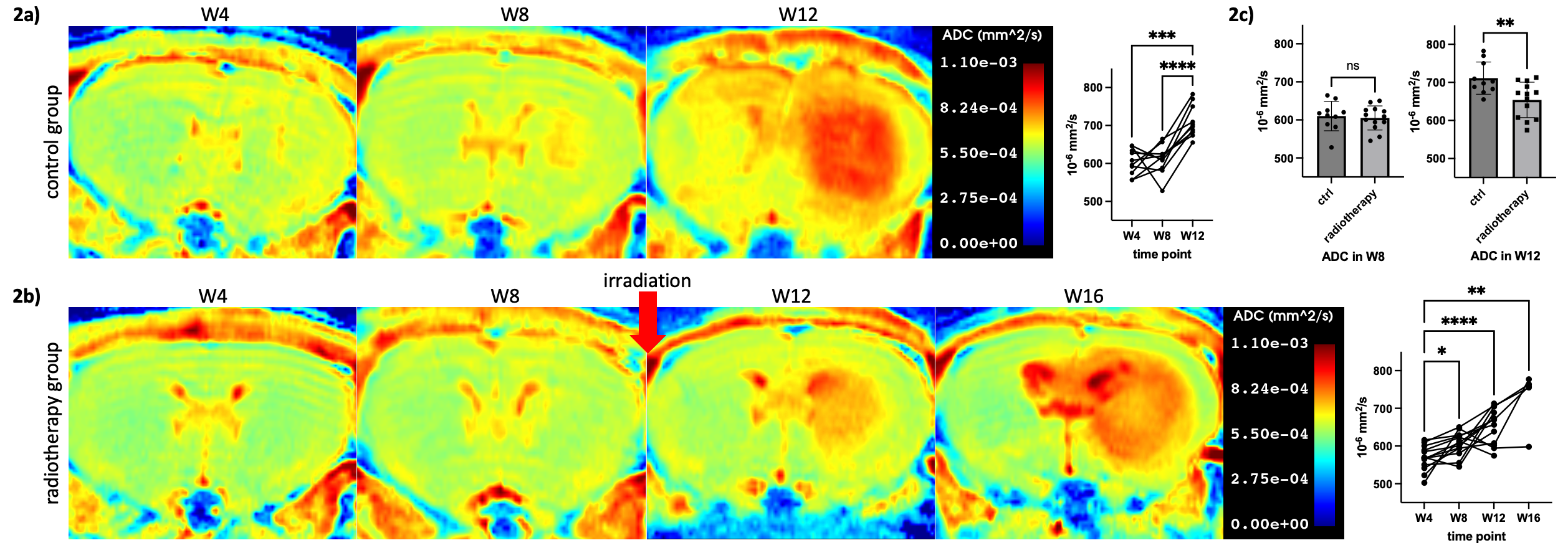
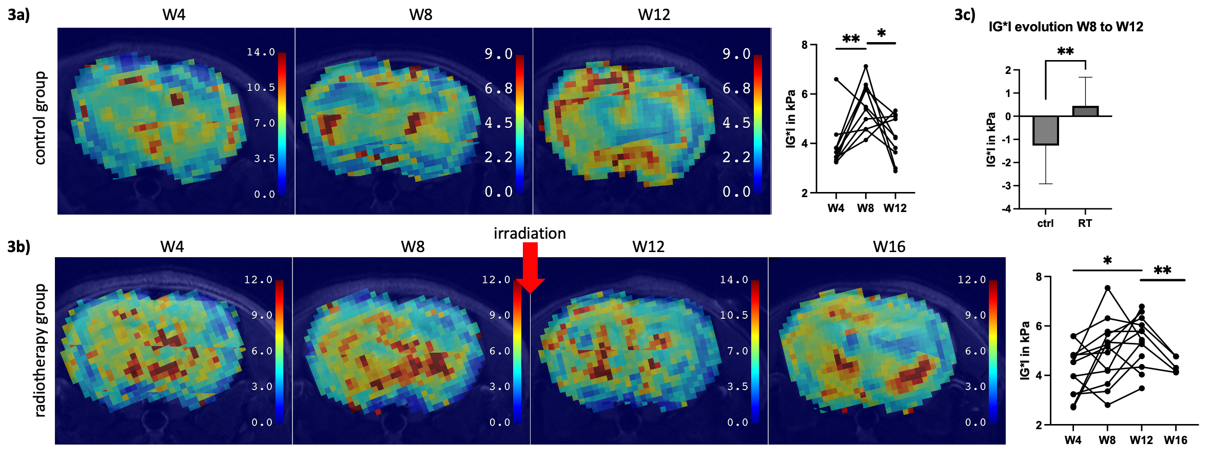
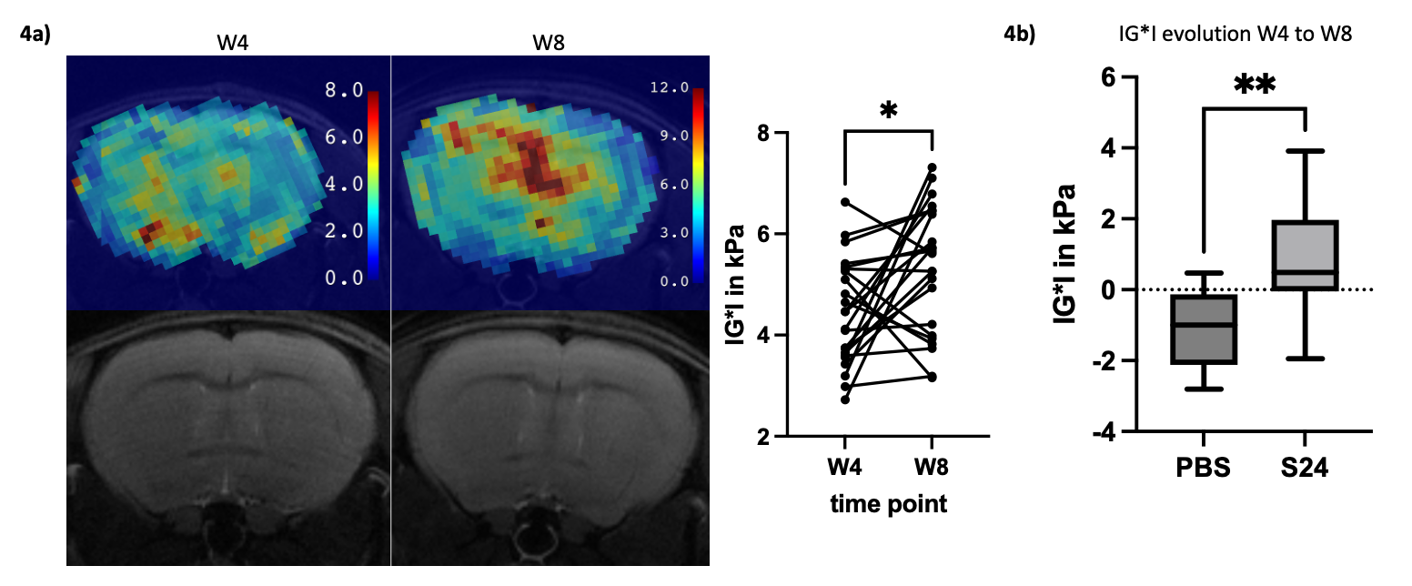
Based on T2w imaging tumor growth in week 12 was significantly decelerated after irradiation, however a tumor regress or other T2w signal alterations in the treated animals were not observable (figure 1). With regards to the tumor core the ADC value increased progressively over time. However, the irradiation led to a significant deceleration of this process (figure 2). In MRE a steady tumor core stiffness increase up to week 8 was observed resulting in a better delineation from the surrounding parenchyma. The increase in tumor stiffness was followed by a progressive softening in untreated tumors with heterogeneous, stiffer clusters remaining in the tumor periphery in week 12 (figure 3a). This process was decelerated, but not prevented by radiotherapy, as a delayed tumor stiffness drop in week 16 was observed (figure 3b). To assess if tumor cell invasion could be monitored the ADC and IG*I were determined in the corpus callosum, an anatomical structure demonstrated to be invaded by tumor cells 40-60 days after tumor implantation (5). While in week 8 post-implantation no T2w signal alteration or significant changes in the ADC were detectable, a clear increase in tissue stiffness could be observed by MRE (figure 2a). Furthermore, when comparing tumor-bearing animals to sham injected mice, significant differences in corpus callosum stiffness were detected (figure 2b).Discussion
The determination of IG*I in the corpus callosum at early time points (week 8) shows that MRE is superior to conventional MRI-sequences and the ADC with regards to the detection of tumor cell invasion. We hypothesize that the observed increase in corpus callosum stiffness from week 4 to week 8 reflects the development of the tumor microtube network during the migration and invasion process, as previously described for this model (6).Visually no clear distinction in T2w (e. g. different signal intensity) between irradiated and untreated tumors could be made. Yet, the tumor core in treated animals showed a slower ADC increase and a delayed stiffness drop when compared to untreated tumor-bearing mice. This might reflect the ablation of unconnected tumor cells while the connected tumor cells have been shown to be resistant to radiotherapy (3,7). To verify these hypotheses, we are currently performing correlative LSM and histopathology.Conclusion
In summary, the determination of biomechanical tissue properties and the ADC enables new insights into the tumor microenvironment during longitudinal tumor growth and under therapeutic pressure that could so far not be monitored with conventional imaging.Acknowledgements
No acknowledgement found.References
(1) Ellingson, Benjamin M et al. “Pseudoprogression, radionecrosis, inflammation or true tumor progression? challenges associated with glioblastoma response assessment in an evolving therapeutic landscape.” Journal of neuro-oncology vol. 134,3 (2017): 495-504. doi:10.1007/s11060-017-2375-22.
(2) Muthupillai, R et al. “Magnetic resonance elastography by direct visualization of propagating acoustic strain waves.” Science (New York, N.Y.) vol. 269,5232 (1995): 1854-7. doi:10.1126/science.75699243.
(3) Osswald, Matthias et al. “Brain tumour cells interconnect to a functional and resistant network.” Nature vol. 528,7580 (2015): 93-8. doi:10.1038/nature160714.
(4) Renier, Nicolas et al. “iDISCO: a simple, rapid method to immunolabel large tissue samples for volume imaging.” Cell vol. 159,4 (2014): 896-910. doi:10.1016/j.cell.2014.10.0105.
(5) Schubert, Marc C et al. “Deep Intravital Brain Tumor Imaging Enabled by Tailored Three-Photon Microscopy and Analysis.” bioRxiv, Jan. 2023, p. 2023.06.17.545350, doi.org/10.1101/2023.06.17.5453506.
(6) Venkataramani, Varun et al. “Glioblastoma hijacks neuronal mechanisms for brain invasion.” Cell vol. 185,16 (2022): 2899-2917.e31. doi:10.1016/j.cell.2022.06.054.
(7) Jung, Erik et al. “Tumor cell plasticity, heterogeneity, and resistance in crucial microenvironmental niches in glioma.” Nature communications vol. 12,1 1014. 12 Feb. 2021, doi:10.1038/s41467-021-21117-3.
Figures