0052
Cell-Specific Mapping of MR Spectroscopic Signatures: A Pilot Study in a Murine Glioma Model1Department of Bioengineering, University of Illinois at Urbana-Champaign, Urbana, IL, United States, 2Beckman Institute for Advanced Science and Technology, University of Illinois at Urbana-Champaign, Urbana, IL, United States, 3Neuroscience Program, University of Illinois at Urbana-Champaign, Urbana, IL, United States, 4Department of Biochemistry, University of Illinois at Urbana-Champaign, Urbana, IL, United States, 5Cancer Center at Illinois, University of Illinois at Urbana-Champaign, Urbana, IL, United States, 6Department of Electrical and Computer Engineering, University of Illinois at Urbana-Champaign, Urbana, IL, United States
Synopsis
Keywords: Spectroscopy, Spectroscopy
Motivation: Effective monitoring of tumor progression and therapeutic efficacy should benefit from new in vivo imaging capability to resolve cell-specific contributions at tissue level.
Goal(s): To develop a new MRSI-based approach to resolve tumor cell-specific components at individual imaging voxels leveraging the spectral dimensions.
Approach: We proposed a multiscale experimental and computational MRSI framework that learns cell-specific spectroscopic signatures from glioma cell lines and resolves intravoxel nontumor and tumor-specific components in vivo using the learned signatures.
Results: Results from cellular mixtures and glioma-bearing mice demonstrated the potential of our method. Time-dependent, spatially-resolved tumor cell maps can be obtained, showing tumor growth in vivo.
Impact: The proposed approach marks a potential new paradigm to map cellular complexity at tissue level leveraging additional imaging dimensions and machine learning. It holds the promise to provide new tools for tumor grading, progression monitoring and treatment assessment.
Introduction
MR spectroscopic imaging (MRSI) affords non-invasive molecular level tumor diagnosis and characterization.1-3 The potential of high-resolution MRSI for mapping altered metabolite levels in brain tumors has been shown.3,4 However, interpretation of such molecular variations can be challenging with the complex cellular composition in the tumor microenvironment and significant partial volume effects. Different cell types within the tumor microenvironment have their own altered metabolite profiles compared to healthy tissues across different disease stages, which have different biological/clinical implications. We introduce here a new MRSI-based, label-free approach to map tumor cells at the tissue level in vivo. Specifically, we leveraged cell cultures to learn cell-type-specific spectroscopic signatures, and developed a computational strategy to separate the tumor-cell-specific component from the overall spectroscopic signals using the learned features and subspace fitting.5,6 We evaluated our method using cellular mixtures and time-dependent in vivo data from glioma-bearing mice generated by an advanced MRSI acquisition developed for ultrahigh-fields7, producing promising tumor cell fraction maps and successfully tracking tumor progression.Methods
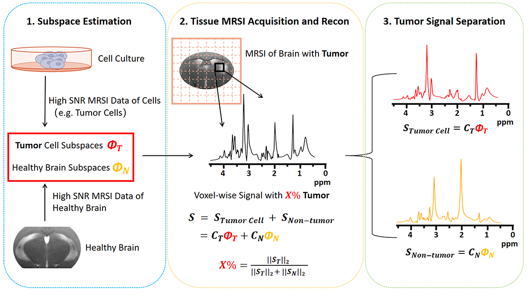
The key features of our approach include an integrative experimental and computational strategy to extract cell-specific spectroscopic signals from cell cultures and computationally resolving tumor and nontumor components in tissues leveraging the additional spectral dimensions in MRSI and subspace modeling. Figure 1 illustrates the conceptual steps. More methodological details are provided below.Learning Cell-Specific Subspaces
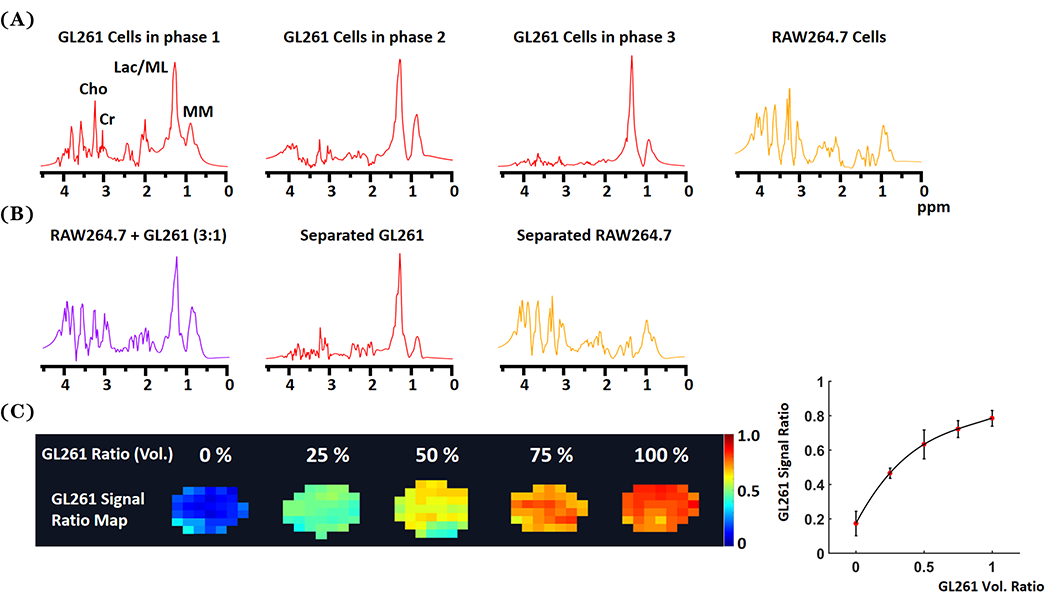
Special classes of cells can be cultured or derived from tissue samples. As a proof-of-concept, we cultured GL261 (glioma) and RAW264.7 (macrophage) cell lines in DMEM medium and selectively harvested them at distinct growth phases: Phase 1 (cell density ~70%), Phase 2 (density ~90%), and Phase 3 (density exceeding 90%). We collected cell-culture-specific features using a relaxation-enhancement CSI acquisition with unique SNR benefits at ultrahigh-fields. A 9.4T system was used in this work with TR = 1500 ms, TE = 24 ms, FOV = 16×16 mm2, slice thickness = 1.5 mm, matrix size = 18×18, and two averages. A special denoising method was applied to the data8, followed by SVD to extract cell-specific subspaces for GL261 and RAW264.7, respectively (Fig. 2).
Mapping Tumor Cells in Glioma-bearing Mice
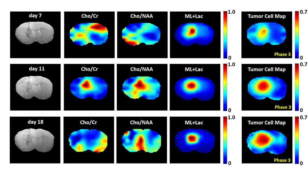
To demonstrate our ability to resolve tumor cell component in vivo, GL261 cells were implanted into mouse brains to induce glioma.9 One mouse (clear evidence of tumor growth) was selected for longitudinal imaging at the 7th, 11th, and 18th days. The same RE-CSI acquisition was used. Data were reconstructed using the SPICE-based method6,10 with a combination of cell-specific and healthy tissue subspaces (learned from normal brains) to separate the tumor cell signals and estimate their “fractions” at individual voxels.
Results
Cellular Mixtures
We first validated our method by creating cell phantoms with mixtures of GL261 and RAW264.7 at various volume ratios (i.e., 0%, 25%, 50%, 75%, 100% of GL261). CSI data were collected from these phantoms and reconstructed using our method and respective cell-specific subspaces. Figure 2A presents representative spectroscopic profiles for GL261 (at different phases) and RAW264.7. Figure 2B illustrates the separation outcome for a cellular mixture with 25% GL261. A positive correlation with monotonic trend between the calculated GL261 tumor cell fractions and actual volume ratios were observed in Fig. 2C. The bias observed due to noise projection can be further reduced with better subspace and reconstruction strategies, and can also be calibrated.
In Vivo Imaging
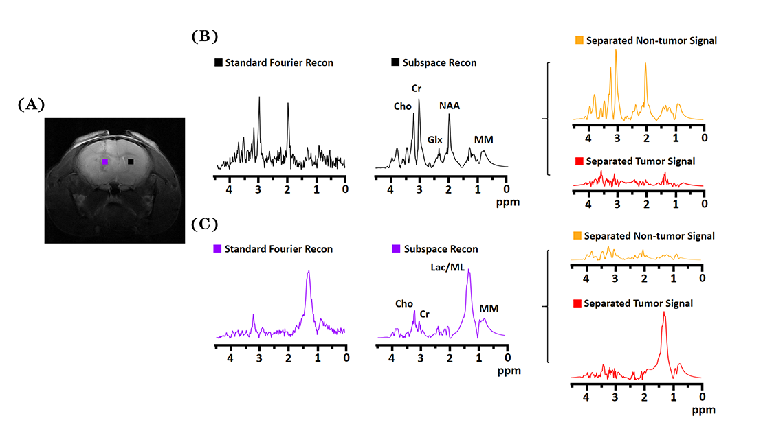
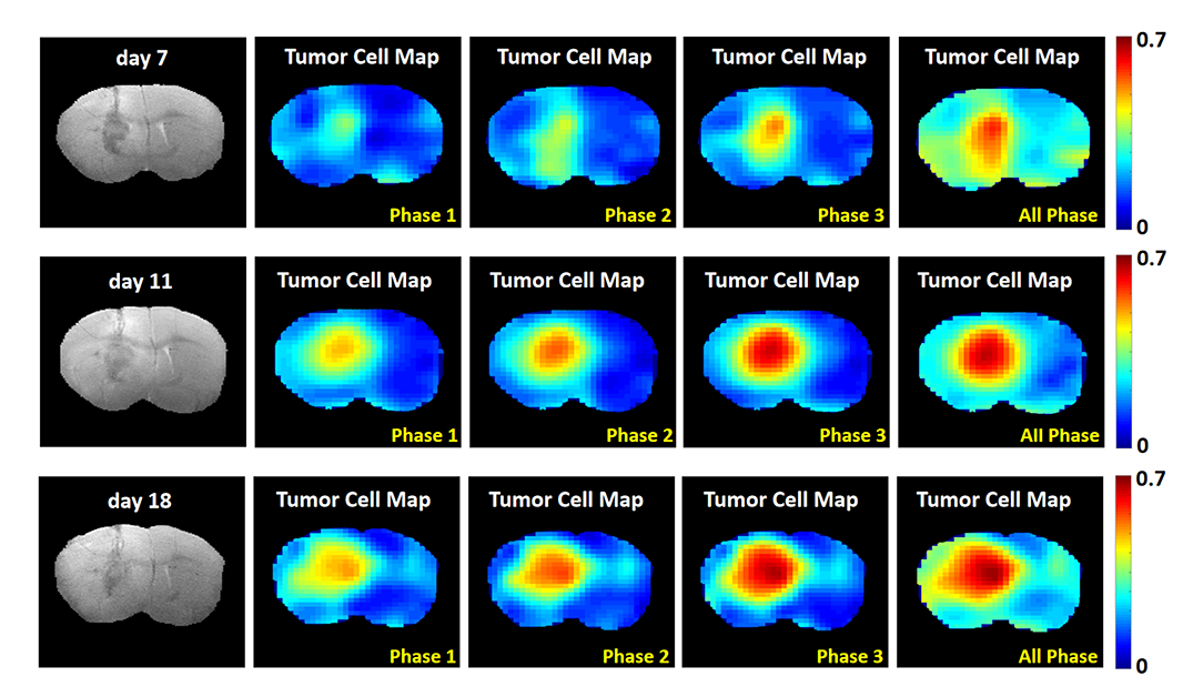
Figure 3 shows clear tumor cell signal separation results from tumor-bearing mouse, for one voxel in the tumor region and one in the contralateral normal-appearing region. Tumor cell distribution maps over time are shown in Fig. 4, revealing a clear trend of increasing tumor cell fraction during progression. Meanwhile, maps of individual metabolites/ratios are harder to interpret due to confounding factors such as tissue injury at early days. We also compared the tumor cell maps generated using GL261 subspaces from cell culture data acquired at different growth phases (Fig. 5). Although dynamic progression of glioma was observed, the actual fraction values calculated depend on which subspace was used, indicating the diversity of tumor compositions and the importance of subspace accuracy and dynamics for future research.
Conclusion
We have proposed a new approach to generate cell fraction maps from spectroscopic imaging data. We validated the proposed method using cell mixtures and glioma-bearing mice. The computed tumor cell maps demonstrated potential in detecting and monitoring tumor growth. While its clinical translatability is still under investigation, we believe our approach holds great promise to enable cell-type-resolved imaging and cellular heterogeneity mapping in tumor tissues.Acknowledgements
This work was supported in part by NIH-NIGMS-R35GM142969 and a Cancer Center at Illinois seed grant.References
1. Horská, A., & Barker, P. B. (2010). Imaging of brain tumors: MR spectroscopy and metabolic imaging. Neuroimaging Clinics, 20(3), 293-310.
2. van Dijken, B. R., van Laar, P. J., & van der Hoorn, A. (2017). Diagnostic accuracy of magnetic resonance imaging techniques for treatment response evaluation in patients with high-grade glioma, a systematic review and meta-analysis. European Radiology, 27, 4129-4144.
3. Aseel, A., McCarthy, P., & Mohammed, A. (2023). Brain magnetic resonance spectroscopy to differentiate recurrent neoplasm from radiation necrosis: A systematic review and meta‐analysis. Journal of Neuroimaging, 33(2), 189-201.
4. Verma, N., Cowperthwaite, M. C., & Markey, M. K. (2013). Differentiating tumor recurrence from treatment necrosis: a review of neuro-oncologic imaging strategies. Neuro-Oncology, 15(5), 515-534.
5. Lam, F., & Liang, Z. P. (2014). A subspace approach to high‐resolution spectroscopic imaging. Magnetic Resonance in Medicine, 71(4), 1349-1357.
6. Lam, F., Peng, X., & Liang, Z. P. (2023). High-Dimensional MR Spatiospectral Imaging by Integrating Physics-Based Modeling and Data-Driven Machine Learning: Current progress and future directions. IEEE Signal Processing Magazine, 40(2), 101-115.
7. Yizun, W., Stanislav S. R., & Lam, F. (2023). High-resolution 1H-MRSI of the brain at 9.4T integrating relaxation enhancement and subspace imaging. In Proc. ISMRM, p. 3691.
8. Ruiyang, Z., Yahang, Li., & Lam, F. (2023). MR spatiospectral reconstruction using plug&play denoiser with self-supervised training. In Proc. ISMRM, p. 0955.
9. Tang, B., Guo, Z. S., & Roy, E. J. (2020). Synergistic combination of oncolytic virotherapy and immunotherapy for glioma. Clinical Cancer Research, 26(9), 2216-2230.
10. Li, Y., Lam, F., & Liang, Z. P. (2017). A subspace approach to spectral quantification for MR spectroscopic imaging. IEEE Transactions on Biomedical Engineering, 64(10), 2486-2489.
Figures