0051
Brain metabolic alteration at a late phase of immune fatigue model mice using parahydrogen-polarized [1-13C] pyruvate MRI.1Information Science and Technology, Hokkaido University, Sapporo, Japan
Synopsis
Keywords: Biomarkers, Hyperpolarized MR (Non-Gas)
Motivation: Long-lasting brain issues including cognitive impairments after infections have become a worldwide problem after COVID-19.
Goal(s): Our goal was to detect brain metabolic alteration at a late phase of immune fatigue.
Approach: Parahydrogen-polarized 13C MRI of pyruvate metabolism was applied in the brain of 3 days poly I:C treated mice.
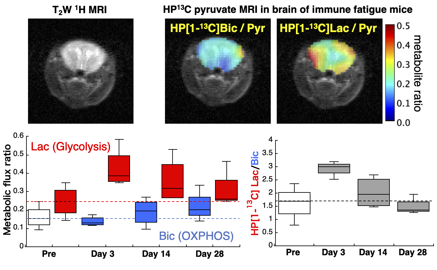
Results: Alteration of brain pyruvate metabolism toward glycolysis was observed in both acute phase at day 3 and late phase at day 14 of immune fatigue model mice, which was correlated with diminished dopamine signal marker and nighttime moving distance in open-field test.
Impact: Our demonstration of a detectable alteration of brain pyruvate metabolism by parahydrogen-polarized 13C MRI at a late phase of immune fatigue mice can be a useful biomarker of cognitive impairments after infections such as brain fog of long-COVID.
INTRODUCTION
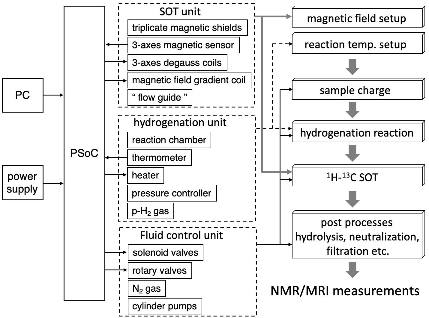
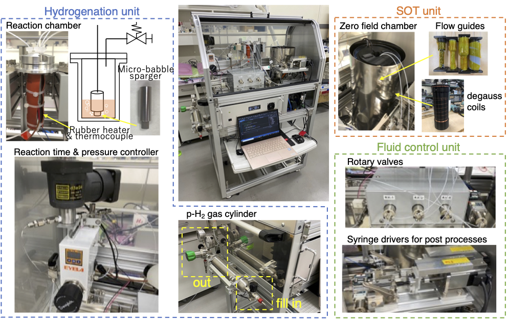
Since December 2019, long-lasting symptoms after acute phase of COVID-19 infection have become a worldwide social problem. The long COVID-19 symptoms include taste and smell disorders, depression, anxiety disorders, cognitive impairments, and myocarditis, many of which are difficult to objectively assess and only limited diagnostic methods have been established. Hyperpolarization is a technique to transiently enhance the MRI signal of 13C or other nuclear spins more than 10,000 times, allowing non-invasive and real-time observation of metabolic reaction in the body. Clinical trials of hyperpolarized 13C MRI of pyruvate metabolism, prepared by dissolution dynamic nuclear polarization (dDNP), in patients with cancer and cardiac diseases have been conducted or on going over 15 facilities worldwide. We previously reported that metabolic reprogramming in the liver of immune fatigue model mice can be observed by hyperpolarized 13C MRI of pyruvate metabolism1. Recently, we developed a unique parahydrogen-induced hyperpolarization (PHIP)-based 13C hyperpolarizer system as a cost-effective alternative of the dDNP-type hyperpolarizer (under review). In this study, we investigated if the brain metabolic alteration in immune fatigue, i.e. pseud infection, model mice can be detectable as a biomarker of long-lasting brain issue such as cognitive impairment using PHP-polarized [1-13C] pyruvate MRI.METHODS
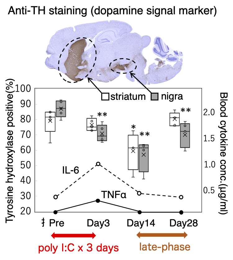
Pseud infection/immunological fatigue model mice were made by intraperitoneal injection of a toll-like receptor 3 (TLR3) ligand polyinosinic:polycytidylic acid (poly I:C, 100μg) into C3H/HeJYokSlc mice for 3 days. Immunohistochemical analysis of tyrosine hydroxylase (TH) as a marker of dopamine signal, lactate dehydrogenase-A (LDH-A), and microglial activation in the brain was conducted on day 0, 3, 14, and 28 after start of poly I:C treatment. Locomotor activity was evaluated as a moving distance per hour during nighttime by the open field test. Hyperpolarized [1-13C]pyruvate solution (80-90mM) was prepared by ParaHydrogen-Induced Polarization Side Arm Hydrogenation (PHIP-SAH) technique2 using our developed fully automated PHIP polarizer system and intravenously injected into the mice (12 µL/g body weight). Two-dimensional chemical shift imaging (CSI) was conducted 25 seconds after start of injection at the head of mouse using a lab-made multinuclear 1.5T preclinical MRI system. Typical CSI parameters are as follows; FOV 32x32 mm, matrix 16x16, TR 75ms, FA 10°, spectral width 122 ppm for 128 spectral data points, centric k-space acquisition. Metabolic images of hyperpolarized [1-13C]pyruvate, lactate, and bicarbonate were reconstructed by a code written in MATLAB and analyzed using ImageJ software.RESULTS AND DISCUSSION
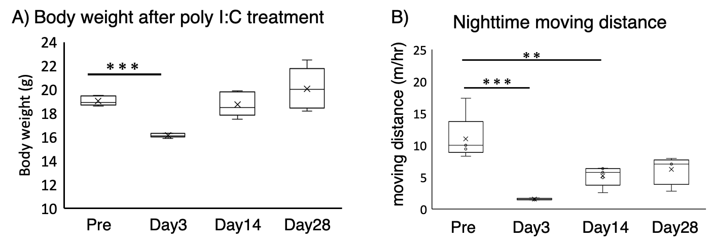
Three days poly I:C treatment induced pseud infection symptoms similar to COVID-19 or influenza infections in an acute phase (around day 3). After the end of poly I:C treatment, the transiently decreased body weights of mice were fully recovered and the increased levels of blood inflammation cytokines IL-6 and TNFα were also normalized until day 14. However, nighttime moving distance of poly I:C treated mice significantly decreased on day 3 and 14, which did not recover to control level even 4 weeks after poly I:C treatment. Immunohistochemical analysis revealed that expressions of dopamine signal marker TH in striate body and substantia nigra and LDH-A in whole brain kept decreased on day 14, and microglial activity stained by aint-Iba1 antibody also significantly increased on day 14. These results suggest that some kind of damage remains in the brain of immune fatigue mice after the end of acute phase. Using the developed PHIP-based 13C hyperpolarizer system, 80-90 mM of hyperpolarized [1-13]pyruvate solution with 13C polarization of 4-5% at the time of MRI measurements can be generated within 2 min after initiation of parahydrogenation reaction. Non-invasive 2D chemical shift imaging (CSI) of hyperpolarized [1-13C]pyruvate metabolism showed the glycolytic shift in brain pyruvate metabolism, decreased flux to bicarbonate and increased flux to lactate, on day 3 and 14 after poly I:C treatment compared to non-treated control mice.CONCLUSION
We have demonstrated the long-lasting alteration of brain metabolism in a late phase of poly I:C-induced immune fatigue mouse model using parahydrogen-polarized 13C MRI of pyruvate metabolism. Our observation suggests that hyperpolarized 13C MRI of pyruvate metabolism can be a useful and non-invasively available biomarker of long-lasting symptom of infectious diseases such as brain fog of long COVID-19.Acknowledgements
This research was supported by AMED (grant no. JP23gm6910009) and the Japan Society for the Promotion of Science (JSPS) KAKENHI (grant no: 22K1843502).References
- Tarasenko TN, Jestin M, Matsumoto S et al. Macrophage derived TNFα promotes hepatic reprogramming to Warburg-like metabolism. J Mol Med (Berl). 2019 Sep;97(9):1231-1243.
- Reineri F, Boi T, Aime S. ParaHydrogen Induced Polarization of 13C carboxylate resonance in acetate and pyruvate. Nat Commun. 2015 Jan 5;6:5858.
- Stewart NJ, Sato T, Takeda N, Hirata H, Matsumoto S. Hyperpolarized 13C Magnetic Resonance Imaging as a Tool for Imaging Tissue Redox State, Oxidative Stress, Inflammation, and Cellular Metabolism. Antioxid Redox Signal. 2022 Jan;36(1-3):81-94.
- Stewart NJ, Nakano H, Matsumoto S et al. Hyperpolarized 13 C Magnetic Resonance Imaging of Fumarate Metabolism by Parahydrogen-induced Polarization: A Proof-of-Concept in vivo Study. ChemPhysChem. 2021 May 17;22(10):915-923.
Figures