0050
Impact of Gender on Alzheimer’s disease: a Behavioral and Metabolic Analysis in AβPP-PS1 Mouse Model1NMR Microimaging and Spectroscopy, Centre for Cellular and Molecular Biology, Hyderabad, India, 2Academy of Scientific and Innovative Research, Ghaziabad, India
Synopsis
Keywords: Alzheimer's Disease, Metabolism, Neurodegeneration, Alzheimer's disease
Motivation: Sex specific status of neurometabolic activity in Alzheimer’s disease is poorly understood.
Goal(s): To assess glutamatergic and GABAergic neurometabolism in different brain regions of male and female AβPP-PS1 mouse model of AD.
Approach: Tracer approach involving infusion of [1,6-13C2]glucose in AβPP-PS1 mouse model of AD together with 1H-[13C]-NMR spectroscopy was used to estimate rate of neuronal Glucose oxidation (CMRGlc(Ox)) in AD mice.
Results: The neuronal metabolic activity reduced in the cerebral cortex, hippocampus and cerebellum of female AβPP-PS1 mice. However, only hippocampal neurometabolic activity was reduced in the male mice. This suggests a differential progression of AD in male and female AβPP-PS1 mice.
Impact: The findings of the study suggest differential progression of AD in male and female AβPP-PS1 mice. These factors require attention for future interventions for AD treatment.
BACKGROUND
Alzheimer's disease (AD) is an age-related neurodegenerative disorder, and the most common form of dementia.1AD is characterized by the presence of amyloid beta plaques and neurofibrillary tangles associated with loss in memory, progressive cognitive impairment and behavioral changes. Epidemiological studies suggest that two-thirds of American AD subjects are women.2 However; it is not clear whether the higher prevalence of AD in women is due to longer life expectancy or increased susceptibility. This study evaluates the memory and neurometabolism in female and male AβPP-PS1 mouse model of AD.METHODS
All animal experiments were performed under protocols approved by the Institutional Animal Ethics Committee of CSIR-CCMB. Male (n=23) and female (n=22) AβPP-PS1 mice of 6 months age were used for the study. Working memory of the mice was assessed using Y-maze3, while the spatial memory was evaluated by the Morris Water Maze (MWM) test.4 For metabolic analysis, the mice were anesthetized with urethane (1.5 g/kg, intraperitoneal), and infused with [1,6-13C2]glucose for 10 min using a bolus rate infusion protocol.5 Blood was collected from the retro orbital sinus, and mice were euthanized by a focused beam microwave irradiation (3kW, 1.2s). The metabolites were extracted from the cerebral cortex, hippocampus and cerebellum. Proton observed Carbon-13 edited NMR spectroscopy was done to determine the concentration of 13C labeled neurometabolites.6The cerebral metabolic rate of glucose oxidation (CMRglc(ox)) in glutamatergic and GABAergic neurons was estimated by 13C labeled trapped into different neurometabolites as described previously.5RESULTS
The escape latency of male AβPP-PS1 (70.8±23.8 s) in MWM test was not significantly different from the male WT mice (61.6±29.8 s). Similarly, the female AβPP-PS1 could reach the escape platform in (59±26.2s) and was not different from the female WT (59.1±27.6s). The spontaneous alternation of male AβPP-PS1 mice(54.5±11.7 %) in Y-maze was similar to the WT(58.5±1.0 %).Similarly, the female AβPP-PS1 mice (65.2±7.6 %)did not show change in the spontaneous alternation WT (63.4±10.5 %). Female AβPP-PS1 mice showed reduction in levels of NAA (5.8±0.3 vs. 6.2±0.3 µmol/g/min; p=0.023)in the cerebral cortex, and glutamine (5.9±0.4 vs. 6.3±0.3 µmol/g/min; p=0.036)in the cerebellum when compared with WT. The neurometabolite homeostasis was unperturbed in male AβPP-PS1 mice.The labeling of GluC4 (1.51±0.13 vs.1.81±0.15 µmol/g; p=0.007), AspC3 (0.15±0.02 vs. 0.19±0.01 µmol/g; p=0.004), GABAC2 (0.17±0.02 vs. 0.19±0.01 µmol/g; p=0.017) and GluC3 (0.28±0.05 vs. 0.34±0.04 µmol/g; p=0.02) from [1,6-13C2]glucose was decreased in the cerebral cortex of female AβPP-PS1 mice when compared with female WT mice (Table 1). The male AβPP-PS1 mice exhibited reduced labeling only in GlnC4 (0.28±0.05 vs. 0.34±0.04 µmol/g; p=0.02) and GABAC4 (0.28±0.05 vs. 0.34±0.04 µmol/g; p=0.02) in the cerebral cortex. In female AβPP-PS1 hippocampus, 13C labeling of GluC4 (p=0.023) and GABAC2 (p=0.05) were decreased. In contrast, male AβPP-PS1 mice showed a reduction in the labeling of GABAC4 (p=0.001), GlnC4 (p=0.016), AspC3 (p=0.021) and GluC3 (p=0.015). In Cerebellum, The female AβPP-PS1 exhibited a reduction in GluC4, GABAC2, AspC3 and GluC3 (p<0.05) when compared to female WT (Table 1). However, Male AβPP-PS1 showed reduction in GlnC4 (p=0.021) alone (Table 1).
The cerebral metabolic rate of glucose oxidation in glutamatergic neurons CMRGlc(Glu)was decreased in the cerebral cortex (AβPP-PS1 0.18±0.02 µmol/g/min, WT 0.22±0.02 µmol/g/min; p=0.009), hippocampus (0.19±0.02 µmol/g/min vs. 0.21±0.02 µmol/g/min; p=0.03), and cerebellum (0.18±0.02 µmol/g/min vs. 0.21±0.02 µmol/g/min; p=0.01),of female AβPP-PS1 (Fig. 3). In contrast, male AβPP-PS1 mice showed reduction in CMRGlc(Glu) in the hippocampus (0.20±0.03 µmol/g/min vs. 0.23±0.04 µmol/g/min; p=0.04) only, while cerebral cortex and cerebellum were unperturbed. The GABAergic neurons exhibit reduction in CMRGlc(GABA) in cerebral cortex (0.04±0.004 µmol/g/min vs. 0.05±0.00 µmol/g/min; p=0.009) and cerebellum (0.06±0.00 µmol/g/min vs. 0.07±0.01 µmol/g/min; p=0.045) of female AβPP-PS1 mice, while male exhibit reduction in GABAergic neurometabolism in the hippocampus (0.05±0.01 µmol/g/min vs. 0.06±0.01 µmol/g/min, p=0.02) only (Fig. 4).
DISCUSSION
The working and spatial memory was intact in male and female AβPP-PS1 mice at 6 months of age. The neurometabolite homeostasis was largely unperturbed except a reduction in the cortical NAA suggestive of neurodegeneration in female APP-PS1 mice. The reduced CMRGlc(Glu) is suggestive of decreased neuronal viability in the cerebellum along with cerebral cortex and hippocampus of female AβPP-PS1 mice. In contrast, only hippocampal neurometabolic activity was reduced in the male mice. These data suggest differential progression of AD in male and female AβPP-PS1 mice.Acknowledgements
This study was supported by Council of Scientific and Industrial Research (CSIR-CCMB: NCP/MLP0139).
References
1. WHO (2017) Global action plan on the public health response to dementia 2017–2025
2. Alzheimer’s Association (2017) Alzheimer’s disease facts and figures. Alzheimers Dement13:325–373
3. Holcomb et al (1998) Accelerated Alzheimer-type phenotype in transgenic mice carrying both mutant amyloid precursor protein and presenilin 1 transgenes. Nat Med4:97-100.
4. Morris R (1981) Spatial Localization Does Not Require the Presence of Local Cues. Learning and Motivation12:239-260.
5. Tiwari et al (2013) Glutamatergic and GABAergic TCA cycle and neurotransmitter cycling fluxes in different regions of mouse brain. J Cereb Blood Flow Metab33:1523-1531.
6. deGraaf RA, Brown PB, Mason GF, Rothman DL, & Behar KL(2003). Detection of [1,6-13C2]-glucose metabolism in rat brain by in vivo 1H-[13C]-NMR spectroscopy. Magnetic resonance in medicine, 49(1), 37–46.
Figures
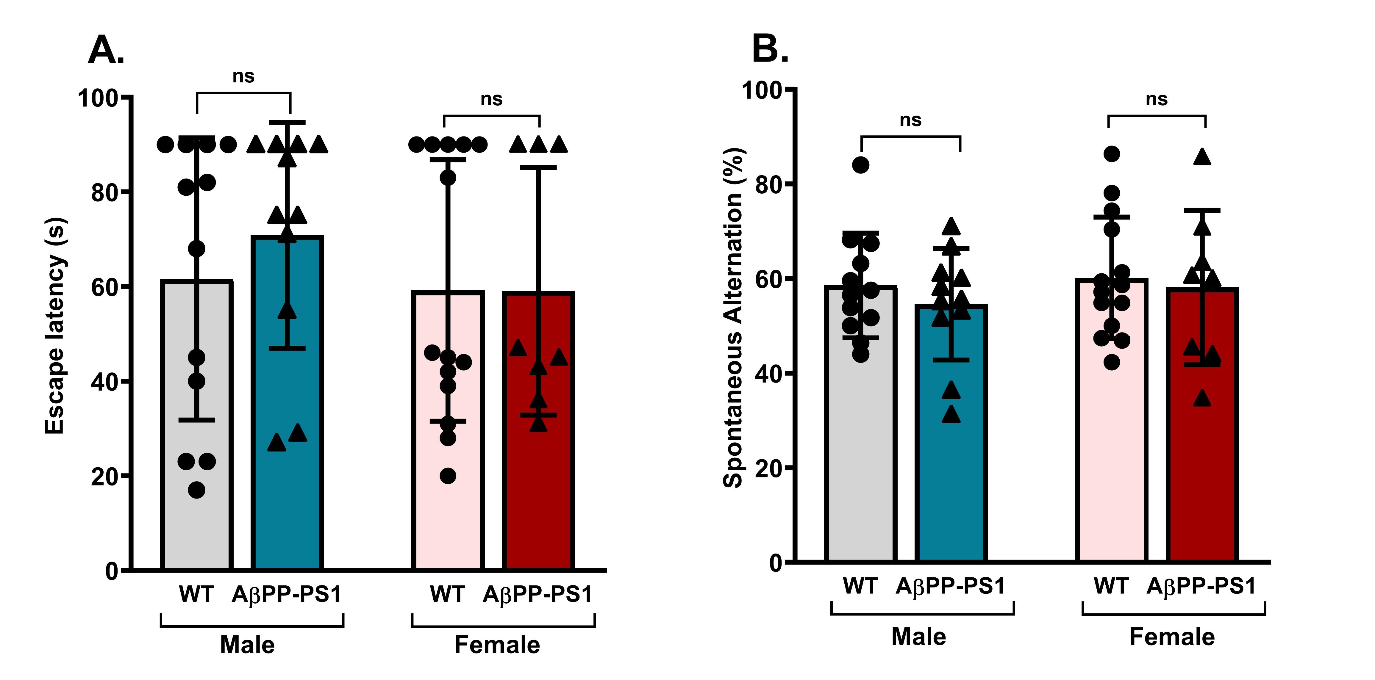
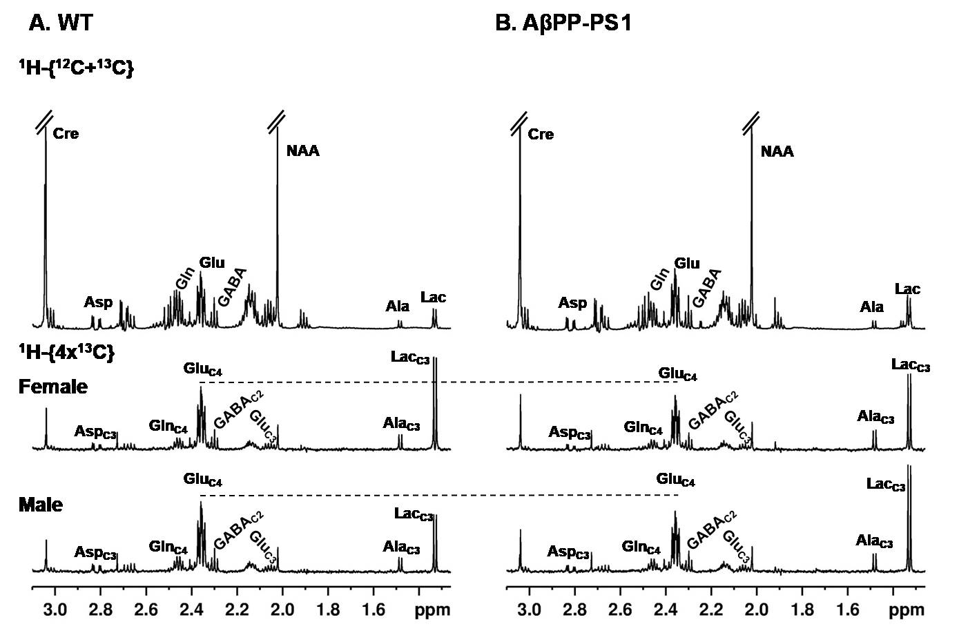
Fig. 2 Representative 1H-[13C]-NMR spectra of hippocampal metabolites from A. WT and B. AβPP-PS1 mice showing 1H resonances of metabolite (upper panel) and 13C labeled metabolites(lower panel) from 10 min of [1,6-13C2]glucose infusion.
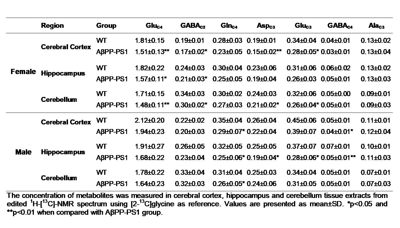
Table 1. 13C concentration (µmol/g) of neurometabolites from [1,6-13C2]glucose in female and male mice.
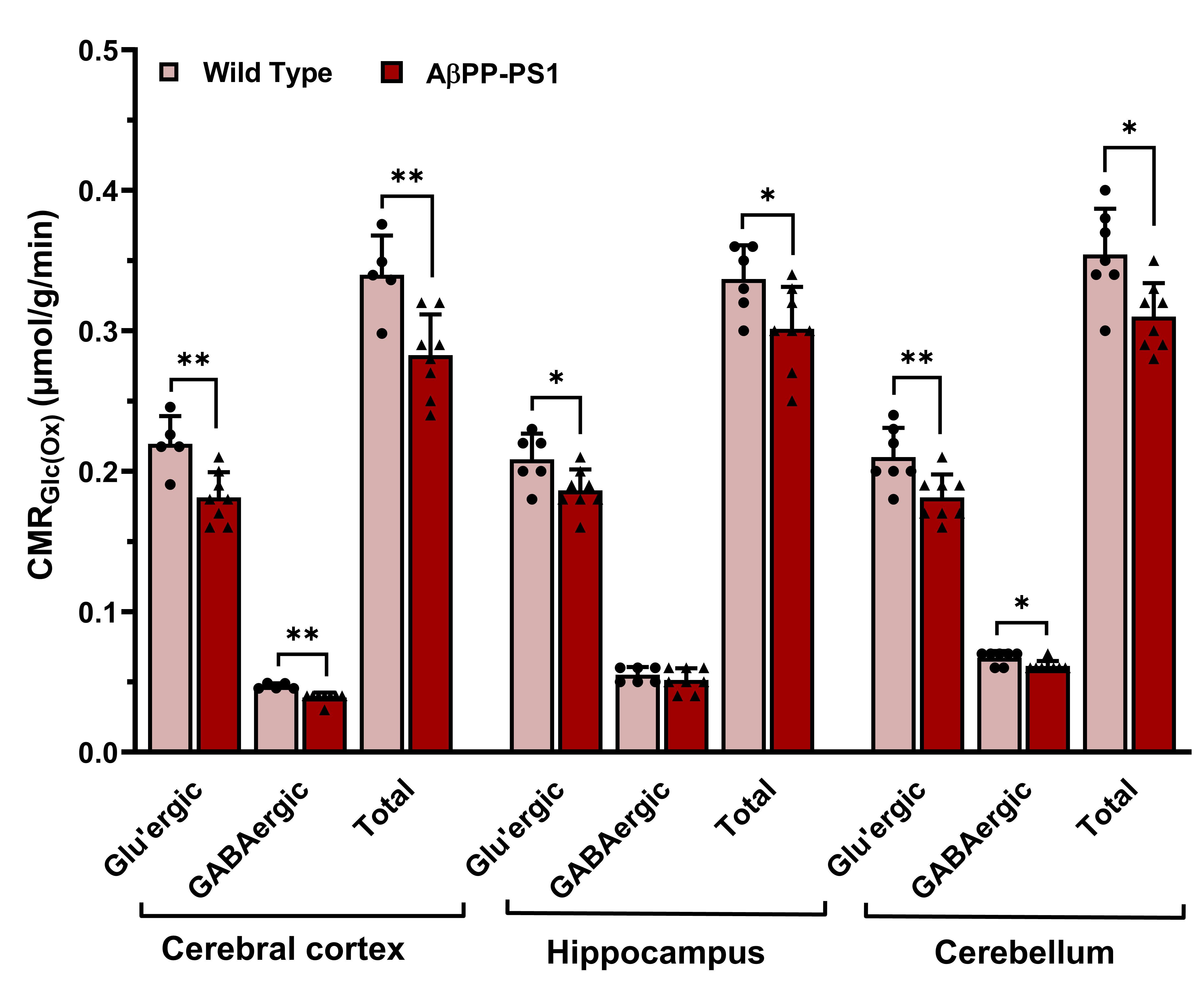
Fig. 3 Cerebral metabolic rate of glucose oxidation in different brain regions of 6 months female AβPP-PS1 and WT mice. Values are presented as mean±SD. *p<0.05 and **p<0.01 when compared with AβPP-PS1 group.
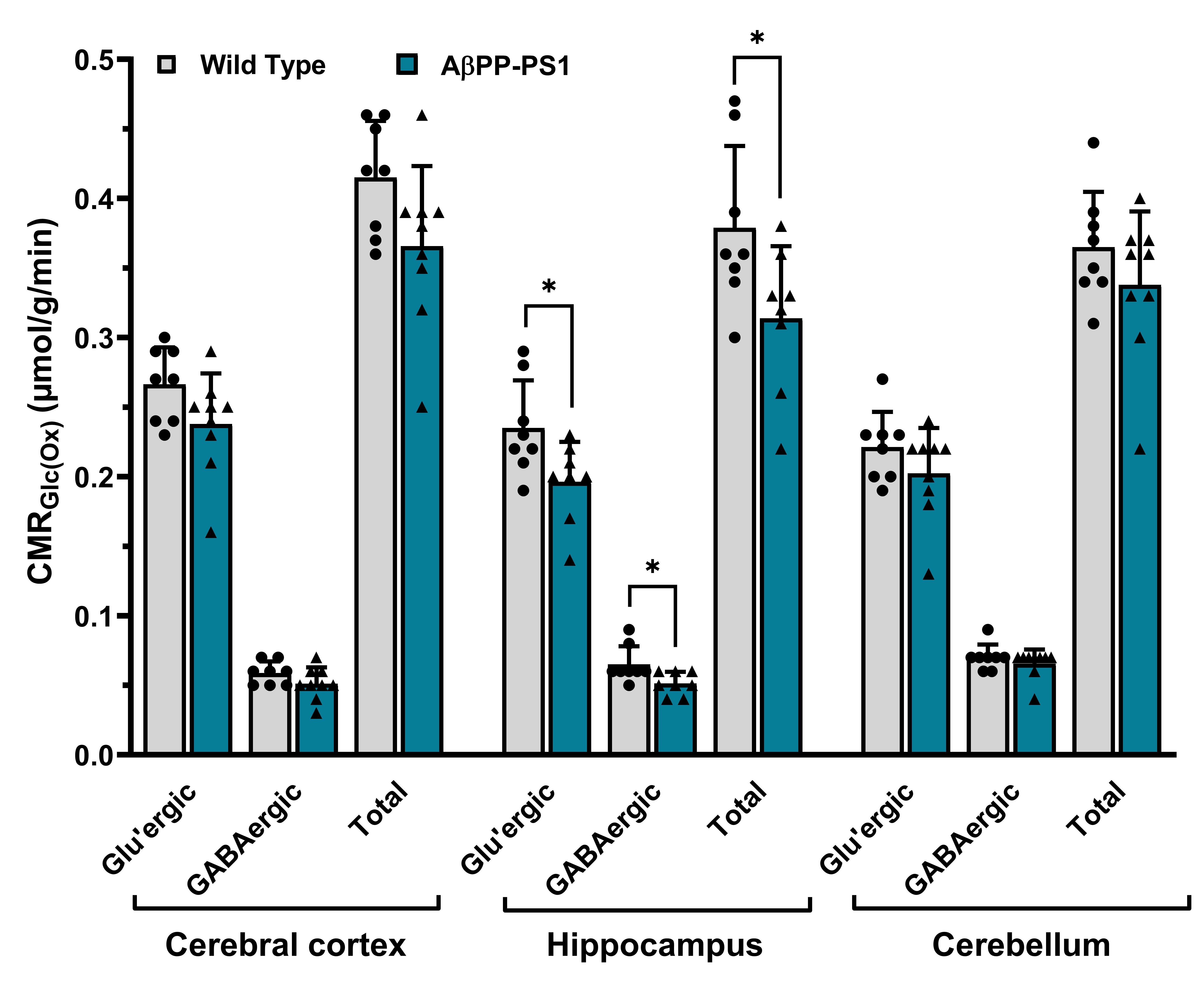
Fig. 4 Cerebral metabolic rate of glucose oxidation in different brain regions of 6 months male AβPP-PS1 and WT mice. Values are presented as mean±SD. *p<0.05 and **p<0.01 when compared with AβPP-PS1 group.