0049
Cross-species comparison: imaging and mapping gastric motor functions in humans and rats using contrast-enhanced rapid MRI1Biomedical Engineering, University of Michigan, Ann Arbor, MI, United States, 2Mechanical Engineering, University of Michigan, Ann Arbor, MI, United States, 3Electrical and Computer Engineering, University of Michigan, Ann Arbor, MI, United States, 4Biomedical Engineering, Electrical and Computer Engineering, University of Michigan, Ann Arbor, MI, United States
Synopsis
Keywords: Digestive, Digestive, Gastrointestinal, Stomach
Motivation: Direct and granular cross-species comparisons of gastric motor functions remain scarce in the literature.
Goal(s): This study aims to establish functional similarities and distinctions of the stomach between humans and rats, and lay the foundation for integrating preclinical findings into clinical gastrointestinal studies.
Approach: Using comparable MRI protocols, we examined the interspecies parallels and distinctions in their functions as pressure and peristaltic pumps.
Results: Similarities were confirmed with high-resolution spatial maps, including intragastric pressure gradient and spatial distribution of peristaltic amplitude and frequency, despite their differences in scale. We highlighted the pronounced variance in initialization and spatial coordination of peristaltic contractions across species.
Impact: This work serves as the first one to map and compare gastric motor events with comparable MRI protocols, laying the foundation for preclinical rat research to clinical translation using contrast-enhanced gastrointestinal MRI.
Purpose
Rats are widely used as models for human gastrointestinal research due to their anatomical parallels1. Despite being monogastric with seemingly similar anatomy, both the rat and human stomachs perform multiple functions: behaving as a compliant pressure pump to accommodate and propel ingesta, and a peristaltic pump for mixing and emptying ingesta2. However, direct and granular functional comparisons at the organ level between the two species remain scarce. Recent studies have demonstrated the viability of contrast-enhanced rapid MRI for imaging the gastrointestinal tract in humans and rats. In this study, we bridged the gap between humans and rat models, and used gastrointestinal MRI to provide a more accurate mapping and comparison of gastric motor functions. Our objective is to establish cross-species similarities and distinctions and lay the foundation to connect preclinical and clinical studies of digestive systems using MRI.Methods
We synthesized a test meal to label food inside the stomach for both rats and humans. The test meal was labeled with gadolinium for rats or manganese from natural ingredients for humans3, 4. After the meal intake, we scanned the gastrointestinal tract in ten Sprague-Dawley rats and ten healthy human subjects. For rats under anesthesia (0.5% isoflurane and 0.01 mg/mL dexmedetomidine, 0.025 mg/kg/h, SC), we acquired T1-weighted 2D gradient echo images (TR = 10.50 ms, TE = 1.44 ms, FA = 25◦, 20 slices, FOV = 64mm × 42mm, voxel size = 0.5 × 0.5 mm, 1.5 mm slice thickness, respiration gated, <3 s per volume, 4 hours in total) using 7-Tesla Agilent MRI3. For humans, we scanned 3D Spoiled Gradient Echo (TR = 3.02 ms, TE = 1.33 ms, FA = 12◦, FOV = 360 × 360 mm, 200 mm thick slab, voxel size = 1.9 × 2.8 × 3.8 mm, free-breathing, ~3 s per volume, rate-5 acceleration, 30 - 45 mins in total ) using 3-Tesla GE MRI4.For both rats and humans, we segmented the intragastric volume and morphed a mesh-based surface template to enclose the segmented volume at each moment in time3, 4. We characterized the gastric morphology and resolved its changes over different time scales, ranging from seconds to hours. In particular, we quantified the slow decrease or increase of local surface area as surrogate measures of tonic contraction or relaxation of gastric muscle, respectively. We detected the rapid displacement of every surface point and characterized the onset, frequency, amplitude, and coordination of peristaltic contractions.
Results
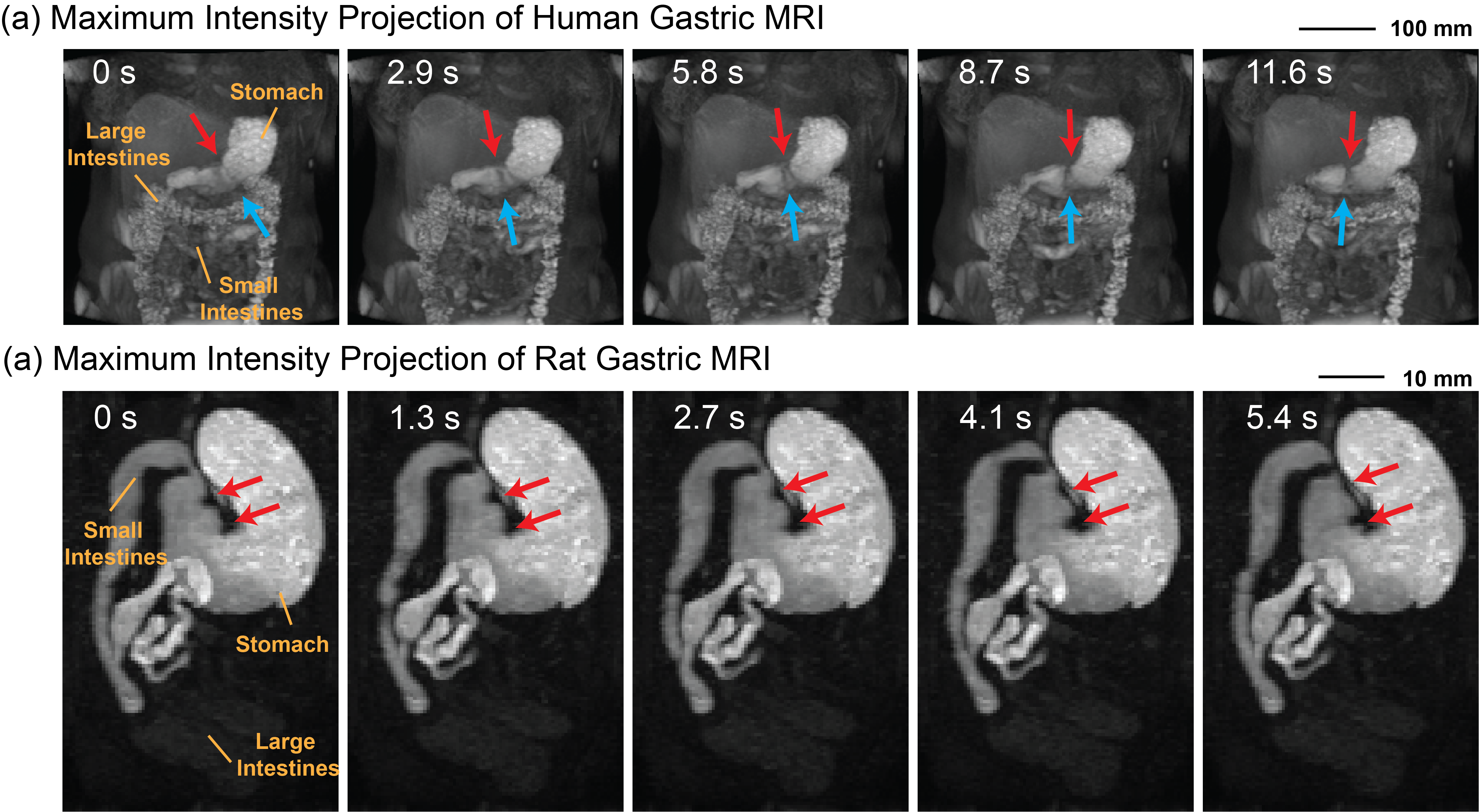
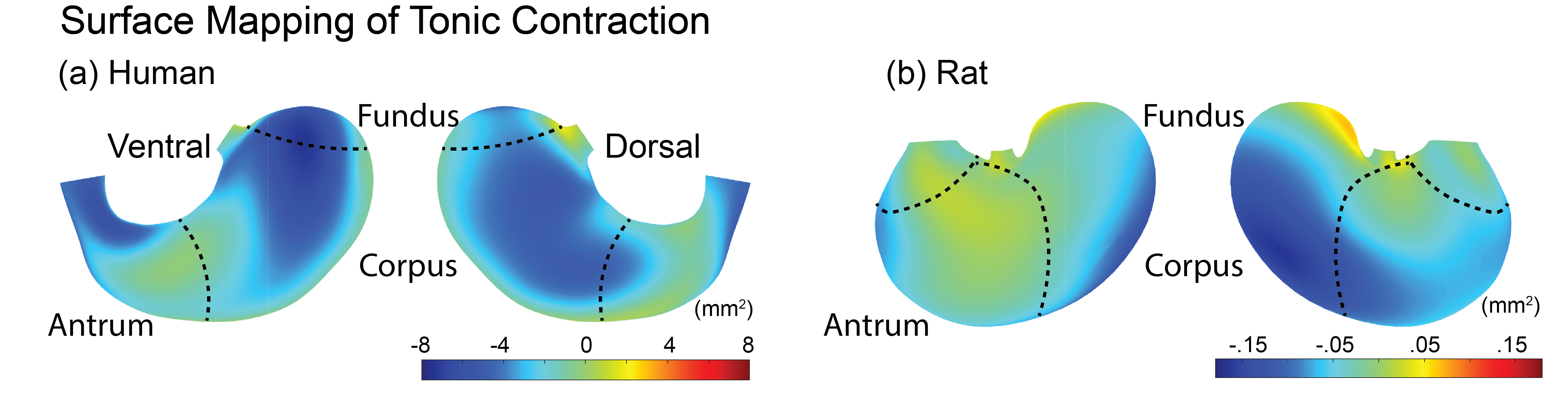
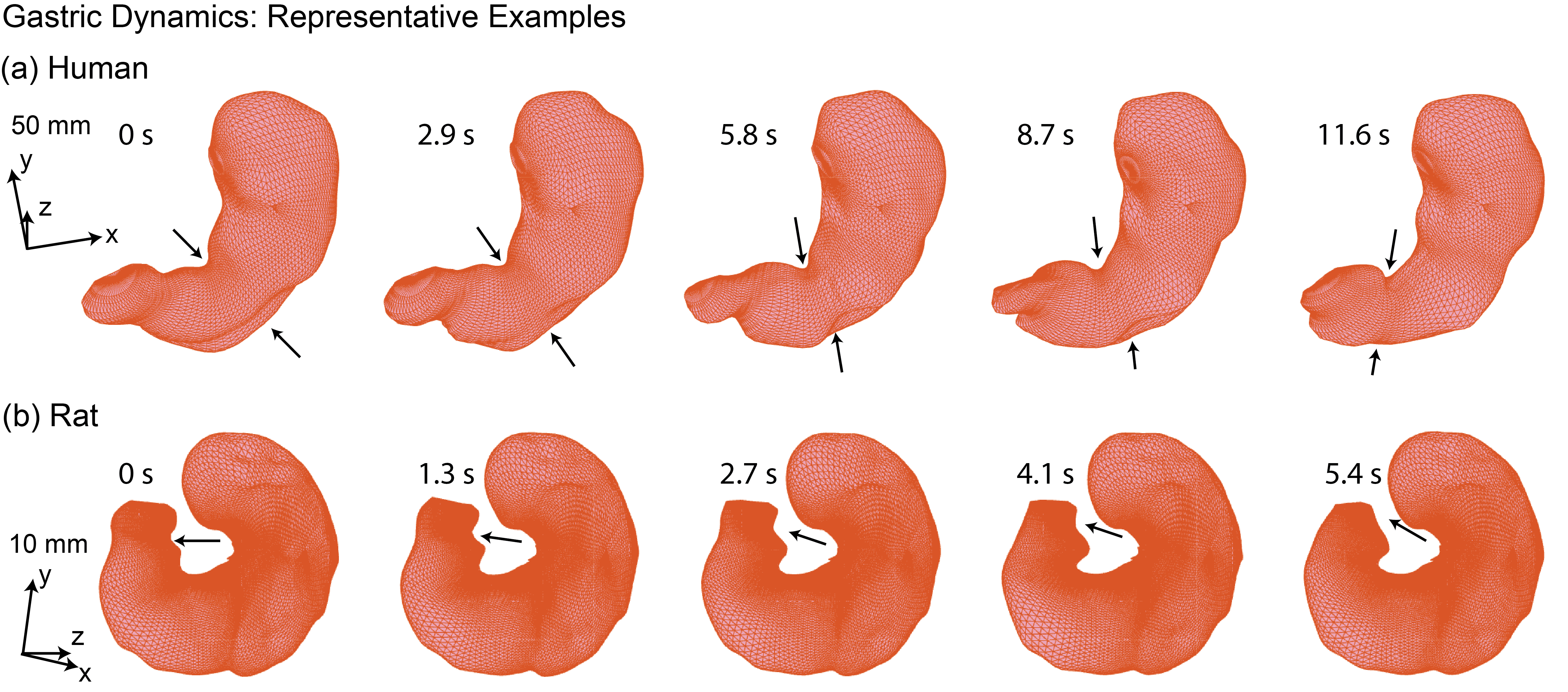
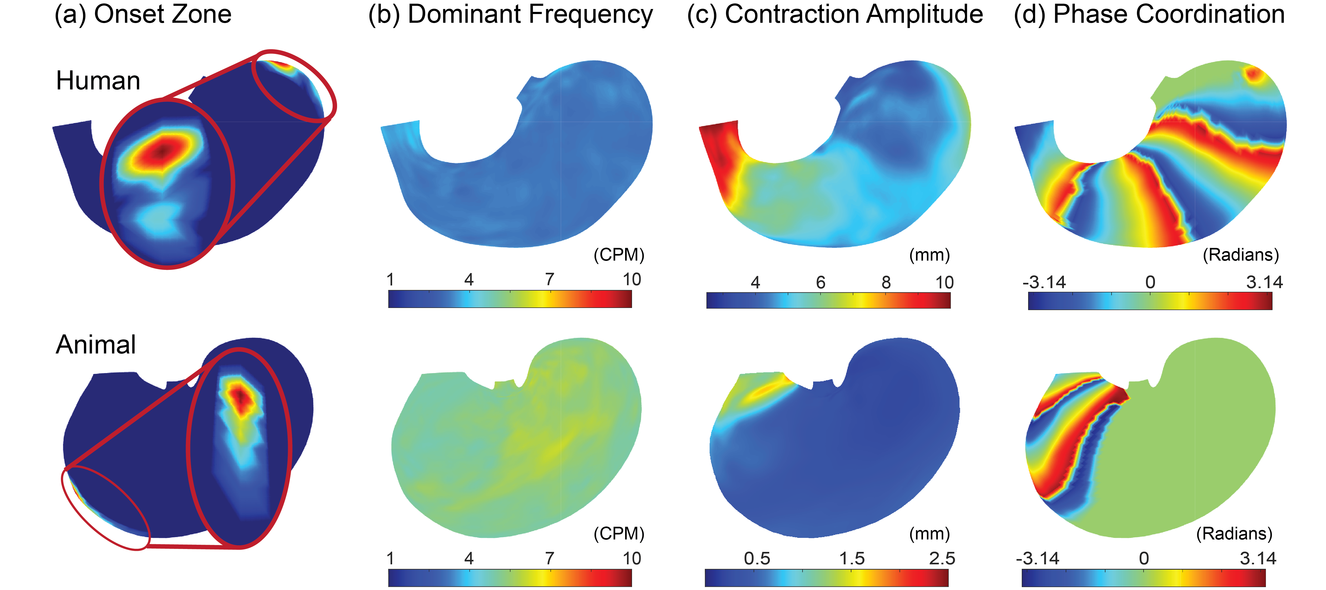
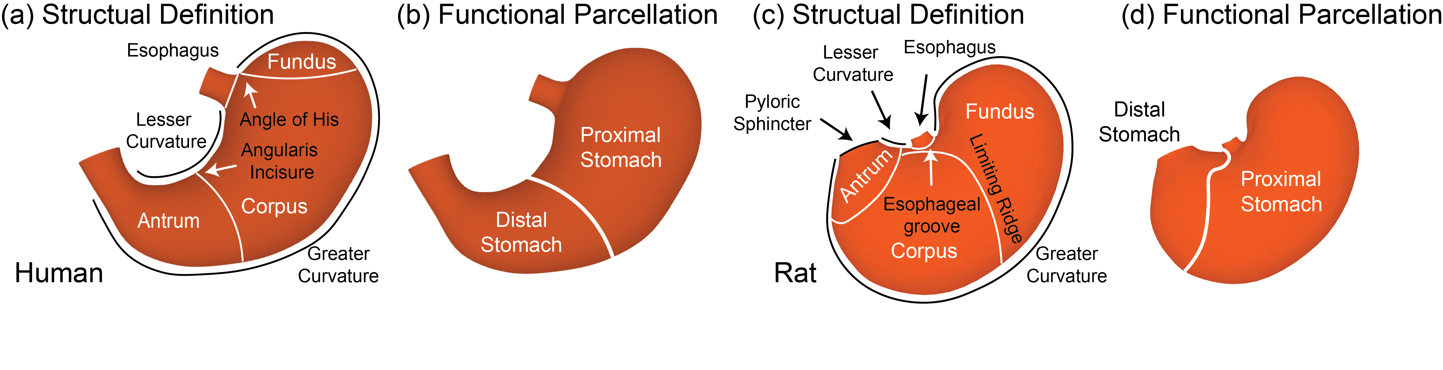
Dynamic T1w images deliver rich information about the stomach and other compartments of the gastrointestinal tract (Figure 1). From the segmented stomach volume, we deduced the shape and dynamics of the gastric wall and mapped the characteristics of both tonic and peristaltic contractions that collectively support digestion.The stomach slowly shrinks its surface area over time by 5.91±0.02 % in rats and by 12.51±0.03% in humans. In both species, the shrinkage is most pronounced in the proximal regions (Figure 2), hypothetically setting up intragastric pressure to propel food towards the middle and distal regions for mixing and grinding. In a shorter time scale, the gastric wall moves as a traveling wave of contractions, driving moment-to-moment morphological changes (Figure 3). This traveling (or peristaltic) wave originates from different onset zones in humans versus rats (Figure 4. a). Its frequency is 3.31±0.08 cycles per minute (CPM) in humans and 5.30±0.18 CPM in rats (Figure 4. b). Its amplitude is increasingly greater towards the distal end of the stomach (Figure 4. c) for both species. The peristaltic wave is phase-coupled across locations in the distal stomach for rats, whereas it is coordinated across a much greater region in the human stomach (Figure 4. d). Based on the characteristics of tonic and peristaltic contractions, the stomach can be divided into two functional regions: the proximal and distal stomach, which appear to be notably different from the conventionally defined structural regions: the fundus, corpus, and antrum (Figure 5).
Conclusion
This work is the first of its kind to use comparable MRI protocols for mapping gastric motor events in both humans and rats, and establish cross-species relationships and distinctions on tonic and peristaltic contractions. Similarities were confirmed with high-resolution spatial maps, including intragastric pressure gradient, and spatial distribution of peristaltic amplitude and frequency, despite their differences in scale. We note the significant interspecies differences in both initialization and spatial coordination of peristaltic contractions. Our findings lay the foundation for preclinical (rat) to clinical (human) translation using gastrointestinal MRI.Acknowledgements
This work was supported by NIH OT2OD030538, OT2OD023847, R01DK131524, and R01AT011665. The authors acknowledge Owen MacKenzie for his help in editing the abstract, and Ashley Cornett for her support in subject recruitment and other logistics.References
1. Di Natale, Madeleine R., et al. "Functional and anatomical gastric regions and their relations to motility control." Neurogastroenterology & Motility (2023): e14560.
2. Goyal, Raj K., Yanmei Guo, and Hiroshi Mashimo. "Advances in the physiology of gastric emptying." Neurogastroenterology & Motility 31.4 (2019): e13546.
3. Wang, Xiaokai, et al. "Diffeomorphic Surface Modeling for MRI-Based Characterization of Gastric Anatomy and Motility." IEEE Transactions on Biomedical Engineering (2023).
4. Wang, Xiaokai, et al. "Mapping human gastric motility using contrast-enhanced MRI with a natural test meal." The International Society of Magnetic Resonance in Medicine (2023).
Figures