0048
Establishment, behavioral, structural and functional characterization of a hindlimb amputation model in mice with multimodal MRI and MRS1RG Translational Imaging, Central Institute of Mental Health, Medical Faculty Mannheim, University of Heidelberg, Mannheim, Germany, 2Pharmacology institute, Medical Faculty, University of Heidelberg, Heidelberg, Germany
Synopsis
Keywords: Small Animals, Spectroscopy
Motivation: Limb amputation frequently leads to pain in residual limb and phantom sensations, but there is no comprehensively described mouse model for translational research.
Goal(s): To extensively phenotype a mouse model of limb amputation to investigate contributing factors of pain.
Approach: Behavioral characterization and multimodal in vivo brain imaging (Voxel-based Morphometry, resting-state functional Magnetic Resonance Imaging and MR-spectroscopy).
Results: VBM showed reduction in primary somatosensory and visual areas (ipsilateral-hemisphere). Functional analysis showed potential neurocompensatory mechanisms and reorganization (left hemisphere). Metabolic data indicated reduced glutamate in the left somatosensory area, and increased N-acetylaspartate in the right somatosensory area.
Impact: We phenotyped a mouse model of limb amputation and showed that sensory and motor areas are involved in the manifestation of pain, which strengthens previous evidence and guides future research.
Introduction
Following limb amputation, patients frequently suffer pain in the residual limb, phantom pain and phantom sensations. These can be spontaneous or evoked by stimulating body parts adjacent and distant to the amputated limb. Considerable evidence suggests that these symptoms occur due to reorganizational changes in primary sensory and motor cortices. However, it has been debated whether maladaptive cortical plasticity or preserved function of the representation of the limb contribute to pain1. As modeling limb amputation is possible only in animals, we established a hindpaw amputation mouse model.Methods
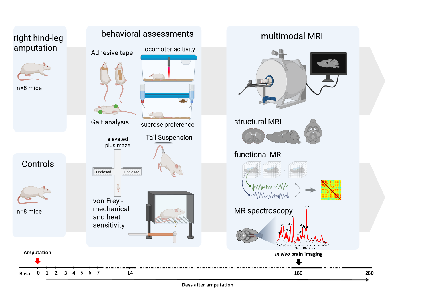
Sixteen mice (8 Amp, 8 Controls) were measured at a 9.4T animal scanner equipped with a circular polarized cryogenic mouse coil (Fig. 1). Anesthesia was a combination of 0.5% isoflurane and medetomidine (0.1 mg/kg/h). Respiratory and cardiac signals were monitored (100Hz) to correct functional data for physiological noise. The MRI acquisition protocol included a high-resolution 3D-scan (T2-weighted RARE, (0.078)²×0.156mm³ resolution), MR-spectroscopy in the left and right primary somatosensory area (PRESS, 2µl-voxel, TR/TE=4000/10ms, frequency-shift -2ppm, respectively) and resting-state fMRI (rsfMRI) (T2*-weighted EPI-FID, TR/TE 1300/18ms, (0.18mm)²*0.5mm, 400 acquisitions, 21 slices). An additional field map was acquired to correct for geometrical distortions. Analyses were performed using SPM12, in-house MATLAB scripts, FSL and LCModel.VBM: Preprocessing steps included brain extraction, bias correction, coregistration, segmentation and nonlinear normalization using DARTEL. Prior knowledge images were created in a previous study2. Normalized and modulated gray matter images as well as resulting jacobian determinant images were analyzed in a second-level GLM using total intracranial volume as covariate.
MRS: Individual frequency and phase correction were done over coils and averages as well as eddy current correction using an unsuppressed water-signal. Spectra were quantified by LCModel using a calibrated phantom basis dataset. Metabolite concentrations were referenced to the unsuppressed water signal, assuming mean water concentration of 46.106mol/L, and corrected for relaxation effects (T1/T2met=1500/300ms, T2water=45ms)3.
rsfMRI: Preprocessing included correction for field inhomogeneities and movement, physiological noise4 regression, slice-timing correction and spatial normalization to a template using DARTEL-based flow fields from the 3D images, regression of movement parameters and CSF signal, DVARS scrubbing5, and band-pass filtering (0.01–0.1Hz). Regional time courses were extracted using 45x2 unilateral anatomical regions6. Preprocessed rsfMRI data was entered into graph theoretical network analyses.
We also performed a detailed behavioral characterization in a greater sample of mice (N=12/group), including homecage monitoring, gait analysis, video-observations, free-choice, stimulus-dependent and -independent nociceptive tests and affective behavior evaluation. To assess intra- and inter-cortical reorganizations, anterograde and retrograde tracers were injected into the somatosensory hindpaw region.
Results
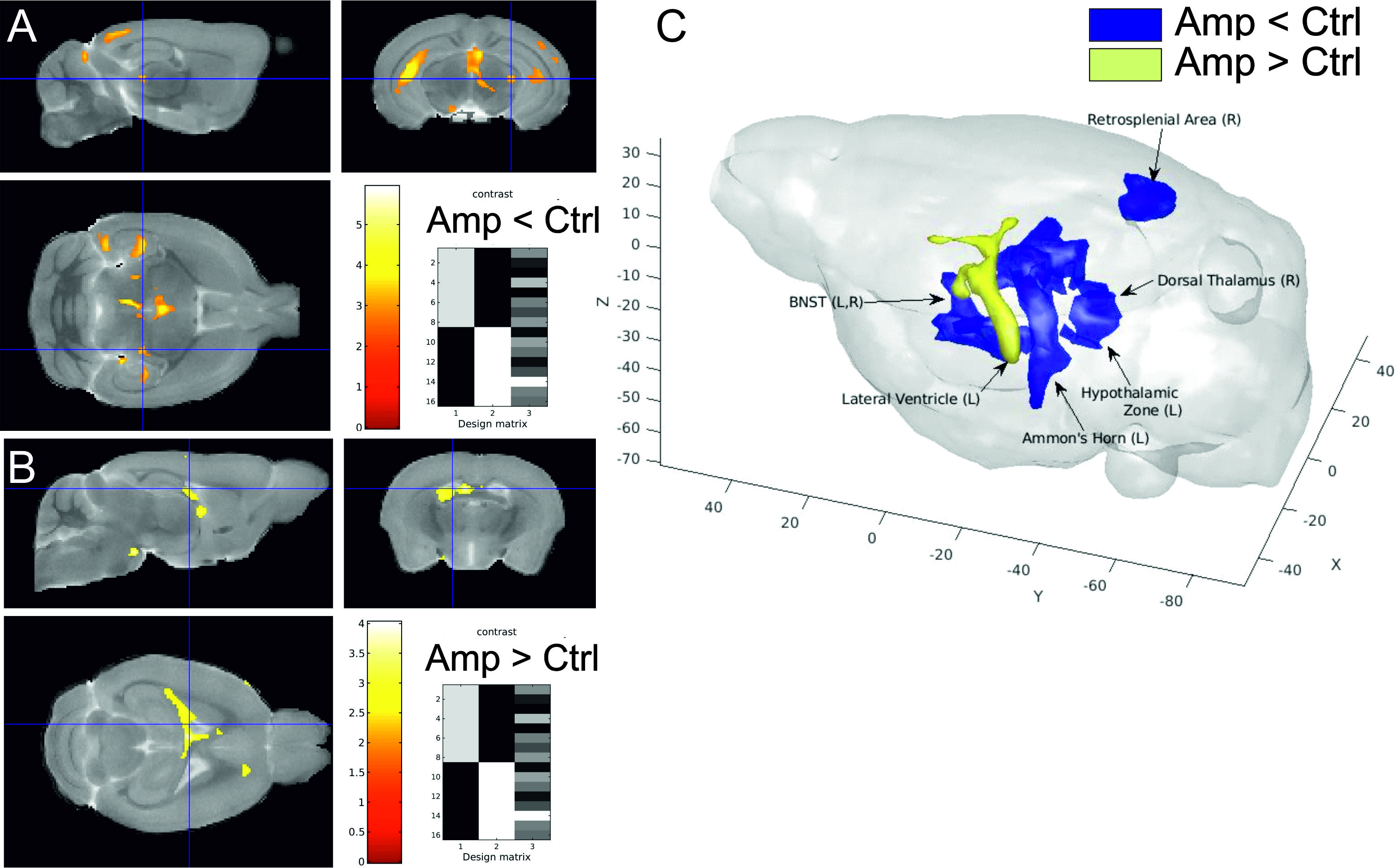
Following right hindpaw amputation, mice showed no apparent general impairment of homecage behavior, despite gait alterations. However, we measured mechanical and thermal hypersensitivity in the contralateral paw and found an increased touch sensitivity in the shoulder/nape region. These changes were not associated with affective behavior alterations.VBM data showed strongly lateralized structural changes with increased left hemisphere ventricle, volume reduction of Ammon’s horn and dentate gyrus (left hemisphere) and bilateral habenula, thalamus and hypothalamus. Regional analysis revealed reduction in the primary somatosensory and visual areas in the right hemisphere. Only the bilateral septal nucleus involved in movement in the context of motivation showed increased volume in the amputee group (Fig. 2).
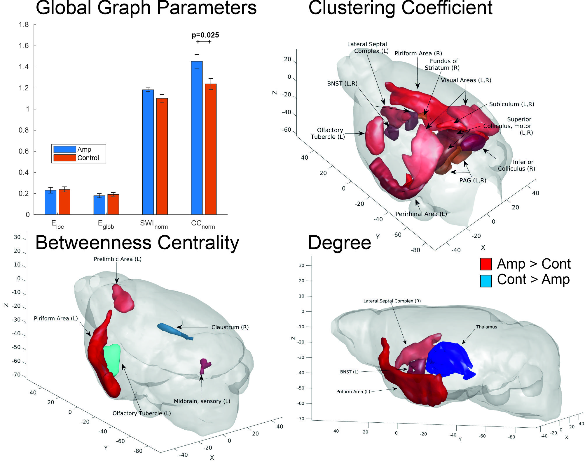
In the functional analysis the right lateral septal nucleus showed an increased number of connections. The functional analysis also demonstrated reorganisation in the left hemisphere signified by a stronger segregated network in the amputated animals, with a higher overall clustering coefficient driven mostly by regions involved in motor and reward functioning. Additionally, the left BNST showed an increased number of connections, while the left thalamus manifested the opposite pattern (Fig. 3).
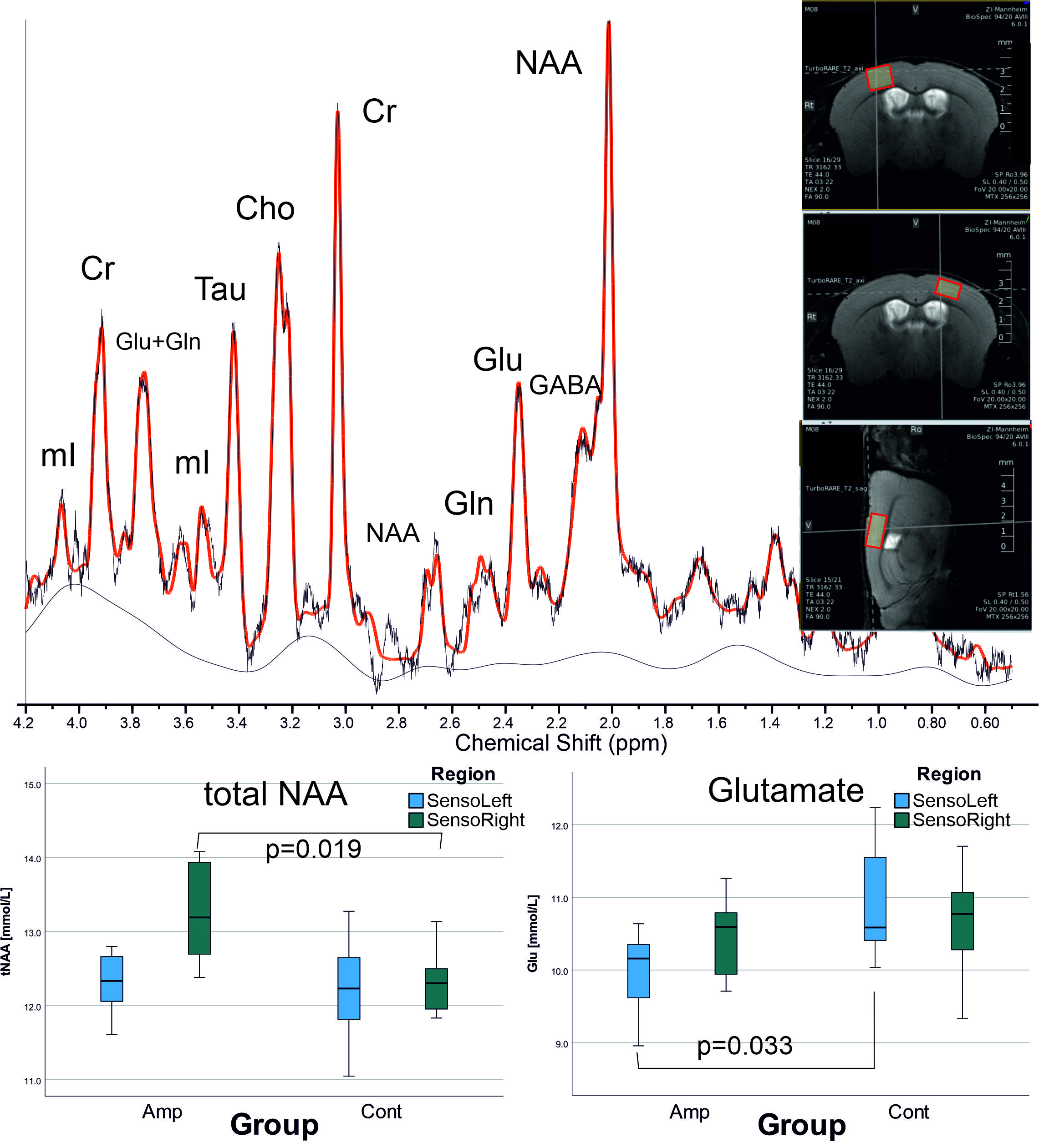
Metabolic data indicated reduced glutamate in the left somatosensory area, likely related to reduced activation after amputation and increased N-acetylaspartate in the right somatosensory area, possibly reflecting higher neuronal density as a marker for ipsilateral side compensation (Fig. 4).
Summary/Discussion
Changes were strongly lateralized in all three modalities. In VBM, only the bilateral septal nucleus (involved in movement in the context of motivation) showed increased volume in the amputee group. In the functional analysis the right lateral septal nucleus showed an increased number of connections, which hints at potential neurocompensatory mechanisms. Metabolic data can be related to reduced activation after amputation and higher neuronal density as a marker for ipsilateral side compensation. This first hindpaw mouse amputation model shows behavioral and brain neuroplastic changes, shedding light into the mechanisms underlying pain-related alterations observed in patients.Acknowledgements
We thank Felix Hörner for his skillfully conducted MR-measurements.References
- Makin TR, Flor H. Brain (re)organisation following amputation: Implications for phantom limb pain. Neuroimage. 2020 Sep;218:116943.
- Biedermann S, Fuss J, Zheng L, Sartorius A, Falfán-Melgoza C, Demirakca T, Gass P, Ende G, Weber-Fahr W. In vivo voxel based morphometry: detection of increased hippocampal volume and decreased glutamate levels in exercising mice. Neuroimage. 2012 Jul 16;61(4):1206-12.
- Bilbao A, Falfán-Melgoza C, Leixner S, Becker R, Singaravelu SK, Sack M, Sartorius A, Spanagel R, Weber-Fahr W. Longitudinal Structural and Functional Brain Network Alterations in a Mouse Model of Neuropathic Pain. Neuroscience. 2018 Sep 1;387:104-115.
- van Buuren M, Gladwin TE, Zandbelt BB, van den Heuvel M, Ramsey NF, Kahn RS, Vink M, Cardiorespiratory effects on default-mode network activity as measured with fMRI. Hum Brain Mapp 30:3031-3042 (2009).
- J. D. Power, A. Mitra, T. O. Laumann, A. Z. Snyder, B. L. Schlaggar, S. E. Petersen, Methods to detect, characterize, and remove motion artifact in resting state fMRI. Neuroimage. 2014 84:320–341.
- Sack M, Zheng L, Gass N, Ende G, Sartorius A, Weber-Fahr W. Interactive tool to create adjustable anatomical atlases for mouse brain imaging. MAGMA. 2021 Apr;34(2):183-187.
Figures