0045
High-fidelity Four-dimensional Abdominal Diffusion-Weighted Imaging Enabled by SCOPER and Multi-Band acceleration (4D-DW-MB-SCOPER)1The Hong Kong Polytechnic University, Hong Kong, China, 2The Chinese University of Hong Kong, Hong Kong, China
Synopsis
Keywords: Image Reconstruction, Radiotherapy
Motivation: 4D-DWI can benefit the treatment planning in radiotherapy (RT) because of its high tumor-to-tissue contrast. Both 4D-DW-PROPELLER-EPI and 4D-DW-SCOPER have been proposed but suffer from long acquisition time, thereby limiting clinical applications
Goal(s): This study aims to develop a new technique to achieve distortion-free 4D-DWI with a practical acquisition time.
Approach: 4D-DW-SCOPER and multiband (MB) techniques were combined, termed 4D-DW-MB-SCOPER. In vivo experiments were performed.
Results: Results indicate that 4D-DW-MB-SCOPER is feasible for achieving distortion-free 4D-DWI within 7 mins for a coverage of 176 mm in the Superior-Inferior (SI) direction, and has the potential to benefit treatment planning in clinical RT.
Impact: The results might offer a new way for clinicians to perform 4D RT planning as well as for patients to have a better treatment outcome. However, the reconstruction time is long for the technique and need to be further investigated.
INTRODUCTION
Four-dimensional diffusion-weighted imaging (4D-DWI) can provide high tumor-to-tissue contrast, thereby potentially benefiting treatment planning for radiotherapy (RT). Two previously proposed motion-resolved 4D-DWI techniques, i.e., 4D-DW-PROPELLER-EPI1 and 4D-DW-SCOPER2, can achieve distortion-free abdominal 4D-DWI, therefore overcoming the distortion problem in the originally proposed 4D-DWI technique3. Compared with 4D-DW-PROPELLER-EPI, 4D-DW-SCOPER joint reconstructs the image from all blades by solving encoding equation through conjugate gradient (CG)-SENSE, resulting in around 22% reduction in acquisition time (i.e., 840s for covering 176 mm in the superior-inferior (SI) direction). However, further acquisition time reduction is still needed to improve its clinical applicability. A widely adopted through-plane acceleration technique, multi-band imaging (MB), can be used for maximizing productivity in various DWI and functional MRI applications4-7, with a slice acceleration by a factor of Rslice. Additionally, the combination of MB with PROPELLER reconstruction has been shown to reduce acquisition time by approximately 40%8. In our study, we propose a novel 4D-DWI technique by integrating MB into 4D-DW-SCOPER, termed 4D-DW-MB-SCOPER, to achieve distortion-free 4D-DWI with a more reasonable acquisition time.METHODS
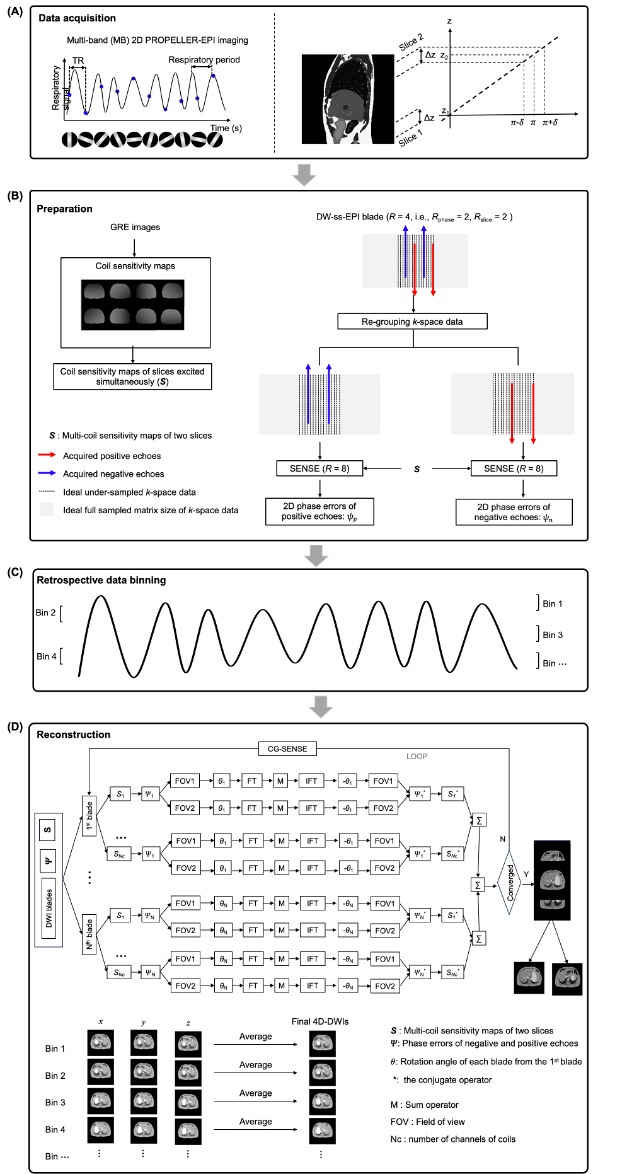
Flowchart of 4D-DW-MB-SCOPER: Figure 1 demonstrates the flowchart of the proposed motion-resolved 4D-DW-MB-SCOPER for reconstructing the 4D-DWI dataset (i.e., A series of 2D datasets at different respiratory bins). First, DW-MB-ss-EPI blades with Rslice=2, and GRE images were acquired (Figure 1(A)). Then, coil sensitivity maps were estimated by GRE images, and the phase errors were estimated for each blade (Figure 1(B)). Subsequently, all blades were sorted into several amplitude bins based on K-B amplitude binning9(Figure 1(C)). Because each sorted blade contained the aliased data acquired from two locations, they shared the same binning index according to the recorded respiratory information. Then, the 4D-DWI data in each diffusion direction were reconstructed using the MB-SCOPER method and aliased slices were separated. The reconstructed 4D-DWI data from the three diffusion directions were averaged to produce the final 4D-DWI dataset (Figure 1(D)).In vivo experiment:
Acquisition: Abdominal data was acquired from a healthy volunteer from the mid-thorax to mid-pelvis at a 1.5 T MRI scanner (SIGNA Artist, GE Healthcare) using a 45-channel phase-array coil. Free-breathing T2W-MB-ss-EPI blades (b-value=0 s/mm2, Rphase=2, Rslice=2) and DW-MB-ss-EPI blades (b-value=500 s/mm2, x diffusion direction, Rphase=2, Rslice=2) were acquired using a 2D LAP-PROPELLER sequence with the parameters as follow: TR=1500ms; FOV=380×380mm2; matrix size=128×32; target in-plane resolution=2.97×2.97mm2; rotation angle=111.25o; slice thickness=8.00mm; slice number=22; and number of repeated acquisitions (Nr)=80. The respiratory waveform was synchronously recorded during blade acquisition (sampling rate of 25 Hz). The 20s-breath-holding single-band GRE images were acquired to calculate coil sensitivity maps with the geometric parameters identical to 4D-DW-MB-SCOPER.
Data binning and reconstruction: Two 4D-DWI datasets were reconstructed from either 4D-DW-MB-SCOPER or 4D-DW-MB-PROPELLER-EPI (i.e., 4D-DW-PROPELLER-EPI using the MB) for comparing the performance with the use of MB. Amplitude bin intervals were set as 0-28%, 17%-39%, 33%-61%, 44%-72%, 67%-83%, and 78%-100% (0-100% corresponding the normalized chest wall motion). K-B amplitude binning9 with pre-set angular difference of 10o was used to sort the blades into six respiratory bins. After data sorting, six-bin 4D-DWI datasets were produced by either conventional PROPELLER-EPI reconstruction or MB-SCOPER for each diffusion direction.
Data analysis: To compare the reconstructed images of 4D-DW-MB-SCOPER and 4D-DW-MB-PROPELLER-EPI, visual assessments and SNR evaluations were performed using a two-sample paired t-test.
RESULTS
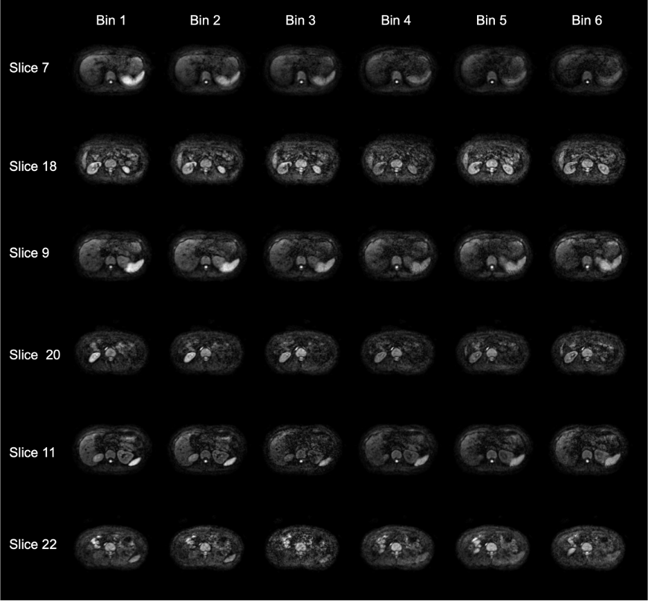
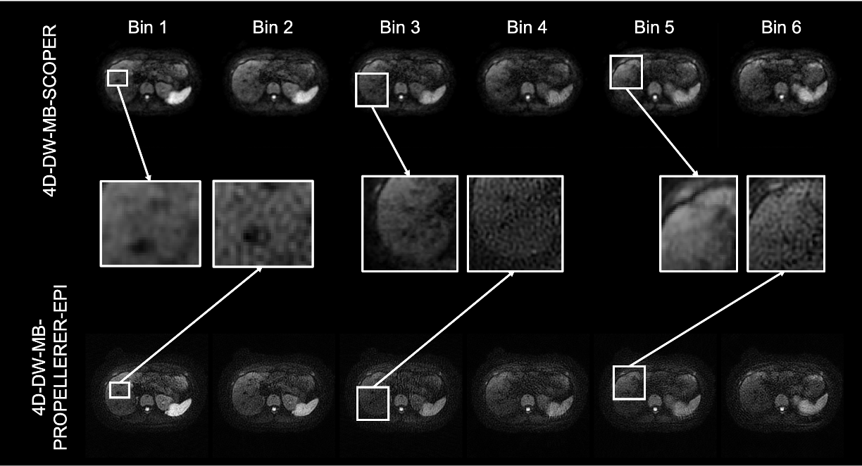
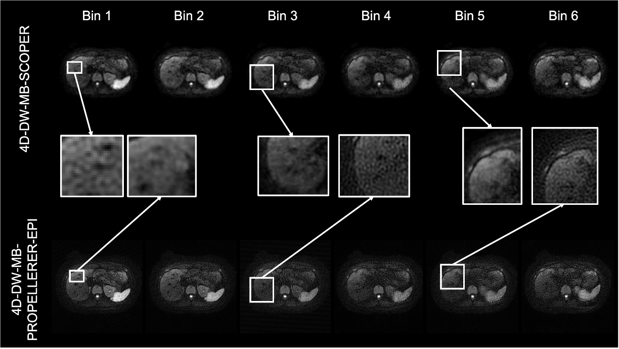
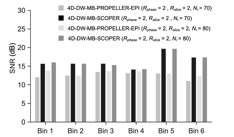
Figure 2 shows that 4D-DW-MB-SCOPER is feasible for achieving distortion-free images with a 7-min acquisition time and coverage of 176 mm in the SI direction, resulting in 62.5% and 50% reductions of acquisition time compared with 4D-DW-PROPELLER-EPI and 4D-DW-SCOPER, respectively. Visual assessments indicate that 4D-DW-MB-SCOPER has better SNR performance than 4D-DW-MB-PROPELLER-EPI at both Nr of 70 (Figure 3) and 80 (Figure 4). In addition, 4D-DW-MB-PROPELLER-EPI has lower SNR than 4D-DW-MB-SCOPER at Nr of 70 (p=0.021) and 80 (p=0.014) (Figure 5). Also, SNR increases in general with the increase of blade number for 4D-DW-MB-PROPELLER-EPI, whereas almost remains steady for 4D-DW-MB-SCOPER (Figure 5).DISCUSSION
Unlike 4D-DW-MB-PROPELLER-EPI, the 10-blade increase does not influence the performance of 4D-DW-MB-SCOPER, verifying its robustness and is consistent with the SCOPER-EPI study in brain10. Recent work shows a 32-channel coil has been developed for Unity MR-LINAC11, allowing 4D-DW-MB-SCOPER in clinical applications. One limitation of the 4D-DW-MB-SCOPER is the phase discontinuity of the slice at the marginal of FOV, which needed to be improved further. Another limitation is the prolonged post-processing time for extensive FOV image reconstruction; thus, to further the clinical feasibility, advanced methods are needed to faster the converge of iterative reconstruction in the future.CONCLUSION
In conclusion, the preliminary in vivo results demonstrate that 4D-DW-MB-SCOPER can achieve distortion-free 4D-DWI within a considerably reduced acquisition time, thereby potentially benefiting clinical RT.Acknowledgements
The work was in part supported by grants from Hong Kong Research Grant Council (GRF17106820, GRF17125321, GRF14206723, and ECS24213522).References
1. Wang L, Li T, Cai J, Chang HC. Motion-resolved four-dimensional abdominal diffusion-weighted imaging using PROPELLER EPI (4D-DW-PROPELLER-EPI). Magnetic Resonance in Medicine. Published online 2023.
2. Wang L, Li T, Liu C, Cai J, Chang HC. Motion-Resolved Four-Dimensional Abdominal Diffusion-Weighted Echo-Planar Imaging Using Scoper Reconstruction (4D-DW-SCOPER-EPI). In: ; 2023.
3. Liu Y, Zhong X, Czito BG, et al. Four-dimensional diffusion-weighted MR imaging (4D-DWI): a feasibility study. Medical physics. 2017;44(2):397-406.
4. Feinberg DA, Setsompop K. Ultra-fast MRI of the human brain with simultaneous multi-slice imaging. Journal of magnetic resonance. 2013;229:90-100.
5. Peng S, Guo Y, Zhang X, et al. High-Resolution DWI with Simultaneous Multi-Slice Readout-Segmented Echo Planar Imaging for the Evaluation of Malignant and Benign Breast Lesions. Diagnostics. 2021;11(12):2273.
6. Obele C, Bhat H, Chandarana H. Simultaneous Multi-Slice Accelerated Free-Breathing Diffusion-Weighted Imaging in Abdomen and Pelvis.
7. Hoge RD, Badhwar A, Doyon J, Ostry D. Improving Sensitivity and Specificity in BOLD fMRI Using Simultaneous Multi-Slice Acquisition. MAGNETOM Flash. 2015;63:65-69.
8. Norbeck O, Avventi E, Engström M, Rydén H, Skare S. Simultaneous multi-slice combined with PROPELLER. Magnetic Resonance in Medicine. 2018;80(2):496-506.
9. Wang L. Evaluation of k-space uniformity and blade number-optimized (K-B-optimized) binning method for abdominal 4D-DW-Propeller-EPI. In: ; 2022.
10. Liu X, Cui D, Dai E, Hui ES, Chan Q, Chang HC. Self-calibrated and Collaborative Propeller-EPI Reconstruction (SCOPER) for High-Quality Diffusion-Tensor Imaging. In: International Society for Magnetic Resonance in Medicine (ISMRM 27th Annual Meeting & Exhibition, 2019. International Society for Magnetic Resonance in Medicine.; 2019.
11. Zijlema SE, Tijssen RH, Van Dijk L, et al. Improving the imaging performance of the 1.5 T MR-linac using a flexible, 32-channel, on-body receive array. Physics in Medicine & Biology. 2020;65(21):215008.
Figures