0044
Distortion-Free Fat/Water Separated Body Diffusion-Weighted Imaging using Spatio-Temporal Joint Reconstruction1Department of Radiology, Stanford University, Stanford, CA, United States, 2Department of Bioengineering, Stanford University, Stanford, CA, United States, 3Department of Electrical Engineering, Stanford University, Stanford, CA, United States
Synopsis
Keywords: Pulse Sequence Design, Data Acquisition
Motivation: DWI is effective for cancer imaging, but conventional EPI suffers from geometric distortion and chemical shift artifacts. Conventional fat suppression techniques are sensitive to the large B0 and B1+ inhomogeneities in the body. Residual fat causes artifacts and is a confounding factor in using DWI for cancer diagnosis.
Goal(s): Perform robust fat/water separation in distortion-free DWI.
Approach: A diffusion-weighted EPTI acquisition and joint reconstruction method is used. Separation is performed using chemical shift encoding along the temporal dimension. A distortion-less FSE-based phase navigator is used to resolve shot-to-shot phase.
Results: The proposed method is validated in vivo in the brain, head&neck, and breast.
Impact: Using the proposed navigated EPTI sequence, we demonstrated fat/water separated DWI that is robust to B0 variation in the body. This will enable more reliable use of DWI to assess cancer and other abnormalities, complementing or replacing contrast-enhanced imaging.
Introduction
Diffusion-weighted imaging (DWI) is an imaging technique for cancer diagnosis. However, the commonly-used EPI images suffer from geometric distortion and chemical shift artifacts. Distortion artifacts can be effectively resolved by using spatio-temporal acquisition methods1,2,3, which distribute samples in ky-t space and generate distortion-free images at a series of echo times. Conventional fat-suppression methods, such as water-selective excitation4 and fat saturation5, are often applied in DWI acquisition, but are sensitive to B0 and B1 inhomogeneities that are prominent in body imaging. Residual fat signal not only results in image artifacts but can also potentially reduce the diffusion contrast between benign and malignant tissues and affect their apparent diffusion coefficient (ADC) estimates, given that fat has an extremely low ADC 6,7. In this work, we propose a method that generates distortion-free, fat/water separated diffusion-weighted images that is robust over B0 field inhomogeneities commonly observed in body.Methods
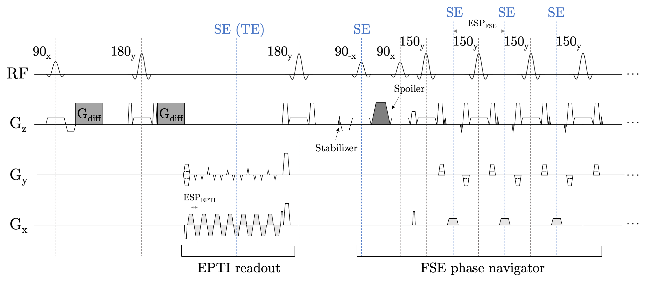
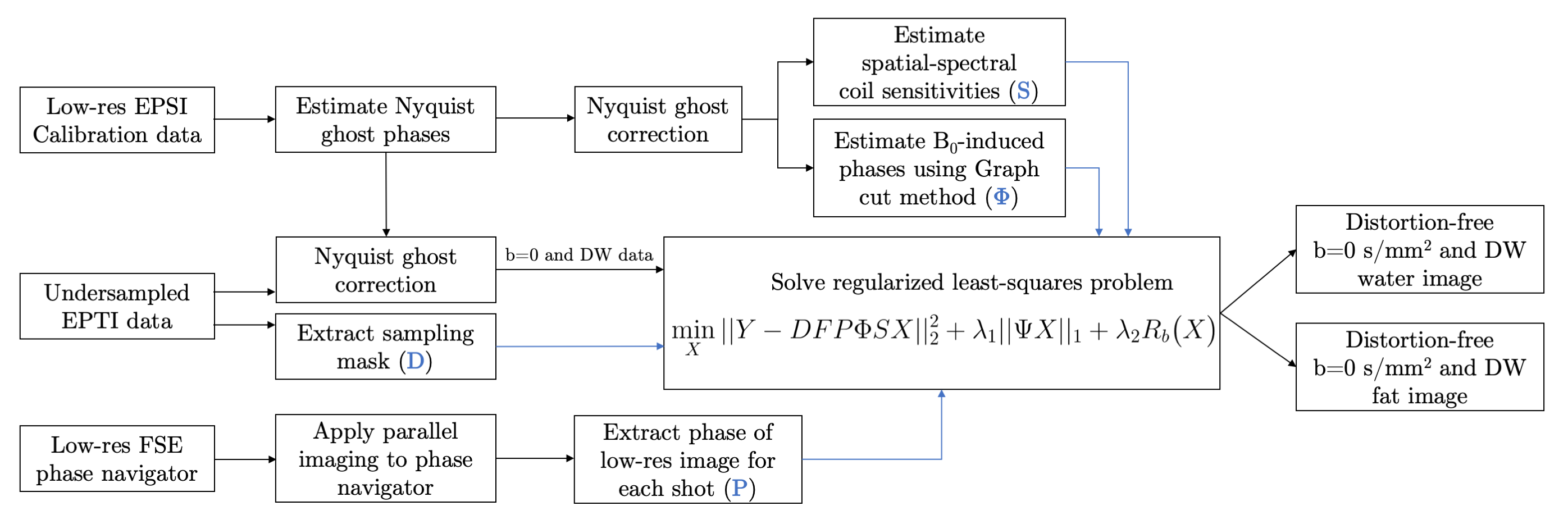
The sequence and the sampling pattern in ky-t space are shown in Figure 1. The Stejskal-Tanner pulsed gradient spin echo8 is followed by the echo planar time-resolved imaging (EPTI) readout3. Echo planar spectroscopic imaging (EPSI)1 was used for the acquisition of calibration data. To resolve the shot-to-shot motion-induced phase in DWI, a phase navigator is acquired after a second refocusing pulse using a fast spin-echo (FSE) readout9. Compared with the conventional EPI phase navigator, the FSE-based phase navigator avoids distortion and chemical shift artifacts, thus is more accurate and better registered with the distortion-free images from the main readout.The reconstruction pipeline is shown in Figure 2. Similar to previous work10, the reconstruction and separation can be performed jointly. The problem is formulated as a least-squares problem with l1-regularization on the 3D wavelet transform in the xyf domain, and locally low-rank (LLR) regularization along xyf dimension.
Acquisitions were performed at 3T (Signa Premier, GE Healthcare). Four healthy volunteers (two head&neck, two breast) and two patients with biopsy-proven breast cancer were scanned following IRB approval and informed consent. Resolution 1.9x1.9x5mm. The main EPTI readout was acquired with rBW ±250kHz, ETL 48 and echo spacing ranging from 0.58 to 0.70ms. We used 24 calibration lines in ky-t, 16 shots, b-value = 600s/mm2 for head&neck scans and 800s/mm2 for breast scans. FSE-based phase navigator was acquired with refocusing flip angle 150°, rBW ±8kHz, ETL 8, undersampling rate along ky (Rpe) = 3. For breast acquisitions, partial fat suppression was applied using an adiabatic spectral inversion (ASPIR) with an inversion time (TI) of 45ms. Total scan time is 4:12 for head&neck and 4:40 for breast. The vendor provided water-only 4-shot DW-EPI with same resolution was used for comparison.
Results
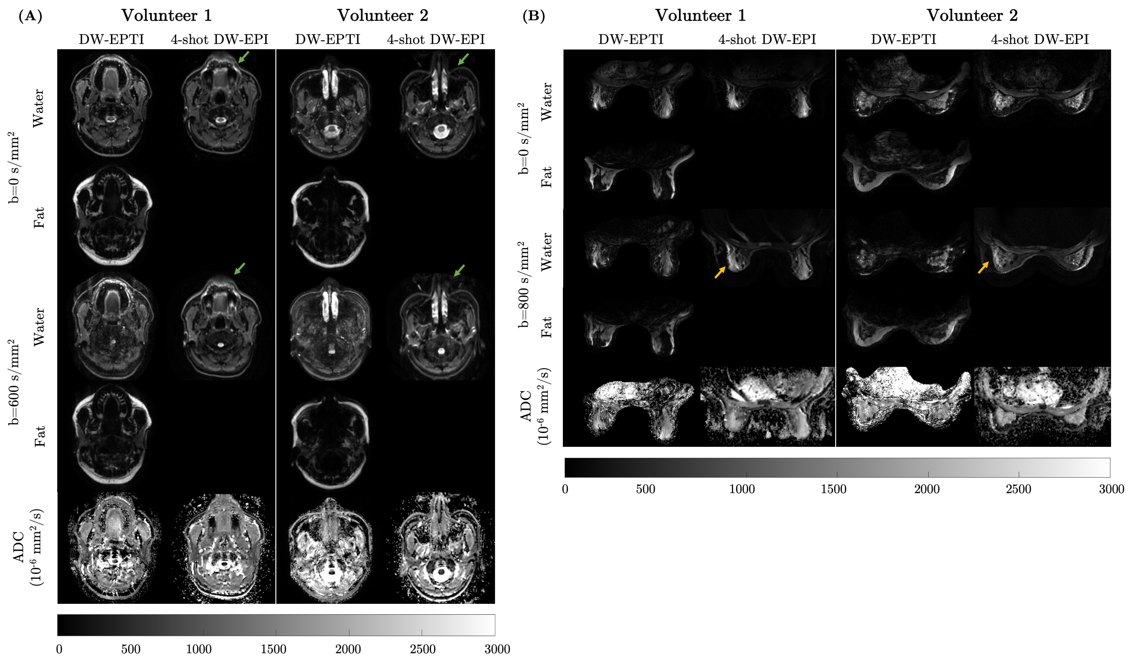
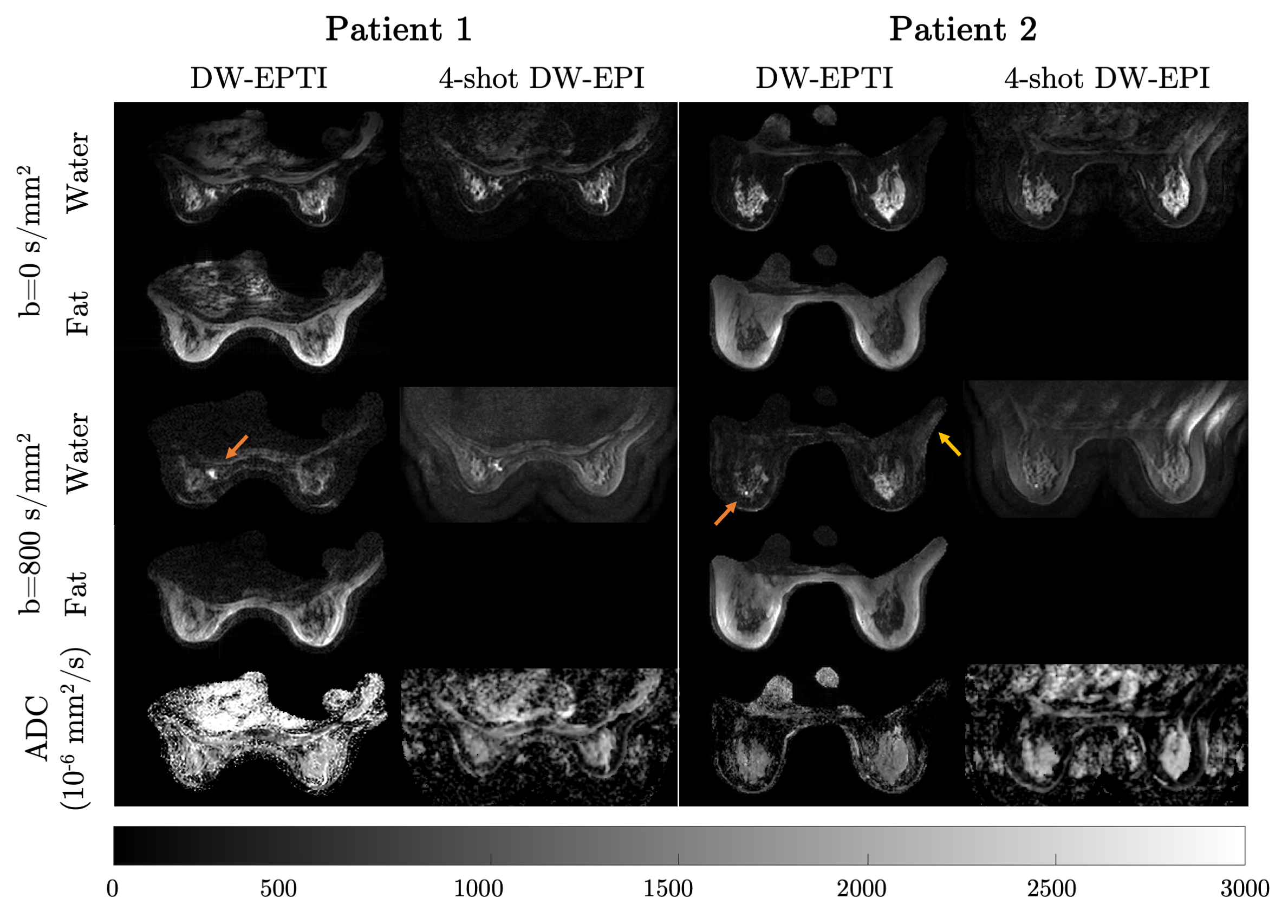
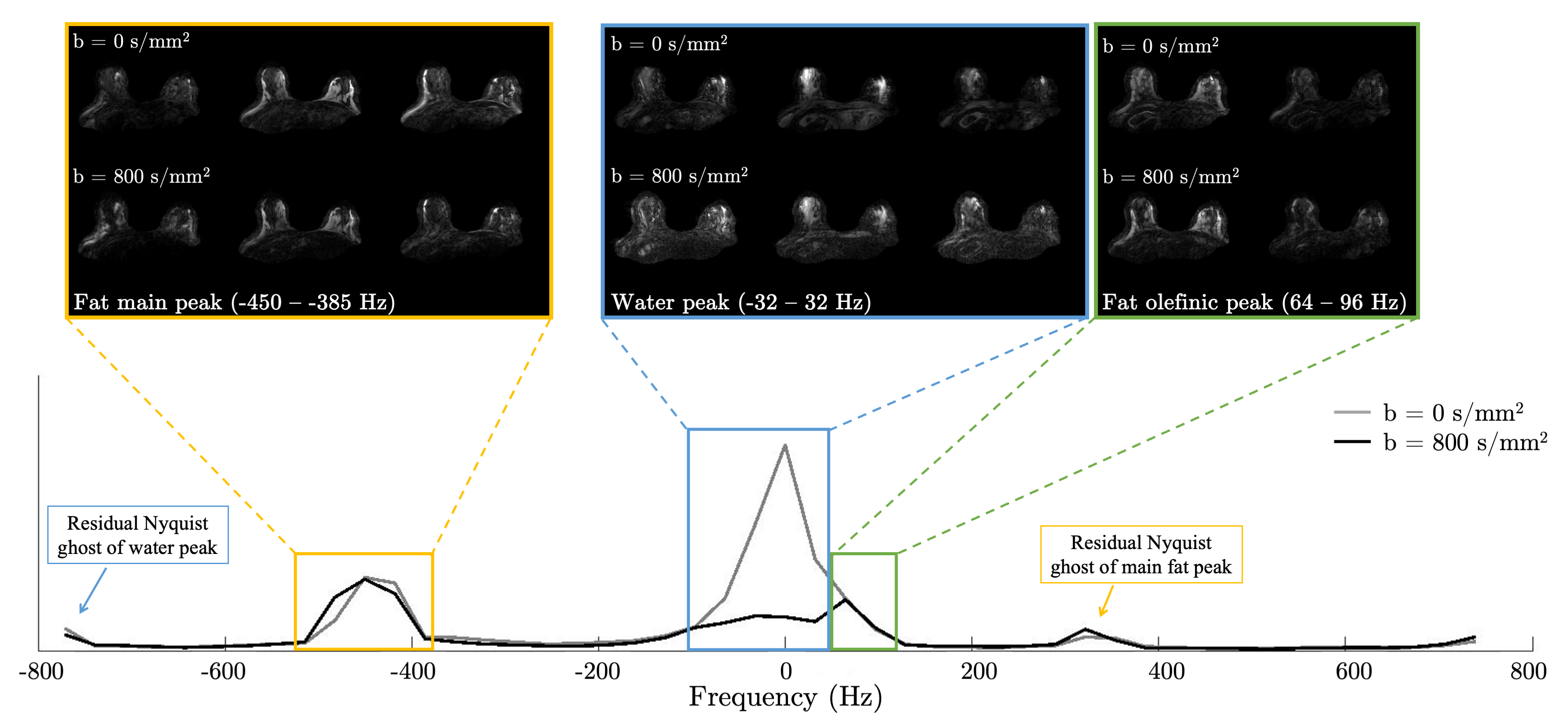
Figures 3 and 4 shows the results of DW-EPTI compared with 4-shot DW-EPI on four healthy volunteers (two on head&neck, two on breast) and two breast cancer patients. DW-EPTI images shows successful fat/water separation and reduced the image distortion compared to DW-EPI. Lesions appeared as a hyperintense contrast on both 4-shot DW-EPI and the proposed water-only DW-EPTI images.An example of the reconstructed spectrum across the entire image on a healthy breast volunteer is shown in Figure 5. Compared to the existing methods, more spectral components, including water and multiple peaks of fat, can be separated. This is particularly useful in the suppression of the olefinic peak of fat11, which is far from the fat main peak and normally cannot be suppressed through the conventional spectrally selective methods.
Discussion
We presented a method that can obtain distortion-free diffusion-weighted images and perform fat/water separation using spatio-temporal joint acquisition and reconstruction. The method is applied in vivo to head&neck and breast imaging. The images show reduced distortion compared to the DW-EPI sequence and successful fat/water separation, which has the potential to improve robustness of body DWI to B0 variations.An FSE-based phase navigator was used to resolve the shot-to-shot motion-induced phase without distortion or chemical shift. Some drawbacks of the FSE-based phase navigator include reducing the signal by a factor of 2 to achieve phase insensitivity, lower time efficiency, and higher specific absorption rate (SAR).
In breast imaging, a partial fat suppression, either ASPIR or fat-sat, can be applied, and our method is able to separate the residual fat from the water signal. This is important for achieving a reliable fat/water separation because, at b=800s/mm2, with the ADC of fat 10-20 times lower than that of fibroglandular tissue, fat signal is much brighter than that of fibroglandular tissue. This dynamic range makes the reconstruction challenging for water.
Conclusion
Distortion-free DWI with fat/water separation in body can be obtained using spatio-temporal acquisition and joint reconstruction methods.Acknowledgements
NIH R01-EB009055, NIH R01-CA249893. Research support from GE Healthcare. Karolinska Neuro MR Physics group for pulse programming assistance.References
1. Mansfield P. Spatial mapping of the chemical shift in NMR. Magnetic Resonance in Medicine. 1984 Sep;1(3):370-86.
2. In MH, Posnansky O, Speck O. High-resolution distortion-free diffusion imaging using hybrid spin-warp and echo-planar PSF-encoding approach. NeuroImage. 2017 Mar 1;148:20-30.
3. Wang F, Dong Z, Reese TG, Bilgic B, Katherine Manhard M, Chen J, Polimeni JR, Wald LL, Setsompop K. Echo planar time‐resolved imaging (EPTI). Magnetic resonance in medicine. 2019 Jun;81(6):3599-615.
4. Kaldoudi E, Williams SC, Barker GJ, Tofts PS. A chemical shift selective inversion recovery sequence for fat-suppressed MRI: theory and experimental validation. Magnetic resonance imaging. 1993 Jan 1;11(3):341-55.
5. Meyer CH, Pauly JM, Macovskiand A, Nishimura DG. Simultaneous spatial and spectral selective excitation. Magnetic resonance in medicine. 1990 Aug;15(2):287-304.
6. Partridge SC, Singer L, Sun R, Wilmes LJ, Klifa CS, Lehman CD, Hylton NM. Diffusion-weighted MRI: influence of intravoxel fat signal and breast density on breast tumor conspicuity and apparent diffusion coefficient measurements. Magnetic resonance imaging. 2011 Nov 1;29(9):1215-21.
7. Baron P, Dorrius MD, Kappert P, Oudkerk M, Sijens PE. Diffusion‐weighted imaging of normal fibroglandular breast tissue: influence of microperfusion and fat suppression technique on the apparent diffusion coefficient. NMR in Biomedicine: An International Journal Devoted to the Development and Application of Magnetic Resonance In vivo. 2010 May;23(4):399-405.
8. Stejskal EO, Tanner JE. Spin diffusion measurements: spin echoes in the presence of a time‐dependent field gradient. The journal of chemical physics. 1965 Jan 1;42(1):288-92.
9. Haase A, Frahm J, Matthaei D, Hänicke W, Bomsdorf H, Kunz D, Tischler R. MR imaging using stimulated echoes (STEAM). Radiology. 1986 Sep;160(3):787-90.
10. Zhou X, Lee PK, Hargreaves BA. Water/Fat separation with spatio-temporal EPI-based acquisition and reconstruction in body imaging. The 32nd Annual Meeting of ISMRM, Toronto, ON, Canada, 2023
11. Hernando D, Karampinos DC, King KF, Haldar JP, Majumdar S, Georgiadis JG, Liang ZP. Removal of olefinic fat chemical shift artifact in diffusion MRI. Magnetic resonance in medicine. 2011 Mar;65(3):692-701.
Figures