0041
Image reconstruction for an 8-element loop-dipole rotating RF coil array (RRFCA) using a novel calibration-free GRAPPA-based method1University of Queensland, Brisbane, Australia, 2King's College London, London, United Kingdom, 3University of Tasmania, Hobart, Australia
Synopsis
Keywords: Image Reconstruction, Image Reconstruction
Motivation: SENSE-based reconstruction is challenging for clinical imaging when rotating the RRFCA into multiple positions; therefore, a novel calibration-free GRAPPA-based method was developed.
Goal(s): To effectively reconstruct k-space data acquired from the RRFCA, enhancing image quality compared to a conventional stationary array without a scan time penalty.
Approach: Conventional GRAPPA was extended by uncovering a subset of the radial grid to cope with the rotation of the RRFCA. Numerical and human brain images were used for validation.
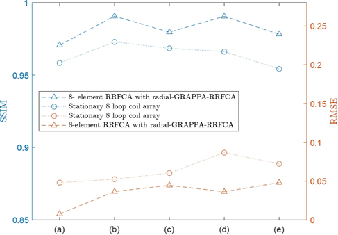
Results: Image quality was improved using the proposed method. Up to 58% reduction in RMSE and 2.5% increase in SSIM was achieved while maintaining scan time.
Impact: The RRFCA utilising our novel calibration-free, GRAPPA-based, radial image reconstruction method provides a clinically relevant parallel imaging technique. In the future, our approach may incorporate compressed sensing to further reduce motion artifacts, particularly in applications like cardiac and dynamic MRI.
Introduction
An eight-coil rotating radiofrequency coil array (RRFCA) has recently been developed1 that offers physical decoupling amongst all coil elements and provides superior $$$B_1$$$ homogeneity and a reduced specific absorption rate (SAR) compared to a static coil array. A drawback, however, is that current image reconstruction methods for the RRFCA are either SENSE-based2,3, requiring rotational sensitivity maps that are hard to accurately obtain, or have restrictions on rotation speed4 which undermines the performance of the RRFCA. In this work, a novel GRAPPA-based5,6 algorithm, radial-GRAPPA-RRFCA, is proposed that reliably reconstructs images from radial k-space without the need for additional calibration data, explicit dependence on sensitivity maps, or gridding operations. This is achieved without incurring a time penalty compared with a static coil array. The algorithm has been validated using both phantom and human brain images.Methods
The RRFCA consist of four groups of loop-dipole coil combinations as described by Li et al.1. The $$$B_1^-$$$ field was achieved using Sim4Life (ZMT, Zurich, Switzerland) and the radial-GRAPPA-RRFCA algorithm was developed using Matlab (MathWorks, Natick, MA).Radial-GRAPPA-RRFCA algorithm
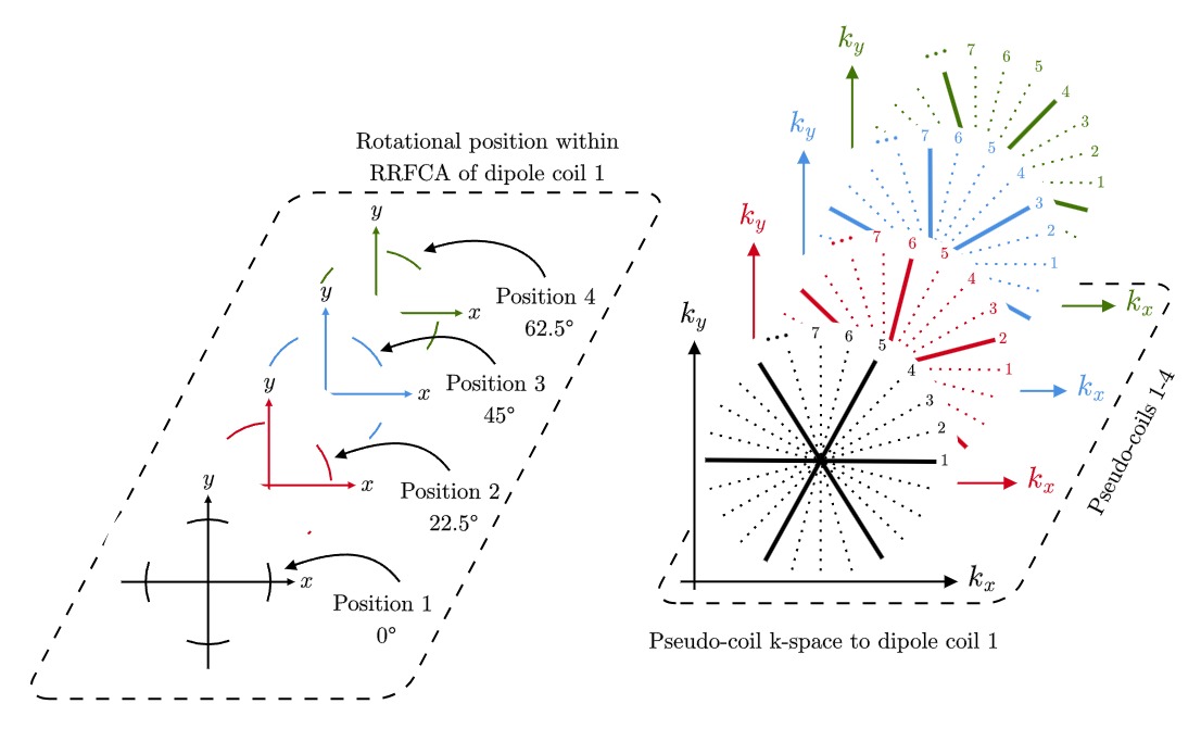
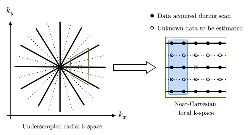
The act of rotating the RRFCA produces an inherent geometric misalignment of sampled data within k-space. For each of the eight coils that make up the RRFCA, a set of pseudo-coils are defined in radial k-space at each rotation; see Figure 1. The proposed algorithm first corrects for this discrepancy by optimally aligning all non-reference coils, based on their angular displacement from the reference coil under reconstruction. While in the reference frame of a particular coil, the radial k-spaces are approximated as a collection of near-Cartesian grids, as shown in Figure 2. Within the local k-space, the acquired data $$$\hat{\Psi}$$$ and $$$S_{cp}$$$ acts as calibration data within the defined kernel, obtaining weights $$$W_{local}=[w_{cp}]$$$ via the matrix inversion of
$$\hat{\Psi}=\sum_{c=1}^{N_c}\sum_{p=1}^{N_p}w_{cp}S_{cp}\ ,$$
where $$$N_c$$$ and $$$N_p$$$ are the number of coils and number of acquired points within the kernel, respectively.
The geometric properties of the radial grid were exploited to formulate a subset of k-space that we termed – relative shift space. Within relative shift space interpolation methods are used to approximate weights that lie outside the collection of near-Cartesian grids. With weights $$$W$$$ now assigned to all acquired data points $$$S$$$, unsampled data points may be estimated via the matrix product $$$\Psi=WS$$$. This allows the RRFCA to obtain a more complete coverage of the region of interest without time penalty. A perturbation expansion method was used to analyse the error introduced by the alignment procedure, given by $$$\Delta=\mathcal{O}\left(\theta^2/h\right)$$$, where $$$\theta$$$ is the angle between acquired radial k-space lines and h is the radial separation between data points.
As the unsampled data was estimated without the necessity for gridding operations, image reconstruction should also be performed without the need for gridding operations. Therefore, a Hankel transform reconstruction (HTR) method was developed for this purpose, given by
$$\rho\left(r,\theta\right)=\frac{1}{4\pi Q}\sum_{m}\sum_{q} e^{im\theta}i^mS_{mq}\left(k_q\right)J_m\left(rk_q\right)k_q\ .$$
The HTR method produces coil-by-coil images $$$\rho$$$ directly from radial k-space without gridding operations. The parameters $$$Q$$$ and $$$k_q$$$ are the total number of data points along a spoke and index value, respectively, $$$r$$$ is the image space radii and $$$J_m$$$ denotes a Bessel function of the first kind.
Results
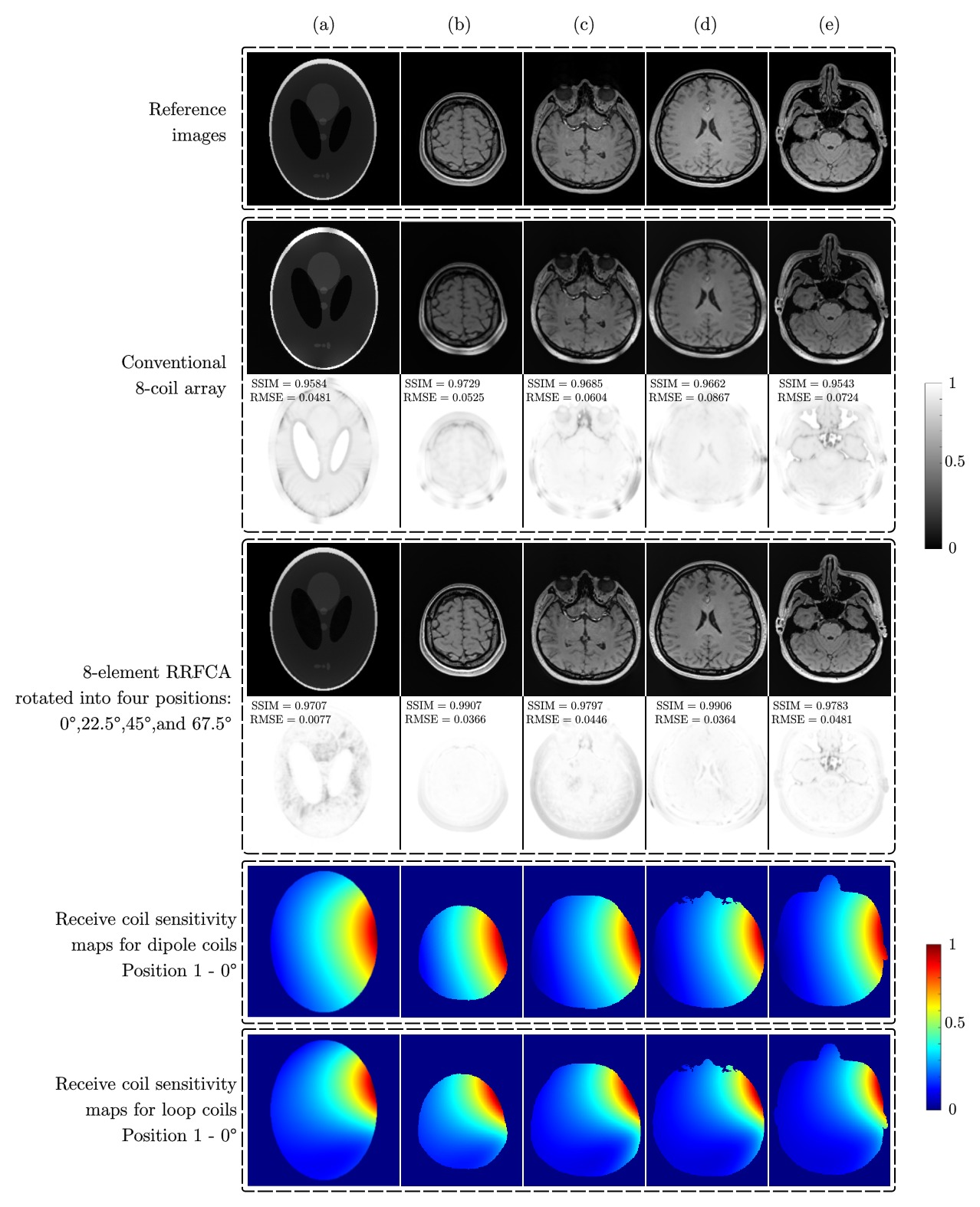
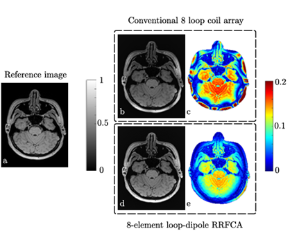
A reference scan using a 3T Siemens Magnetom Prisma Fit based on Li et al.1 was used to compare the RRFCA, under sampled by a factor of four at each position, with a conventional stationary 8-coil array. Under sampling the RRFCA by the same factor as the number of rotations and implementing the proposed reconstruction method, radial-GRAPPA-RRFCA, ensured no scan time penalty. Figure 3 shows results obtained using the RRFCA in conjunction with the proposed method on a numerical phantom (Figure 3a) and four axial brain slices obtained from two human subjects (Figure 3(b-e)). The RRFCA produced higher image quality than the conventional stationary 8-coil array in each case (see Figure 4) achieving up to a 58% decrease in RMSE and a 2.5% increase in SSIM. Error maps are shown in Figure 5 to further demonstrate the validity of the proposed method.Discussion and conclusion
The algorithm proposed in this work, radial-GRAPPA-RRFCA, provides a calibration-free image reconstruction method without explicit dependence on sensitivity maps or gridding operations, thereby allowing the 8-element RRFCA to produce higher-quality results than a stationary 8-coil array without any time penalty. In the future, further experimental validation will be carried out, and broader applications for the RRFCA will be explored. For example, we aim to incorporate compressed sensing methods into radial-GRAPPA-RRFCA to minimise scan time further.Acknowledgements
The first author acknowledges the receipt of a Higher Degree by Research scholarship from the School of Electrical Engineering and Computer Science, University of Queensland that has made this work possible.References
1. Li M, Jin J, Destruel A, Guo L, Weber E, Liu F, Crozier S. Eight element rotating radiofrequency coil array with the combination of dipole and loop coils for efficient B1 shimming and SAR control at 7T. Proc Intl Soc Mag Reson Med 2020;28.
2. Pruessmann KP, Weiger M, Scheidegger MB, Boesiger P. SENSE: sensitivity encoding for fast MRI. Mag Reson Med. 1999;42(5):952-962.
3. Li M, Jin J, Zuo Z, Liu F, Trakic A, Weber E, Zhuo Y, Xue R, Crozier S. In vivo sensitivity estimation and imaging acceleration with rotating RF coil arrays at 7 Tesla. J Mag Res. 2015;252:29-40.
4. Jin J, Weber E, Tesiram Y, Hugger T, Li M, Fuentes M, Ullmann P, Stark S, Junge S, Liu F, Crozier S. Image reconstruction for a rotating radiofrequency coil (RRFC) using self-calibrated sensitivity from radial sampling. IEEE Trans Biomed Eng. 2017;64(2):274-283.
5. Griswold MA, Jakob PM, Heidemann RM, et al. Generalized autocalibrating partially parallel acquisitions (GRAPPA). Magn Reson Med. 2002;47(6):1202-1210.
6. Codella NC, Spincemaille P, Prince M, and Wang Y. A radial self-calibrated (RASCAL) generalized autocalibrating partially parallel acquisition (GRAPPA) method using weight interpolation. NMR in Biomed 2011;24:844–54.
Figures