0039
Time-Resolved Biomechanics using Spectro-Dynamic MRI: Proof of Principle in the Muscles of the Thigh1Department of Radiotherapy, Computational Imaging Group for MR Diagnostics and Therapy, UMC Utrecht, Utrecht, Netherlands, 2Mathematical Institute, Utrecht University, Utrecht, Netherlands
Synopsis
Keywords: Image Reconstruction, Muscle, Time-Resolved, Motion, Strain
Motivation: Measurements of biomechanical tissue properties require time-resolved reconstructions from dynamic experiments. The Spectro-Dynamic MRI framework achieves this by working directly from k-space data.
Goal(s): To develop an experimental setup and reconstruction method with which time-resolved biomechanical information can be measured in vivo.
Approach: An inflatable pressure cuff deformed the thigh muscles of a volunteer. Time-resolved images and strain maps were reconstructed directly from k-space data using the Spectro-Dynamic MRI framework.
Results: Principal strains were obtained for different muscles in the thigh at a temporal resolution of 352 ms. The first principal strain direction could differentiate between muscle structures, indicating different underlying biomechanical properties.
Impact: The reconstruction of time-resolved images and strains using Spectro-Dynamic MRI allows for time-resolved measurements of biomechanical parameters during dynamic loads with a straightforward experimental setup. This information is useful for studying the mechanical behavior of tissues.
Introduction
Measuring biomechanical quantities in a dynamic setting is important in studying the functioning of the musculoskeletal system, and can help in diagnosing pathologies1,2. Therefore, time-resolved methods that can measure biomechanical properties are important research tools.Some elastic tissue properties can be measured with MR elastography (MRE)3, but this method requires a very specific acquisition protocol and a specialized actuator, it assumes the tissue is linear elastic and isotropic, and studies the tissue properties at high frequencies. Phase contrast has also been used to study muscle dynamics, but it usually requires many accurate repetitions of the same motion task, especially when multiple slices are needed4.
Spectro-Dynamic MRI5 is a recently developed method that allows for time-resolved identification of biomechanical tissue properties. It uses k-space data directly, allowing for flexibility in the sampling strategy and temporal resolution. However, it has thus far only been applied to phantom data with piecewise rigid motion. Here, we show a proof-of-principle application of time-resolved Spectro-Dynamic MRI in the human thigh with a straightforward and inexpensive acquisition and reconstruction setup.
Methods
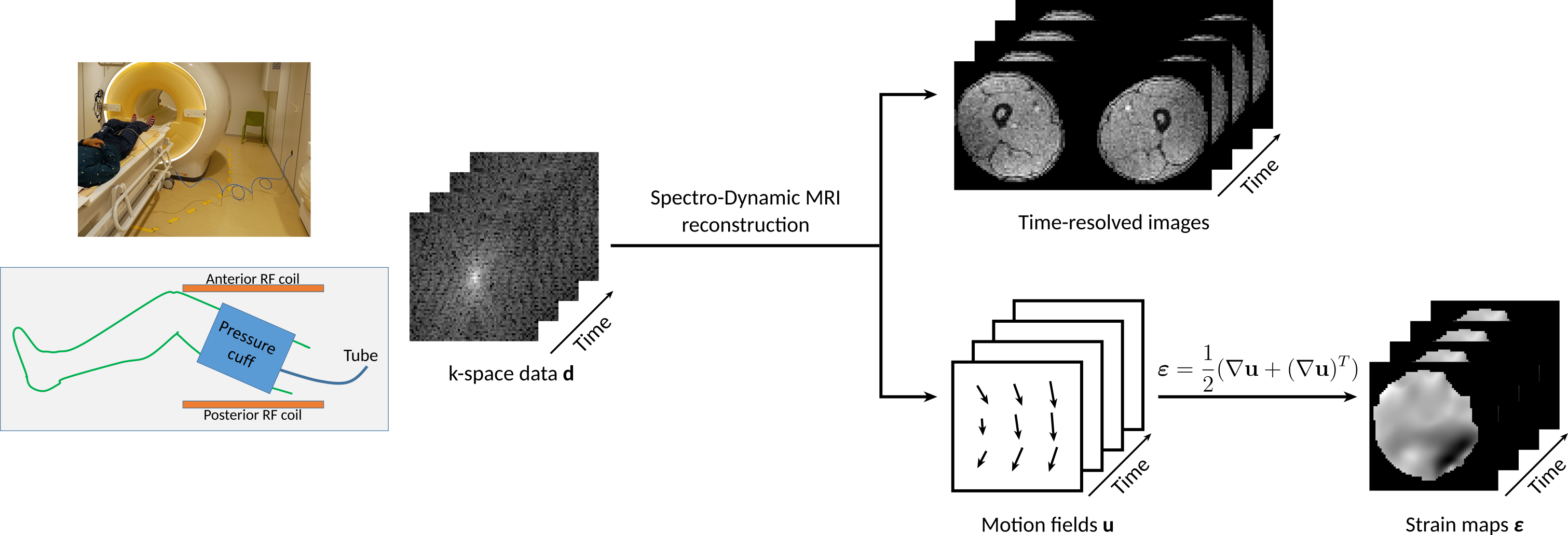
An inflatable pressure cuff was placed around the thigh of a volunteer (Fig. 1). An elongated inelastic tube connected the cuff and the pressure monitor device outside the scanner room. The pressure cuff inflated during the scan, deforming the muscles. The device was turned off at 100 mmHg, after which the pressure gradually decreased.A spoiled gradient echo sequence acquired the data in a slice perpendicular to the legs (TR/TE = 5.5/2.2 ms, FA = 5°, FOV = 384 mm$$$\times$$$192 mm, 3.0 mm$$$\times$$$3.0 mm, 144 dynamics).
The time-resolved displacement fields $$$\mathbf{u}$$$ and k-space data $$$\mathbf{m}$$$ were reconstructed by solving a minimization problem consisting of four terms5: a data consistency term with sampling mask $$$E$$$, a motion model $$$G$$$ relating the k-space data to the deformation fields, a spatial smoothness regularization term using the Laplacian operator $$$L$$$, and a temporal smoothness regularization term using the second order finite difference operator $$$D_{tt}$$$:
$$\min_{\mathbf{m},\mathbf{u}}\|E\mathbf{m}-\mathbf{d}\|_2^2+\lambda_G\|G(\mathbf{m},\mathbf{u})\|_2^2+\lambda_S\|L\mathbf{u}\|_2^2+\lambda_T\|D_{tt}\mathbf{u}\|_2^2.\tag{1}$$
The values of the regularization parameters $$$\lambda_G$$$, $$$\lambda_S$$$, and $$$\lambda_T$$$ were determined empirically. The displacement field was parameterized using cubic B-splines.
The first principal strain value and direction were calculated using the eigendecomposition of the strain tensor $$$\boldsymbol{\varepsilon}=\frac{1}{2}(\nabla\mathbf{u}+(\nabla\mathbf{u})^T)$$$ using the plain strain assumption.
Results
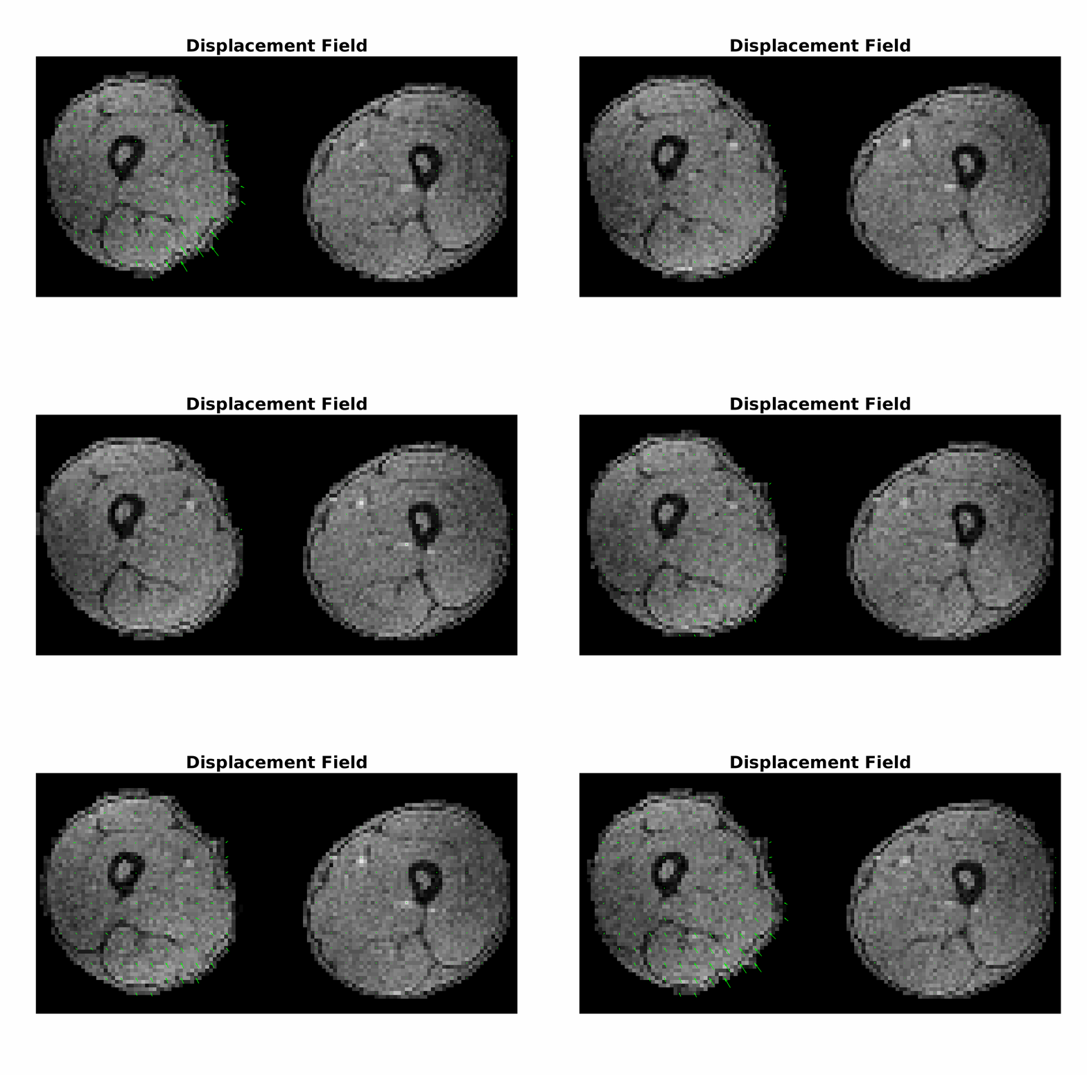
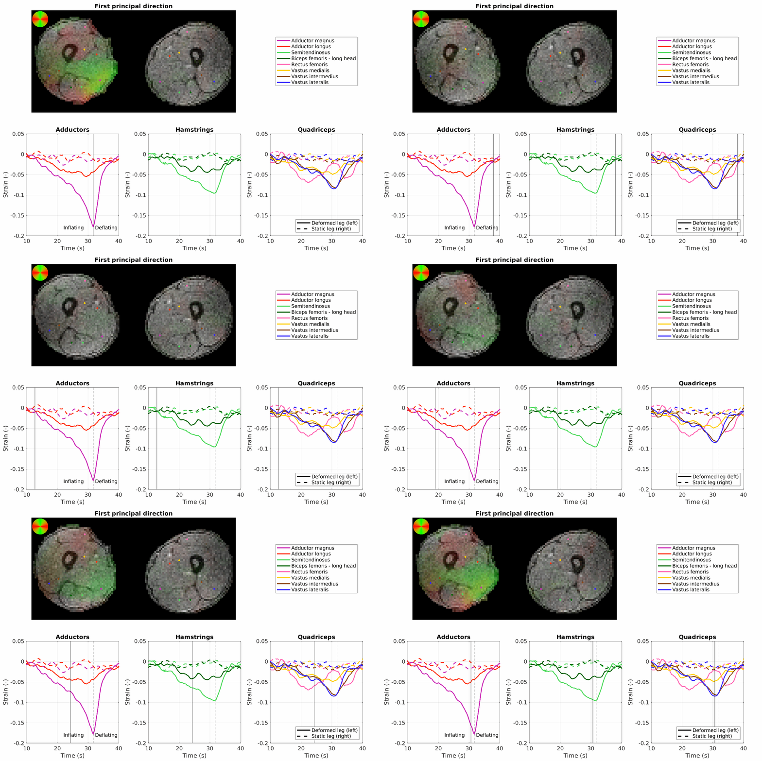
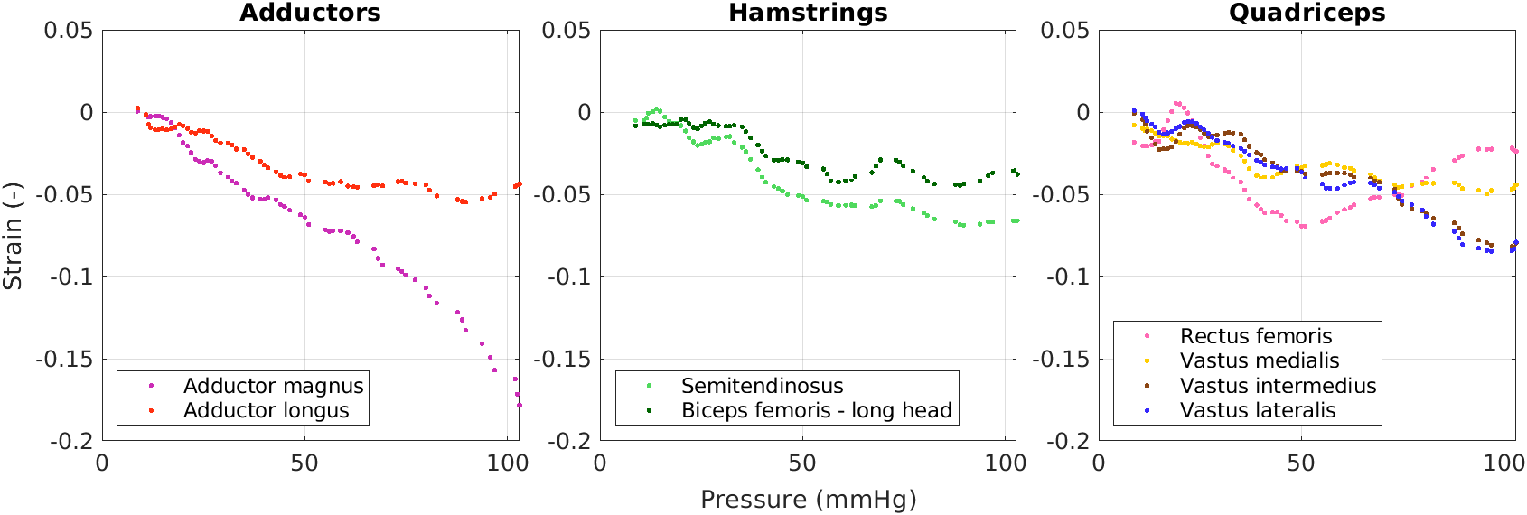
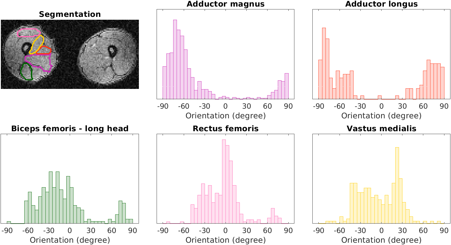
The estimated time-resolved images and motion field (Fig. 2) showed a clear deformation in the left leg caused by the pressure of the inflatable cuff, while the right leg remained stationary. This compression is also evident from the relation between the pressure and the first principal strain (Fig. 3 and Fig. 4). An abrupt change in the direction of the first principal strain can be seen at the border between different muscle groups (Fig. 3), for example between the vastus medialis and the adductor longus (Fig. 5).Discussion
We observed a clear difference in the principal strain direction in different muscles. We expect this to be related to the underlying anatomy of these muscles. The direction of the muscle fibers is closely linked to the direction of the force. These fibers cause an anisotropic behavior of the muscle tissue under compression. Since the fibers’ orientations are different for each muscle, the principal strain directions are expected to be different as well.One limitation of this work is that through-slice motion could not be considered. Since biological tissue is nearly incompressible, any in-plane compression will invariably lead to a through-plane expansion. Furthermore, the fiber direction has a through-plane component, making it difficult to compare the principal strain directions with the fiber orientations. A 3D implementation of the proposed method would solve these issues and will be realized in the future.
Another potential issue is sliding motion between neighboring muscles. As a possible solution, the reconstruction algorithm could be provided with muscle segmentations to allow for discontinuities between muscles.
The reconstruction could be enhanced by adding a constitutive relation connecting the stress and strain of the tissue5. This additional term could function as a regularizer of the motion fields, as well as a source of additional insights regarding the biomechanical behavior of the tissue.
Conclusion
We have demonstrated the reconstruction of time-resolved principal strain maps in the muscles of the human thigh using a straightforward experimental setup. Differences in the measured strain between muscles with different fiber structures suggest that time-resolved measurements of biomechanical tissue properties during dynamic loads using Spectro-Dynamic MRI is possible.Acknowledgements
This work has been financed by NWO, grant number 18897.References
1. Borotikar B, Lempereur M, Lelievre M, Burdin V, Ben Salem D, Brochard S. Dynamic MRI to quantify musculoskeletal motion: A systematic review of concurrent validity and reliability, and perspectives for evaluation of musculoskeletal disorders. PLoS One. 2017;12(12). doi:10.1371/journal.pone.0189587
2. d’Entremont AG, Nordmeyer-Massner JA, Bos C, Wilson DR, Pruessmann KP. Do dynamic-based MR knee kinematics methods produce the same results as static methods? Magn Reson Med. 2013;69(6):1634-1644. doi:10.1002/mrm.24425
3. Low G, Kruse SA, Lomas DJ. General review of magnetic resonance elastography. World J Radiol. 2016;8(1):59-72. doi:10.4329/wjr.v8.i1.59
4. Mazzoli V, Gottwald LM, Peper ES, et al. Accelerated 4D phase contrast MRI in skeletal muscle contraction. Magn Reson Med. 2018;80(5):1799-1811. doi:10.1002/mrm.27158
5. Riel MHC van, Leeuwen T van, Berg CAT van den, Sbrizzi A. Time-Resolved Reconstruction of Motion, Force, and Stiffness Using Spectro-Dynamic MRI. IEEE Trans Comput Imaging. 2023;9:917-927. doi:10.1109/TCI.2023.3324757
Figures