0032
Unveiling Choroid Plexus Changes in Multiple Sclerosis using 7T MRI1Bernard and Irene Schwartz Center for Biomedical Imaging, Department of Radiology, NYU Grossman School of Medicine, New York, NY, United States, 2Center for Advanced Imaging Innovation and Research (CAI2R), Department of Radiology, NYU Grossman School of Medicine, New York, NY, United States, 3Vilcek Institute of Graduate Biomedical Sciences, NYU Grossman School of Medicine, New York, NY, United States
Synopsis
Keywords: Multiple Sclerosis, Multiple Sclerosis, choroid plexus, neurodegeneration, neuroinflammation
Motivation: Conventional imaging limits the assessment of choroid plexus (ChP) inflammatory activity in multiple sclerosis (MS).
Goal(s): Use high-resolution MRI with 0.25mm2 matrix at 7T to assess ChP changes in MS patients compared to controls and explore correlations with lesion volumes.
Approach: In 14 MS patients and 9 controls, ChPs were categorized into vascular and stromal compartments using 7T T2* imaging, and vessel-to-stroma ration was compared between the two groups.
Results: The ChP's vessel-to-stroma ratio quantified by 7T T2*w MRI was lower in MS patients, correlating negatively with lesion volume. Furthermore, the age-related decline was more rapid in MS patients compared to controls.
Impact: This pilot study suggests that the vessel-to-stroma ratio of ChP, as revealed by high-resolution 2D-GRE 7T T2* imaging, could potentially serve as an imaging marker for inflammatory and degenerative changes of ChP in MS patients.
INTRODUCTION
The choroid plexus (ChP) comprises a densely packed epithelium surrounding a highly vascularized stromal core, including fenestrated capillaries and connective tissues [1]. In multiple sclerosis (MS), ChP serves as an entry port for lymphocytes and plays a pivotal role in modulating inflammatory activity [2]. Previous research has associated ChP enlargement, as observed on T1-weighted (T1w) images, with increased neuroinflammation in relapsing-remitting MS (RRMS) [3]. However, 3T T1w images may lack the necessary resolution and contrast to reveal its anatomical details, hindering our comprehension of ChP pathology in MS. In this study, we employed high-resolution 2D gradient echo (GRE) 7T MRI with both T2 and T2* contrast, improving the differentiation of ChP’s vascular and stromal components to better understand the specific pathological changes of ChP in MS.METHODS
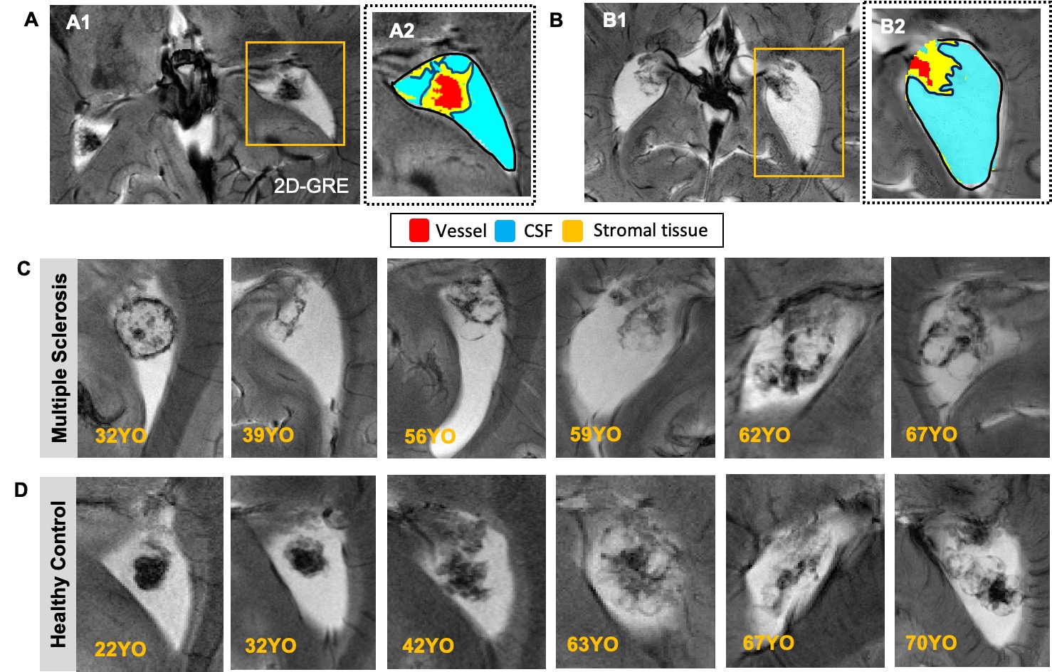
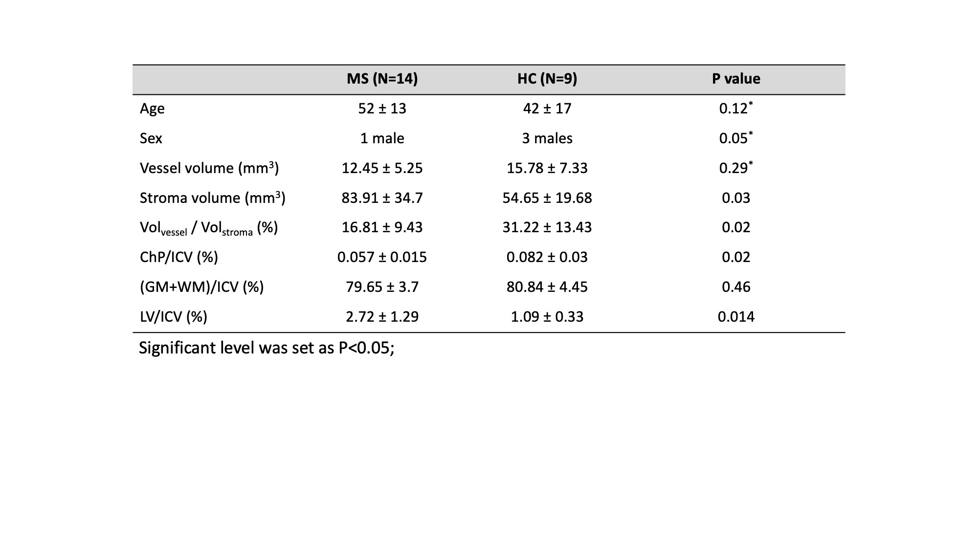
14 RRMS patients (13 females, mean age: 52±13 years) and 9 healthy controls (HC) (6 females, mean age: 42±17 years) were enrolled in this study. Detailed demographic data and measurements were summarized in Table 1. All participants underwent 7T MRI scans employing a 32-channel head coil. The imaging protocol included: 1) T1w MPRAGE: voxel size = 1x1x1mm3; 2) 2D GRE sequence: TR/TE=1250/25ms, FA=40°, voxel size=0.25x0.25x2mm3. FLAIR images from their prior 3T clinical scans were used for lesion segmentation. After co-registering 7T T1w and 2D GRE images (3-parameter rigid body transformation), the lateral ventricle (LV) atrium mask was obtained for further analysis, as ChP primarily resides in this region. The LV atrium was categorized into vascular (hypointense), stromal (iso-intense), and CSF (hyperintense) compartments based on tissue contrast, using the K-Means clustering algorithm (Fig.1).Independent t-tests was performed to reveal group differences in ChP compartment volumes between MS patients and HC. Linear regression analyses were conducted independently for MS and HC to investigate their age-related changes. Furthermore, we investigated the relationship between lesion volume and vessel-to-stroma ratio through linear regression, with age as a controlled variable.
RESULTS
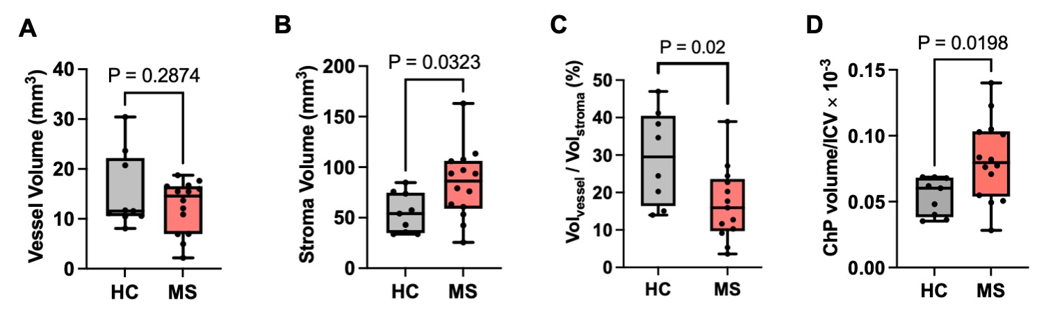
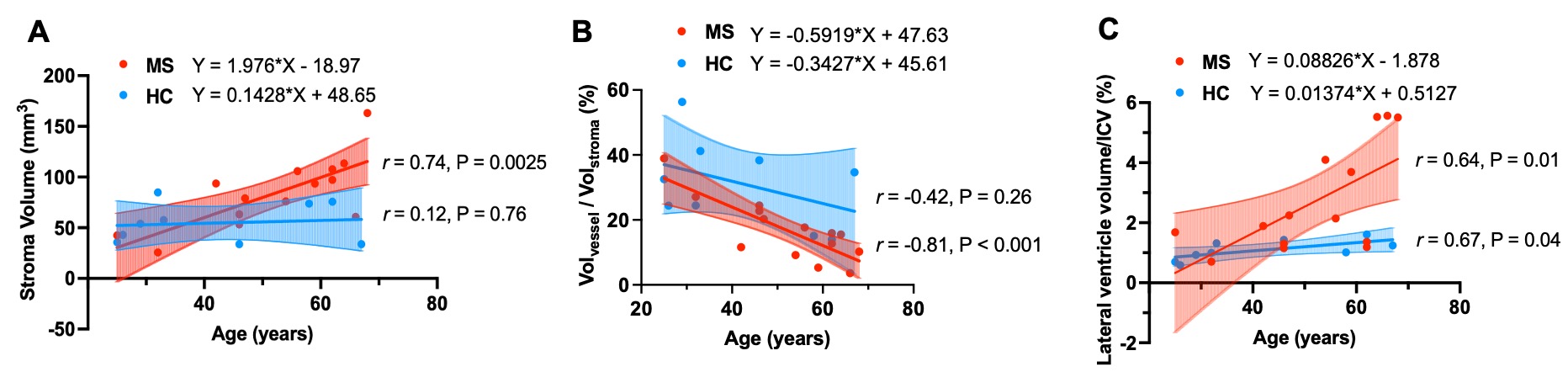
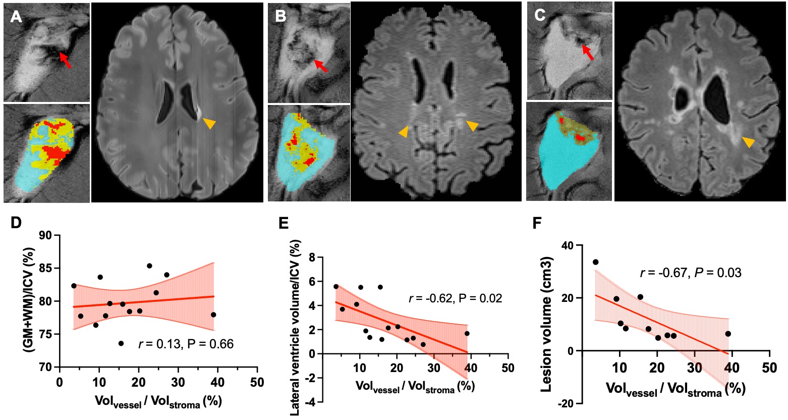
As shown in Fig. 1C-D, we observed the fluid-filled cyst structure, as an indicator of ChP degenerative alteration, occurred earlier in MS compared to HC. MS patients showed significantly higher stromal volume compared to HCs (P<0.05). However, there was no significant difference in the vascular compartment (P=0.28). The vessel-to-stroma ratio was significantly smaller in MS compared to in HC (P=0.0198) (Fig.2).In MS patients, stromal volume demonstrated a positive correlation with age (r=0.74, P=0.0025), exhibiting a faster increase than in HC ( βMS=1.98, βHC=0.14). The vessel-to-stroma ratio displayed a negative correlation with age (r=-0.81, P<0.001), characterized by a faster decline with age (βMS=-0.59 vs βHC=-0.34). No significant correlation was observed in HC (r=-0.42, P=0.26), indicating only a decline tendency. Both HC and MS exhibited age-related LV enlargement (r=0.67, P=0.04, and r=0.64, P=0.01), with MS demonstrating a faster enlargement (Fig.3). Notably, vascular volume, did not correlate with age in either MS or HC (P=0.77 and P=0.23, respectively).
Among the MS subjects (N=14), the vessel-to-stroma ratio exhibited a negative correlation with LV volume (r=-0.62, P=0.02) after adjusting for age. In the subset of 10 MS patients with high quality-FLAIR images, a negative correlation was observed between lesion volume and the vessel-to-stroma ratio (r =-0.67, P=0.03) (Fig.4).
DISCUSSION AND CONCLUSIONS
Compared to HC, MS patients exhibit a smaller vascular but a larger stromal compartment, suggesting the enlarged ChP can primarily be attributed to the increased stromal tissue, possibly a result of inflammation-associated stromal fibrosis[4]. While the precise mechanisms of vascular dysfunction in MS remain partially explored, chronic inflammation and oxidative stress have been proposed as factors to deteriorate vascular endothelial function[5]. This, in conjunction with the inflammatory fibrotic stroma, could potentially lead to CSF homeostatic disruption. Compared to controls, MS patients displayed a heightened level of age-related changes, suggesting a synergistic effect between aging and MS pathology. Furthermore, the inverse relationship between the vessel-to-stroma ratio and lesion volume suggests a possible indicator of disease severity. In MS, the accelerated ChP degeneration may contribute to blood-CSF barrier dysfunction and meningeal inflammation, leading to imbalanced CSF production and absorption.In summary, distinctive ChP alterations in MS patients involve enlarged stromal tissue coupled with slightly diminished vasculature. Collectively, the vessel-to-stroma ratio derived from 7T 2D-GRE imaging has the potential to serve as an effective marker to better elucidate the underlying mechanisms regarding the degenerative ChP changes in MS and their association with disease severity.
Acknowledgements
This work was supported in part by the NIH R21 HD094424 and R01 NS108491 and was performed under the rubric of the Center for Advanced Imaging Innovation and Research (CAI2R, www.cai2r.net), an NIBIB National Center for Biomedical Imaging and Bioengineering (NIH P41 EB017183). We would also like to thank Dr. Ilya Kister from Department of Neurology, NYU Grossman School of Medicine for the comments that greatly improved the work.References
1. Saunders, N.R., et al., The choroid plexus: a missing link in our understanding of brain development and function. Physiol Rev, 2023. 103(1): p. 919-956.
2. Muller, J., et al., Understanding the Role of the Choroid Plexus in Multiple Sclerosis as an MRI Biomarker of Disease Activity. Neurology, 2023. 100(9): p. 405-406.
3. Ricigliano, V.A.G., et al., Choroid Plexus Enlargement in Inflammatory Multiple Sclerosis: 3.0-T MRI and Translocator Protein PET Evaluation. Radiology, 2021. 301(1): p. 166-177.
4. Prineas, J.W., J.D. Parratt, and P.D. Kirwan, Fibrosis of the Choroid Plexus Filtration Membrane. J Neuropathol Exp Neurol, 2016. 75(9): p. 855-67.
5. Boshra, H., et al., Vascular dysfunction and dyslipidemia in multiple sclerosis: are they correlated with disease duration and disability status? Egypt Heart J, 2022. 74(1): p. 9.
6. Geraldes, R., et al., Vascular disease and multiple sclerosis: a post-mortem study exploring their relationships. Brain, 2020. 143(10): p. 2998-3012.
7. Sun, Z., et al., Age-related Vascular Changes in Choroid Plexus Evaluated Using High-resolution USPIO-Enhanced 7T MRI, in ISMRM. 2023.
8. Millward, J.M., et al., Transient enlargement of brain ventricles during relapsing-remitting multiple sclerosis and experimental autoimmune encephalomyelitis. JCI Insight, 2020. 5(21).
Figures