0030
Relaxation-exchange imaging (REXI) for the measurement of trans-barrier water exchange in choroid plexus1State Key Laboratory of Brain and Cognitive Science, Beijing MRI Center for Brain Research, Institute of Biophysics, Chinese Academy of Sciences, Beijing, China, 2University of Chinese Academy of Sciences, Beijing, China, 3Key Laboratory of Biomedical Engineering of Ministry of Education, College of Biomedical Engineering and Instrument Science, Zhejiang University, Hangzhou, China, 4Department of Chemistry, Zhejiang University, Hangzhou, China, 5School of Brain Science and Brain Medicine, Zhejiang University, Hangzhou, China, 6MR Collaboration, Siemens Healthcare, Shanghai, China, 7Interdisciplinary Institute of Neuroscience and Technology, Zhejiang University School of Medicine, Hangzhou, China
Synopsis
Keywords: Neurofluids, Neurofluids, choroid plexus, Blood-CSF barrier, relaxation exchange
Motivation: Scarcity of non-invasive imaging techniques of choroid plexus function hindered our knowledge of the blood-cerebrospinal fluid barrier (BCSFB).
Goal(s): We aimed to measure the trans-barrier water exchange rate in choroid plexus.
Approach: We developed a new imaging method and contrast mechanism, named relaxation-exchange imaging (REXI), and validated its feasibility on both phantoms and rats.
Results: REXI successfully captured the changes in proton exchange rate of urea-water phantoms at varying pH. In-vivo experiments on rats showed the potential of REXI to measure the trans-barrier water exchange in choroid plexus.
Impact: Given the emerging importance of neurofluids and choroid plexus, our novel MRI method REXI provides a way to measure the trans-barrier water exchange in CP and a potential imaging tool to evaluate CP function in future studies.
Introduction
The choroid plexus (CP) located in the ventricles serves as an important exchange interface between blood and cerebrospinal fluid (CSF), where the inward transport of nutrients and outward clearance of metabolites support brain homoeostasis1,2. CP is responsible for CSF generation and is vital as the blood-cerebrospinal fluid barrier (BCSFB). BCSFB dysfunction is now suspected to underlie a broad spectrum of pathophysiological conditions, including neurodegenerative disorders, for example, Alzheimer's disease3,4. However, the lack of non-invasive imaging techniques of CP function precluded our knowledge of BCSFB. Recently, an arterial spin labeling (ASL)-based MRI sequence has been proposed to quantify rates of water delivery from blood to CSF in rodents and human5–7, which utilized an ultra-long echo time (220 ms at 9.4T for mouses) to measure only the signal from labelled blood water that has been exchanged into the CSF.In this study, we proposed a new imaging method and contrast mechanism to measure the trans-barrier water exchange rate in CP, named relaxation-exchange imaging (REXI). REXI relies on the fact that CP tissue (including blood, cells, and interstitial space) has a much shorter T2 than CSF. Inspired by the filter-exchange imaging (FEXI)8–10, REXI also has a filter block (TEf), a mixing block with mixing time tm, and a detection block with multi-echo acquisition (TE) (Figure 1). TEf was selected to largely suppress the magnetization of CP tissue but affect CSF magnetization much less. By increasing the mixing time, the fraction of magnetization with short T2 (T2fast) will also increase due to the inter-compartment water exchange. In addition, a pair of identical crusher gradients was placed before the second-90° pulse and after the third-90° pulse to omit unwanted coherence pathways.
The feasibility of REXI in measuring proton exchange has been validated in the urea-water phantom, in which the pH was adjusted to vary the proton exchange between urea and water. At last, REXI was primarily tested on rats’ brain at different ages to explore the age-dependence of trans-barrier water exchange in CP.
Methods
The two-pool aqueous urea model system11–13 was chosen for the proof-of-concept experiments of REXI. Urea-water phantoms at three different pH (6.78, 7.76, 8.80) with fixed urea-water proton ratio of 30%/70% were used. The longitudinal and transverse relaxation time constants were decreased using Gd-DTPA and MnCl2. To explore aging-induced impairment of BCSFB function, the adult group (~3 month, n = 3) and the aged group (~24 month, n = 3) of Sprague Dawley male rats were involved.All REXI data were acquired on a 9.4T Bruker MRI scanner. Detailed MRI protocol is summarized in Table 1. The scan time of REXI measurements for phantoms/rats is under 12/24 min; and the scan for phantoms was repeated three times. A simplified two-site exchange model (2SXM)14 is applied to fit REXI data with the detailed steps illustrated in Figure 2.
Results and Discussion
For urea-water experiments, the representative image and signal-TE curves of REXI are shown in Figure 3, along with the T2 values for the fast compartment (T2fast) and slow compartment (T2slow) and the fraction of fast T2 compartment (ffast). At various pH, the 2SXM could well fit the ffast-tm curves resulting the exchange rate from fast to slow compartment kfs = 4.08 ± 0.77 s-1 at pH = 6.78, 1.36 ± 0.48 s-1 at pH = 7.76, and 4.92 ± 0.18 s-1 at pH = 8.80. The pH-dependence of kfs well agrees with previous studies11,13 and demonstrates the reliability of REXI in measuring the exchange processes.For rat experiments, the representative images of REXI at varied tm, are shown in Figure 4. The region-of-interested (ROI) of CP was carefully drawn on the high-resolution T2-TurboRARE images and registered back to REXI images. 2XSM showed nice fitting to the REXI data (Figure 4C) with the effective exchange rate k (= kfs + ksf) = 1.55 ± 0.99 s-1 for adult group and k = 0.99 ± 0.55 s-1 for aged group. The decreased trans-barrier water exchange could be a biomarker of CP function declining in aging, though more animals should be included in future studies to validate this finding, and the underlying physiological mechanisms of trans-barrier water exchange should also be further explored.
Conclusion
REXI is a novel method to measure the trans-barrier water exchange between CP and CSF by making use of the large T2 differences between these two compartments. Urea-water phantom results at various pH demonstrated the sensitivity and reliability of REXI in measuring exchange and the primary results on rats’ brain showed the feasibility of REXI in measuring the trans-barrier water exchange in CP.Acknowledgements
This work is supported in part by the National Natural Science Foundation of China (NSFC) (Grant Nos. 82111530201, 82222032, 82172050), the STI2030-Major Projects Q22 of China (Grant No. 2022ZD0206000).References
1. MacAulay, N., Keep, R. F. & Zeuthen, T. Cerebrospinal fluid production by the choroid plexus: a century of barrier research revisited. Fluids Barriers CNS 19, 26 (2022).
2. Saunders, N. R., Dziegielewska, K. M., Fame, R. M., Lehtinen, M. K. & Liddelow, S. A. The choroid plexus: a missing link in our understanding of brain development and function. Physiological Reviews 103, 919–956 (2023).
3. Cousins, O. et al. The blood–CSF–brain route of neurological disease: The indirect pathway into the brain. Neuropathology Appl Neurobio 48, e12789 (2022).
4. Municio, C., Carrero, L., Antequera, D. & Carro, E. Choroid Plexus Aquaporins in CSF Homeostasis and the Glymphatic System: Their Relevance for Alzheimer’s Disease. IJMS 24, 878 (2023).
5. Evans, P. G. et al. Non-Invasive MRI of Blood–Cerebrospinal Fluid Barrier Function. Nat Commun 11, 2081 (2020).
6. Perera, C., Harrison, I. F., Lythgoe, M. F., Thomas, D. L. & Wells, J. A. Pharmacological MRI with Simultaneous Measurement of Cerebral Perfusion and Blood-Cerebrospinal Fluid Barrier Function using Interleaved Echo-Time Arterial Spin Labelling. NeuroImage 238, 118270 (2021).
7. Lee, H. et al. Choroid plexus tissue perfusion and blood to CSF barrier function in rats measured with continuous arterial spin labeling. NeuroImage 261, 119512 (2022).
8. Lasič, S., Nilsson, M., Lätt, J., Ståhlberg, F. & Topgaard, D. Apparent exchange rate mapping with diffusion MRI. Magnetic Resonance in Medicine 66, 356–365 (2011).
9. Nilsson, M. et al. Noninvasive mapping of water diffusional exchange in the human brain using filter-exchange imaging. Magnetic Resonance in Medicine 69, 1572–1580 (2013).
10. Bai, R. et al. Feasibility of filter-exchange imaging (FEXI) in measuring different exchange processes in human brain. NeuroImage 219, 117039 (2020).
11. Vold, R. L., Daniel, E. S. & Chan, S. O. Magnetic resonance measurements of proton exchange in aqueous urea. J. Am. Chem. Soc. 92, 6771–6776 (1970).
12. Bai, R., Benjamini, D., Cheng, J. & Basser, P. J. Fast, accurate 2D-MR relaxation exchange spectroscopy (REXSY): Beyond compressed sensing. The Journal of Chemical Physics 145, 154202 (2016).
13. Stabinska, J. et al. Proton exchange in aqueous urea solutions measured by water‐exchange (WEX) NMR spectroscopy and chemical exchange saturation transfer (CEST) imaging in vitro. Magnetic Resonance in Med 82, 935–947 (2019).
14. Åslund, I., Nowacka, A., Nilsson, M. & Topgaard, D. Filter-exchange PGSE NMR determination of cell membrane permeability. Journal of Magnetic Resonance 200, 291–295 (2009).
Figures
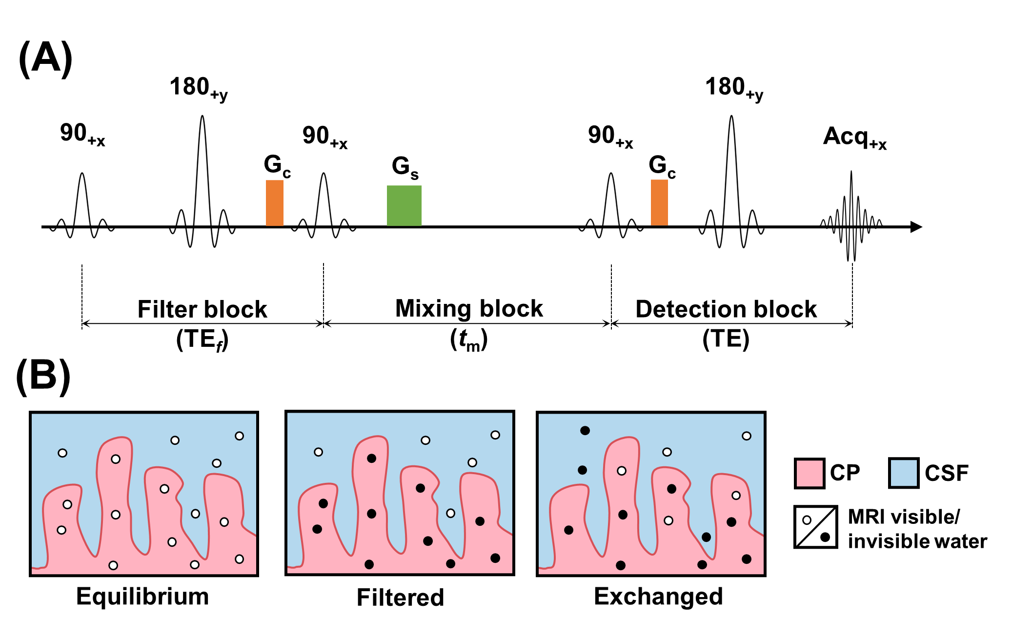
Figure 1. (A) Pulse sequence diagram of REXI, which has a filter block with echo time (TEf) fixed, a mixing block with varying mixing time tm, and a detection block with multi-echo acquisition (TE). The paired crushers (Gc) for coherence pathway selection can replace phase cycling to shorten acquisition time. A spoiler gradient (Gs) is applied to null unwanted transverse magnetization. The pulses in the filter/detection block are non-selective/selective. (B) Illustration of the magnetization evaluation of water molecule in CP and CSF during REXI acquisition.
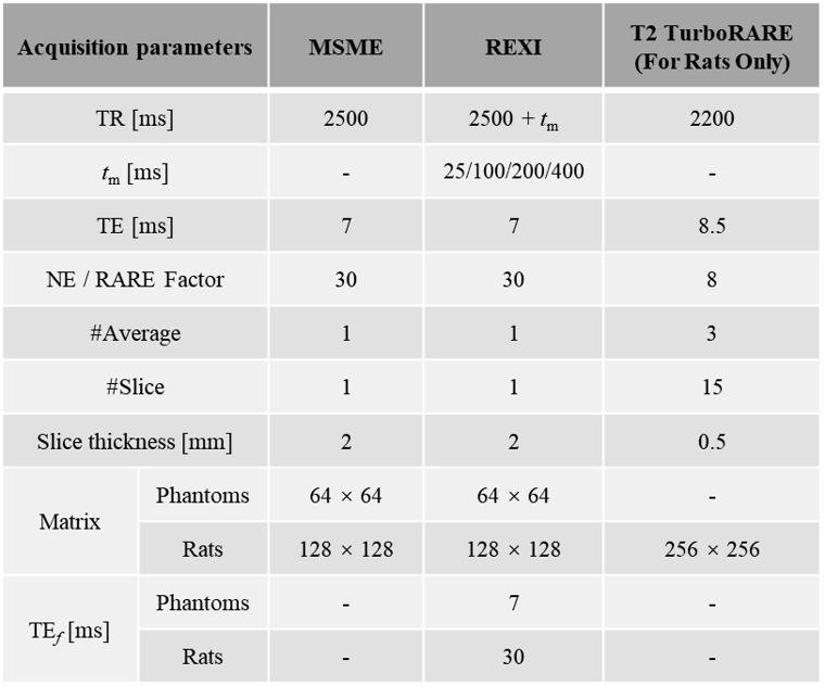
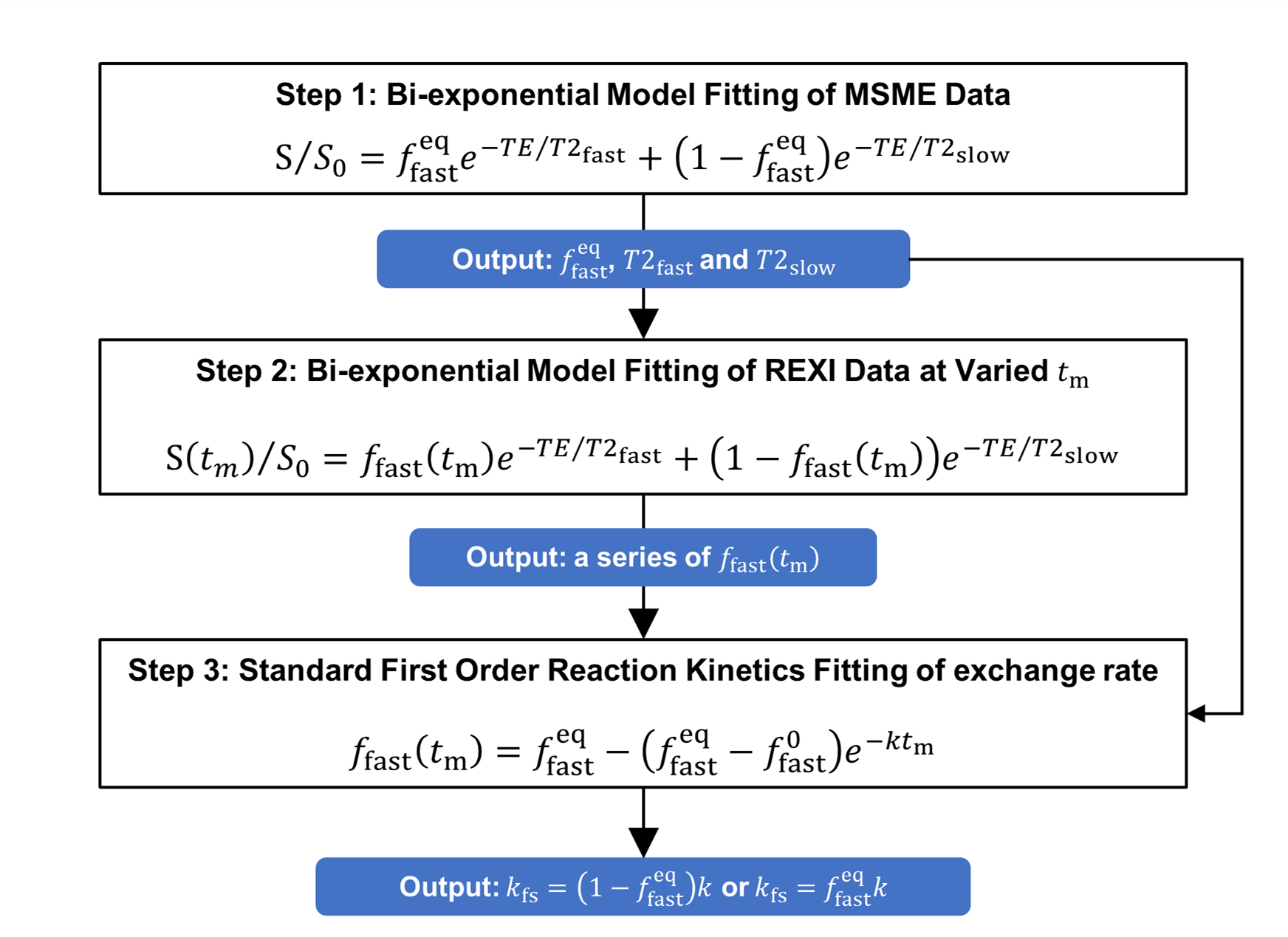
Figure 2. The two-sites exchange model fitting steps of REXI data. T2fast and T2slow are transverse relaxation time constants of the fast compartment (with volume fraction, ffast) and slow compartment (with volume fraction fslow =1- ffast) in the two-pool system. The ffasteq is at equilibrium state, and ffast0 is at tm = 0. The effective exchange rate is k, and the subscript fs means processing from fast to slow compartment. The Multi Slice Multi Echo (MSME) shares the same acquisition parameters with REXI.
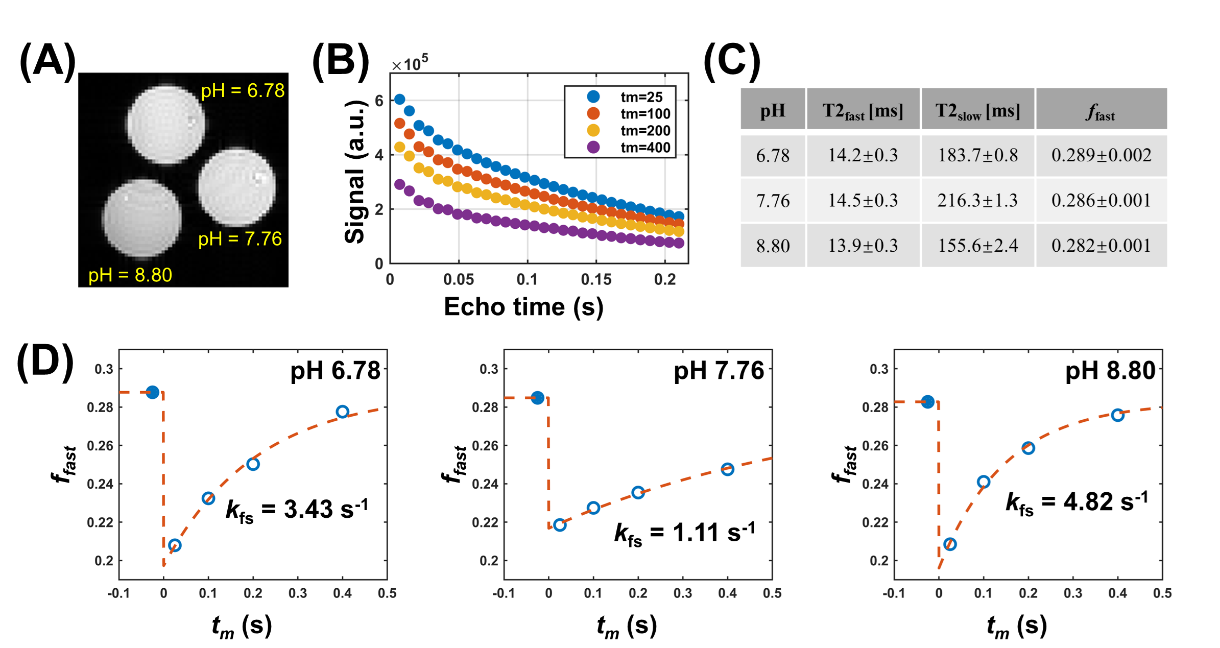
Figure 3. The representative REXI image at tm=25ms and TE = 7ms (A) and signal-TE curves at each tm (B), and the T2fast, T2slow and ffast (volume fraction of fast compartment) at three pH across three repeated scans (C). (D) ffast-tm curves of the entire cross-section ROIs in the urea-water phantoms. In (D), the filled blue circles are the ffasteq and dashed curves are the model fitting results.
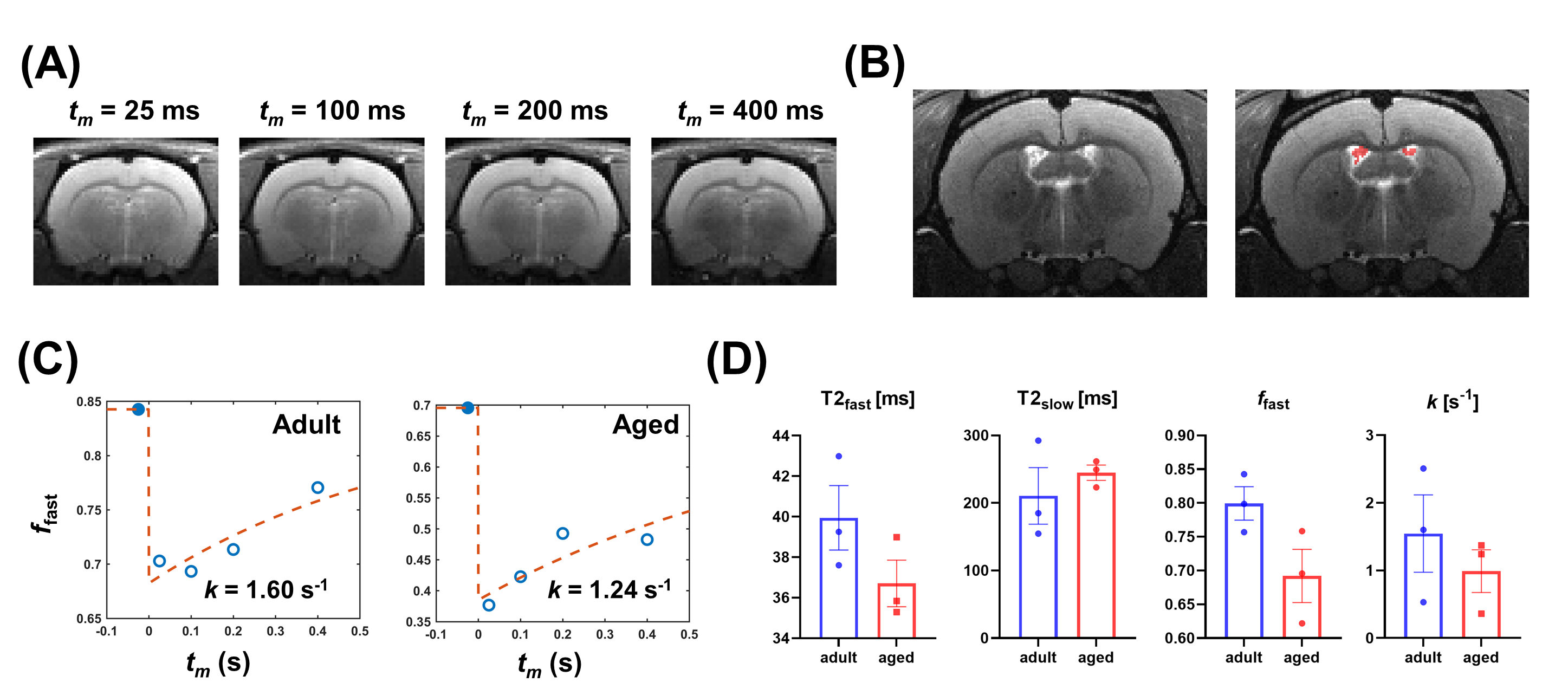
Figure 4. The representative REXI images of TE = 7ms at each tm (A) and T2-TurboRARE image and corresponding ROI of CP (B), and ffast-tm curves of the adult group and aged group (C). (D) Statistical comparison of T2fast, T2slow, ffast, and k = kfs + ksf between the adult and aged groups in CP using unpaired t-test (all P > 0.05).