0029
Choroid Plexus Area and Permeability in Dementia with Lewy Bodies1Department of Biomedical Engineering, School of Medicine, Tsinghua University, Beijing, China, Beijing, China, 2Department of Neurology, Beijing Friendship Hospital, Capital Medical University, Beijing, China, Beijing, China, 3Department of Neurology, Tianjin Huanhu Hospital, Tianjin, China, Tianjin, China
Synopsis
Keywords: Other Neurodegeneration, DSC & DCE Perfusion, choroid plexus; dementia with Lewy bodies
Motivation: The choroid plexus has been demonstrated to play a significant role in the pathophysiology of dementia with Lewy bodies (DLB), however the imaging characteristics are not yet explored.
Goal(s): Our goal was to assess choroid plexus area and permeability based on MRI in DLB patients.
Approach: DLB patients were imaged to acquire choroid plexus area and permeability and compared with patients with Alzheimer’s disease and healthy controls.
Results: DLB patients exhibited larger area and lower fractional plasma volume in choroid plexus than healthy controls. Additionally, they were found to be significantly associated with the mini-mental state examination score.
Impact: The larger choroid plexus area and lower fractional plasma volume detected by T1-weighted MRI and DCE-MRI in dementia with Lewy bodies provide a non-invasive and quantitative metric for advancing the diagnosis and treatment of dementia with Lewy bodies.
Introduction
The choroid plexus has been demonstrated to play a critical role in producing cerebrospinal fluid (CSF) and is closely related to the glymphatic system1, which is involved in the clearance of metabolic waste products and noxious substances2. Previous studies have revealed an enlarged choroid plexus volume in Alzheimer disease (AD) and Parkinson disease (PD) based on MRI, suggesting a possible choroid plexus dysfunction in neurodegenerative disease3,4. Dementia with Lewy bodies (DLB) is the second most common type of neurodegenerative dementia after AD5 and is characterized by the accumulation of pathogenic alpha-synuclein (α-syn) in the brain6. Regarding the role of glymphatic system in clearing the α-syn, amyloid-beta (Aβ) and tau protein7, we could assume that choroid plexus also plays an important role in the disease mechanisms of DLB. To test this hypothesis, we aimed to evaluate the morphological and permeability changes in DLB and compared them with AD and healthy controls (HC).Methods
Study participants: This prospective study was approved by the Ethics Committee, and all participants provided written informed consent. Probable DLB and AD were diagnosed using the criteria McKeith established in 20178 and National Institute on Aging and Alzheimer’s Association criteria9 separately. And HC were recruited from the friends or relatives of the patients. The participants’ demographic characteristics, comorbidities, apolipoprotein E ε4 allele (APOE ε4) genotyping result, Mini-Mental State Examination (MMSE) score, plasma Aβ1-42/Aβ1-40 ratio and brain imaging findings were evaluated.MRI Acquisition: MRI examinations were performed at a 3T scanner (Prisma, Siemens Healthcare, Erlangen, Germany) with a 64-channel head coil. The protocol consisted of a 3D T1-weighted magnetization prepared-rapid gradient echo (MPRAGE) sequence (TR/TE/TI = 1700/3.5/787 ms, 0.8-mm isotropic) and a coronal DCE sequence (TR/TE = 5.2/1.8 ms, voxel size: 1.25×1.25×3 mm3, imaging time = 30 × 12s).
MRI Analysis: The choroid plexus area was measured on the slice demonstrating the largest choroid plexus of MPRAGE images. The mask was manually segmented twice by the same experienced radiologist on two separate days. The final area was obtained by averaging the results of the two delineations. Permeability parameters including the transfer rate of contrast agent (Ktrans) and fractional plasma volume (vp) were calculated from DCE MRI based on the Patlak model10.
Statistical Analysis: Analysis of variance (ANOVA) or Kruskal-Wallis H test was performed for continuous variables and chi-squared test was performed for categorical variables among the three groups. Reproducibility of the two segmentations for choroid plexus area and permeability was quantified by intra-class correlation coefficient (ICC) and Bland-Altman plots. Spearman’s correlation was employed to analyze the correlation between imaging characteristics of choroid plexus and MMSE. P values of less than 0.05 were considered significant.
Results
We finally included 18 DLB patients, 25 AD patients and 24 HC in this study. No significant difference was found in demographic and clinical information except for hypertension, APOE ε4 status and MMSE score (Table 1). Representative MPRAGE images, Ktrans and vp maps of DLB, AD and HC group were shown in Figure 1. Choroid plexus area demonstrated an excellent intraclass correlation coefficient between the two segmentation results (ICC = 0.956; [95% CI: 0.930-0.973], P < 0.001). DLB and AD patients both exhibited a significantly larger choroid plexus area compared to HC (P = 0.035 and P = 0.047) as depicted in Figure 2. Excellent agreement between the two tests was also observed in measuring Ktrans (ICC = 0.876; [95% CI: 0.806-0.922], P < 0.001) and vp (ICC = 0.832; [95% CI: 0.741-0.893], P < 0.001). Figure 3 demonstrated that DLB patients exhibited lower vp compared to the HC group (P = 0.002). And as shown in Figure 4, choroid plexus area was negatively associated with MMSE score (r = -0.41, P = 0.008), while vp was positively associated with MMSE score (r = 0.36, P = 0.019) in DLB and HC.Discussion and Conclusion
As far as we know, our study firstly reported the morphological and permeability changes of choroid plexus in DLB. We found that choroid plexus area was enlarged in DLB patients than that in HC, which may be a compensatory response to reduced CSF production, potentially due to factors such as stromal fibrosis, stromal dystrophic calcification, vessel vasodilation or inflammation11. Our results also suggested that DLB patients exhibited lower fractional plasma volume in choroid plexus compared to HC, which might be related to the reduced microvessel density in DLB12. Additionally, the choroid plexus area and fractional plasma volume were both demonstrated as independent predictors of MMSE score, which could be used as promising imaging biomarkers for clinical prognosis and staging of dementia.Acknowledgements
None.References
1. Lehtinen MK, Bjornsson CS, Dymecki SM, et al. The choroid plexus and cerebrospinal fluid: emerging roles in development, disease, and therapy. J Neurosci. 2013 Nov 6;33(45):17553-9.
2. Natale G, Limanaqi F, Busceti CL, et al. Glymphatic System as a Gateway to Connect Neurodegeneration From Periphery to CNS. Front Neurosci. 2021 Feb 9;15:639140.
3. Choi JD, Moon Y, Kim HJ, et al. Choroid Plexus Volume and Permeability at Brain MRI within the Alzheimer Disease Clinical Spectrum. Radiology. 2022 Sep;304(3):635-645.
4. Jeong SH, Jeong HJ, Sunwoo MK, et al. Association between choroid plexus volume and cognition in Parkinson disease. Eur J Neurol. 2023 Oct;30(10):3114-3123.
5. Sanford AM. Lewy Body Dementia. Clin Geriatr Med. 2018 Nov;34(4):603-615.
6. Guerreiro A, da Costa G, Gomes RA, et al. α-Synuclein aggregation in the saliva of familial transthyretin amyloidosis: a potential biomarker. Amyloid. 2012 Jun;19(2):74-80.
7. Valdinocci D, Radford RA, Siow SM, et al. Potential Modes of Intercellular α-Synuclein Transmission. Int J Mol Sci. 2017 Feb 22;18(2):469.
8. McKeith IG, Boeve BF, Dickson DW, et al. Diagnosis and management of dementia with Lewy bodies: Fourth consensus report of the DLB Consortium. Neurology. 2017 Jul 4;89(1):88-100.
9. McKhann GM, Knopman DS, Chertkow H, et al. The diagnosis of dementia due to Alzheimer's disease: recommendations from the National Institute on Aging-Alzheimer's Association workgroups on diagnostic guidelines for Alzheimer's disease. Alzheimers Dement. 2011 May;7(3):263-9.
10. Buckley DL, Shurrab AE, Cheung CM, et al. Measurement of single kidney function using dynamic contrast-enhanced MRI: comparison of two models in human subjects. J Magn Reson Imaging. 2006 Nov;24(5):1117-23.
11. Silverberg GD, Heit G, Huhn S, et al. The cerebrospinal fluid production rate is reduced in dementia of the Alzheimer's type. Neurology. 2001 Nov 27;57(10):1763-6.
12. Miners S, Moulding H, de Silva R, et al. Reduced vascular endothelial growth factor and capillary density in the occipital cortex in dementia with Lewy bodies. Brain Pathol. 2014 Jul;24(4):334-43.
Figures
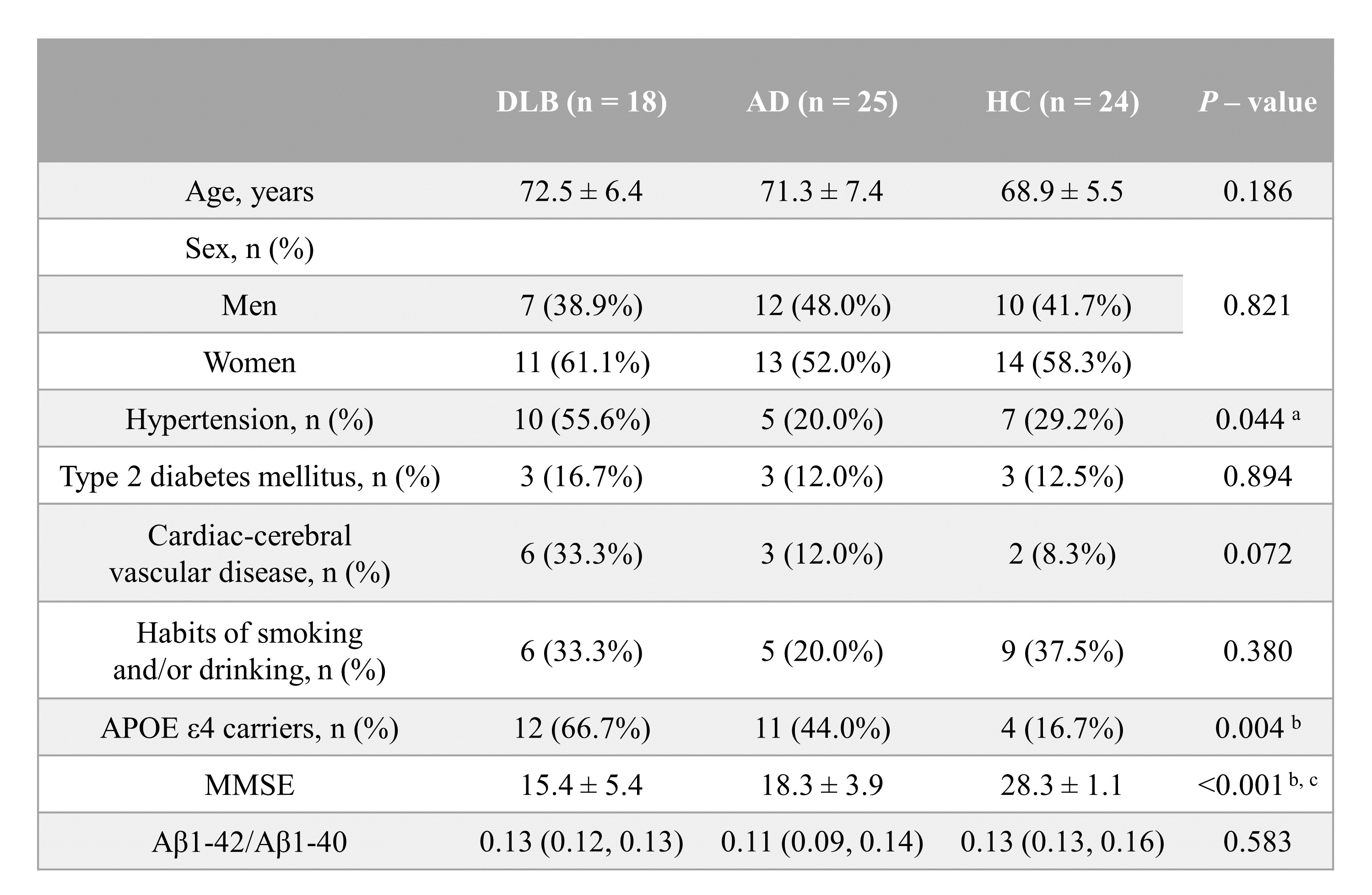
Table 1. Demographic and clinical information of participants. Data are expressed as mean ± SE or median (quartiles) or n (%). a For the comparison between the DLB and AD (Bonferroni-corrected P < 0.05). b For the comparison between the DLB and HC (Bonferroni-corrected P < 0.05). c For the comparison between the AD and HC (Bonferroni-corrected P < 0.05). DLB: dementia with Lewy body; AD: Alzheimer’s disease; HC: healthy controls; APOE ε4: apolipoprotein E ε4 allele; MMSE: mini-mental state examination; Aβ: amyloid-beta.
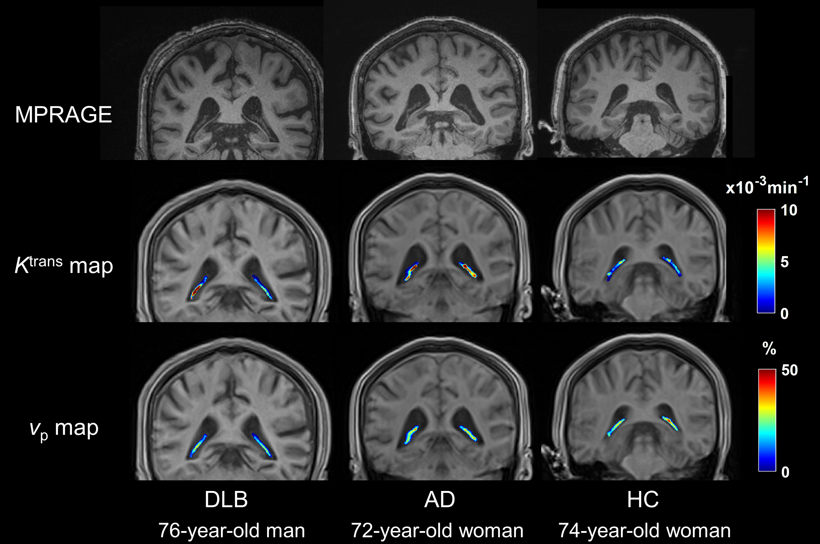
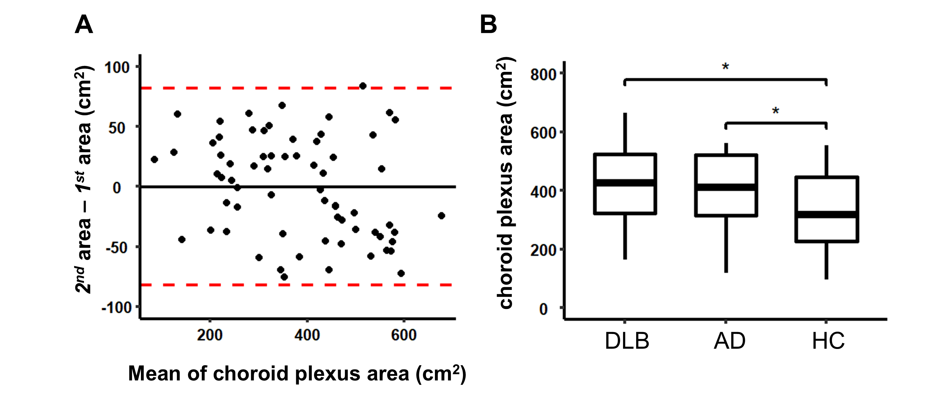
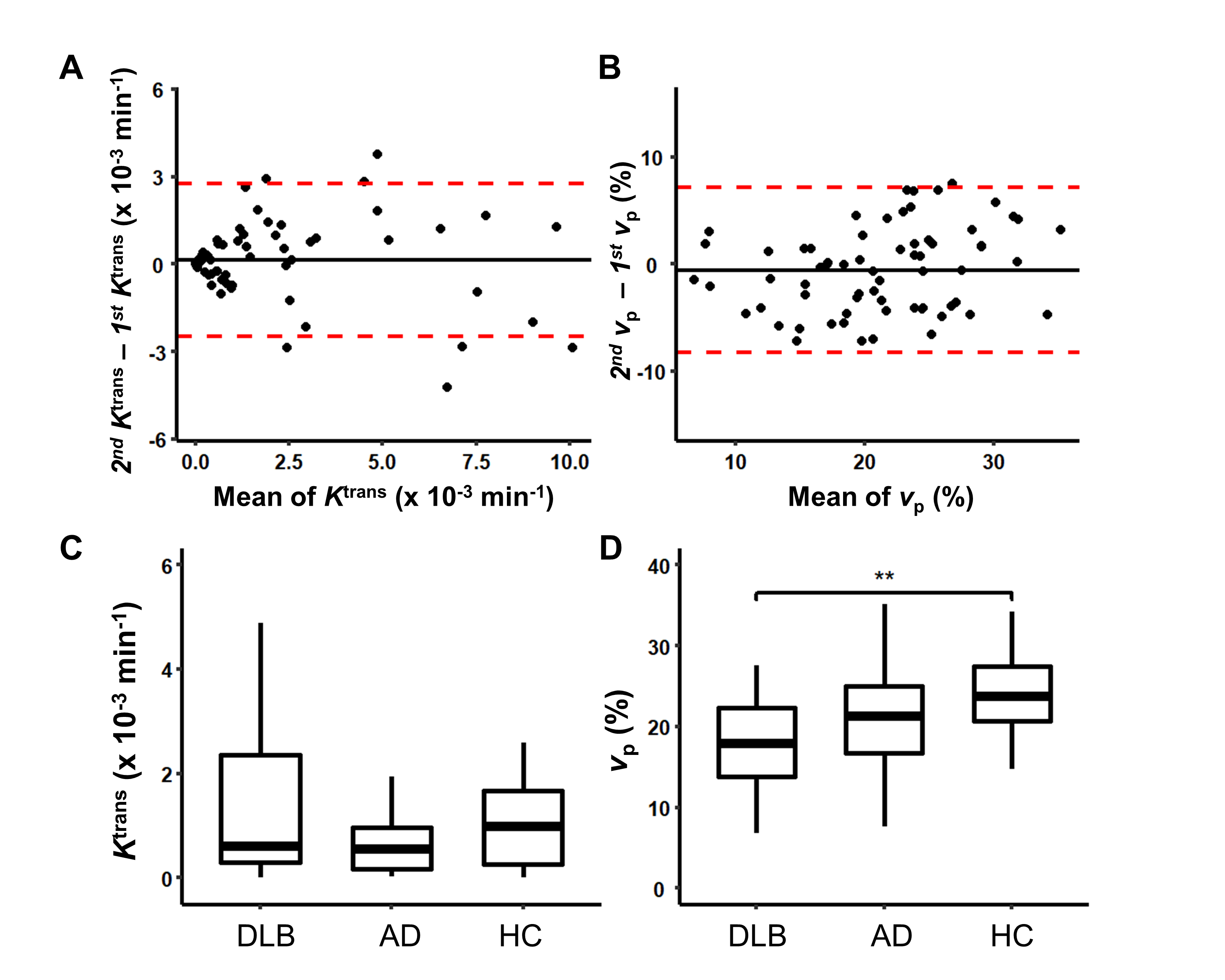
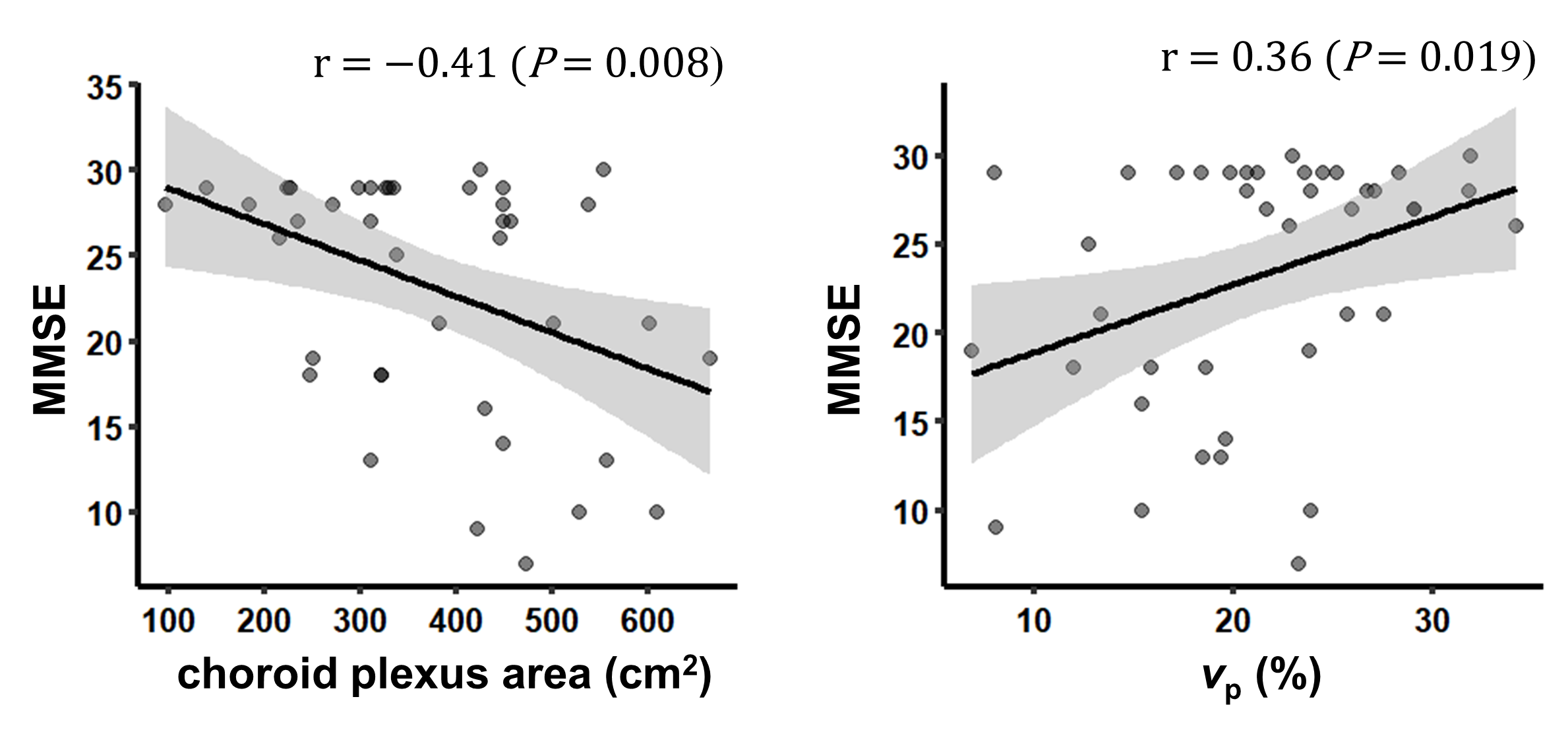
Figure 4. Relationship between the MMSE and choroid plexus area (A), vp (B). Significance was tested by Spearman’s correlation. MMSE: mini-mental state examination; vp: fractional plasma volume.