0019
Predicting Brain Age of Healthy Adults Based on Morphological MRI Parcellation Using Radiomics1Bernard and Irene Schwartz Center for Biomedical Imaging, Department of Radiology,, New York University Grossman School of Medicine, New York, New York, USA, new york, NY, United States, 2Center for Advanced Imaging Innovation and Research (CAI2R), Department of Radiology, New York University Grossman School of Medicine, New York, New York, USA, new york, NY, United States, 3Vilcek Institute of Graduate Biomedical Sciences, New York University Grossman School of Medicine, New York, New York, USA, new york, NY, United States
Synopsis
Keywords: Aging, Aging, aging, structural imaging, radiomics, neuro
Motivation: A machine learning model capable of accurately estimating brain age could have a large clinical impact.
Goal(s): To apply radiomics analysis to morphological MR images and train a machine learning model capable of accurately estimating subjects’ age from radiomics features.
Approach: T1- and T2-weighted brain images of 725 healthy adults were used to extract 18324 radiomics features from bilateral caudate, putamen, and hippocampus, and used to train a stacking regressor machine learning model.
Results: Our machine learning model accurately estimated the subjects’ age with a mean absolute error of 4.77±0.35 years using radiomics features from T1-(45%) and T2-weighted(55%).
Impact: Investigating advanced machine learning methods to accurately estimate brain aging based on commonly used clinical MR images provides vital insights to further improve our understanding of brain changes in both healthy aging and neurodegeneration.
Introduction
Accurately predicting brain age has the potential to elucidate brain changes in both healthy aging and neurodegenerative diseases1,2. Previous work on predicting brain age using structural imaging has achieved mean absolute error (MAE) values of between 5-7 years3-5. Studies that combined structural and functional imaging data have achieved MAE values under 4 years6,7. However, functional imaging data, such as functional MRI (fMRI)) and diffusion-weighted imaging (DWI) scans are still not broadly available and are expensive to utilize (R3).Furthermore, these levels of prediction accuracy are reached only when a large amount of data is available (>23302 cases6,7). Radiomics, a technique based on the extraction of quantitative features from medical images, has emerged as a powerful tool for improving patient outcomes and advancing precision medicine8-12. Radiomics extracts image features from specific regions of interest (ROIs) and then uses machine learning models to associate features with clinical outcomes. For this reason, radiomics requires fewer instances compared to other models trained on the full image, which reduces the degrees of freedom that the model needs to learn13.In this work, we evaluated radiomics as a tool for brain age estimation when data is limited.
Material and Methods
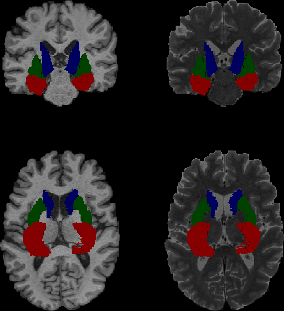
Permission was obtained to access the imaging data acquired through the Human Connectome Project-Aging (HCP-A)14. The dataset included: T1- (MPRAGE) and T2-weighted images of 725 typically healthy adults (age=59±15 years, 319 males) along with a structural parcellation of each subject's brain based on the aprc+aseg atlas (https://surfer.nmr.mgh.harvard.edu/fswiki/FsTutorial/AnatomicalROI).Among the 113 ROIs available in the atlas, we focused the analysis on 6 areas that have been observed to be highly affected in normal aging15: the bilateral hippocampus (H), putamen (P), and caudate (C) (Figure 1)
Feature Extraction and Selection
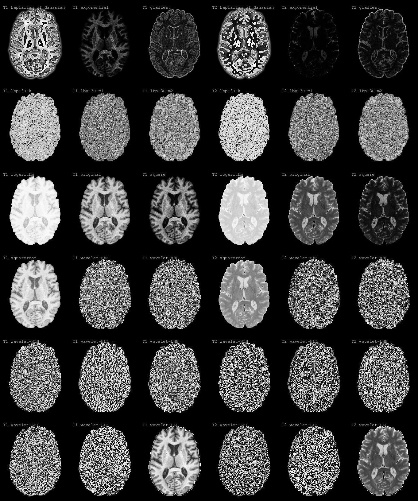
Radiomic features were extracted using the pyradiomics library16 from T1- and T2-weighted images for the six ROIs. The extracted features included first-order statistics, shape-based features, and textural analysis16. Pyradiomics allows for extracting features from several filtered versions of the original T1- or T2-weighted image, such as normalization, filtering, and wavelet decomposition (Figure 2). Image transformations are an essential part of radiomic feature extraction because they enhance the information content of the images and allow for the extraction of more robust and informative features. The total number of features extracted per subject was 18,324.
The feature selection step was conducted using a Sequential Forward Floating Selection (SFFS)11,17 algorithm, varying the final number of features in each subset between 2 and 20 features in 10 steps.
Machine Learning
For this study, we implemented an ensemble stacking regressor18 machine learning model with eight different regressors, including Lasso, random forest, k-nearest neighbors, gradient boosting, AdaBoost, HistGradientBoostingRegressor, and MLPRegressor19. Ensemble stacking is a machine learning technique that combines the predictions of multiple estimators to produce a more accurate prediction18. In particular, the predictions of the individual estimators are used as input to a final estimator, which is typically a linear regression model.
We trained 10 ensemble models, one for every feature selection subset, using a 10-fold cross-validation with a test ratio of 25%. We used the R² or the coefficient of determination and MAE to assess the accuracy of the model in the predictions.
Results and Discussions
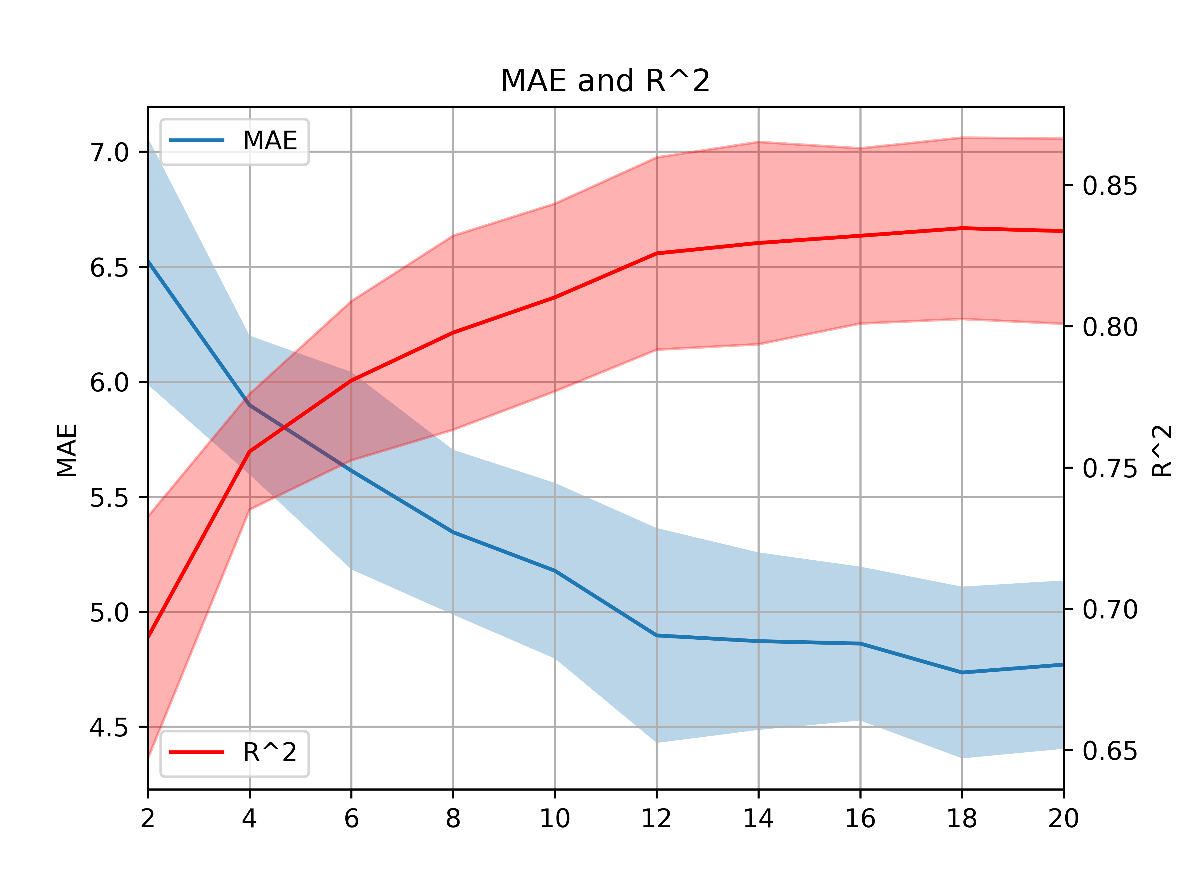
First, we observed lower MAE in predicting brain age for the stacking regressor technique (MAE 4.77±0.35; R2=0.83±0.03) compared to all the other methods individually (Figure 3). This is in line with previous studies supporting the combination of multiple estimators to produce more accurate model predictions, as done in stacking regressors.Our results showed that the performance of the models increases with the number of features used, reaching a plateau after 12 features (MAE: 4.76 ± 0.37; R2 0.83 ± 0.33; Figure 4).
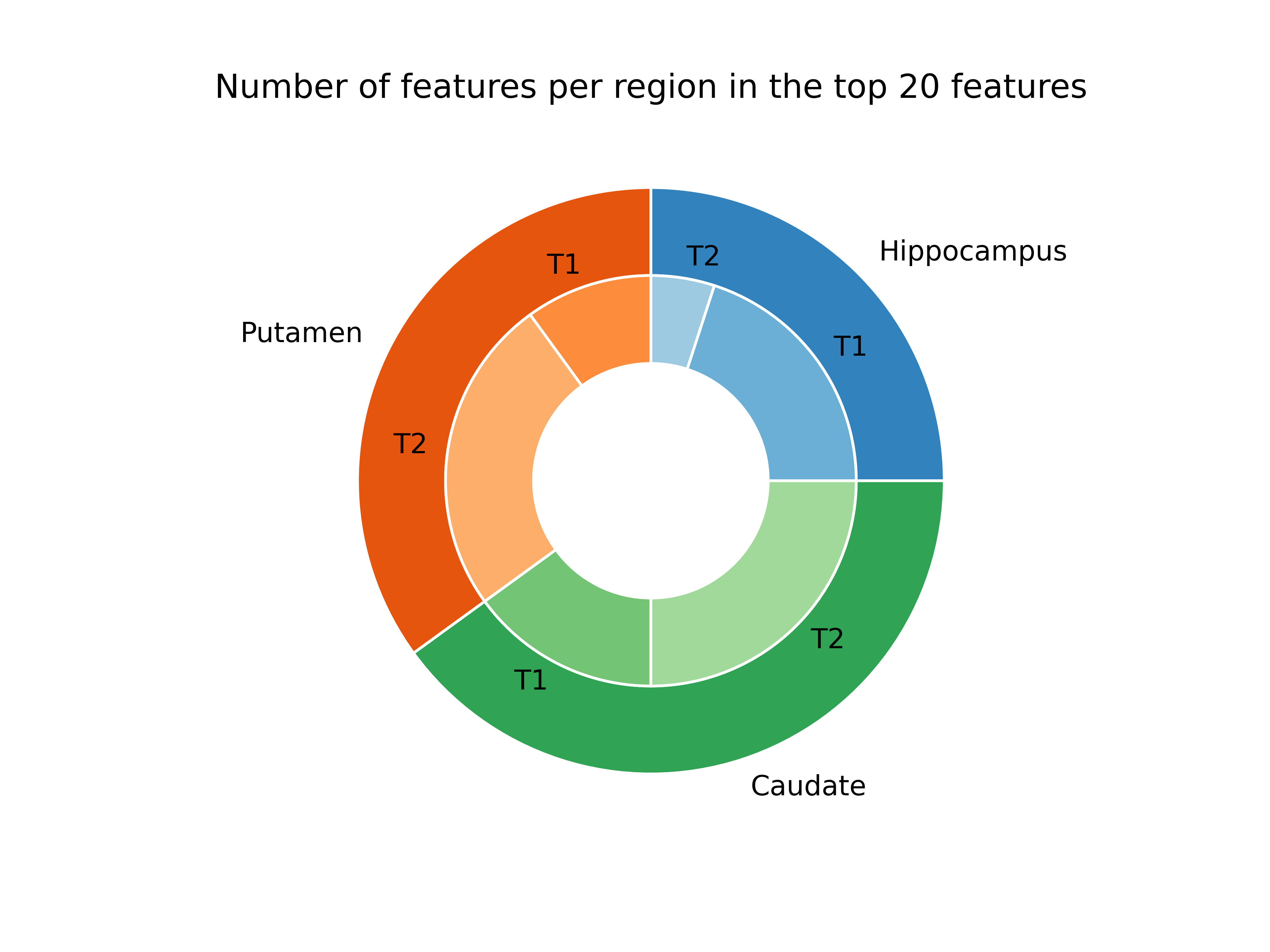
The predictive model using the stacking regressor was obtained by training the model on 20 radiomic features (C=8, P=7, H=5; Fig.5). This supports our hypothesis that these three main brain subcortical areas are enough to provide key information for machine learning-based aging prediction.
Conclusions
Our study shows that radiomic features can be used for the prediction of brain age in healthy adults with a performance comparable to that reported for models trained on considerably larger datasets available in the literature1,2. We aim to extend this approach to patients with different neurodegenerative diseases, by incorporating information from more cortical and subcortical regions.Acknowledgements
This work was performed under the Rubric of the Center for Advanced Imaging Innovation and Research (CAI2R, www.cai2r.net), an NIBIB National Center for Biomedical Imaging and Bioengineering (NIH P41 EB017183).References
- Franke K, Gaser C. Ten Years of BrainAGE as a Neuroimaging Biomarker of Brain Aging: What Insights Have We Gained?. Front Neurol. 2019;10:789. Published 2019 Aug 14. doi:10.3389/fneur.2019.00789.
- Bretzner M, Bonkhoff AK, Schirmer MD, Hong S, Dalca A, Donahue K, Giese AK, Etherton MR, Rist PM, Nardin M, Regenhardt RW, Leclerc X, Lopes R, Gautherot M, Wang C, Benavente OR, Cole JW, Donatti A, Griessenauer C, Heitsch L, Holmegaard L, Jood K, Jimenez-Conde J, Kittner SJ, Lemmens R, Levi CR, McArdle PF, McDonough CW, Meschia JF, Phuah CL, Rolfs A, Ropele S, Rosand J, Roquer J, Rundek T, Sacco RL, Schmidt R, Sharma P, Slowik A, Sousa A, Stanne TM, Strbian D, Tatlisumak T, Thijs V, Vagal A, Wasselius J, Woo D, Wu O, Zand R, Worrall BB, Maguire J, Lindgren AG, Jern C, Golland P, Kuchcinski G, Rost NS; MRI-GENIE and GISCOME Investigators and the International Stroke Genetics Consortium. Radiomics-Derived Brain Age Predicts Functional Outcome After Acute Ischemic Stroke. Neurology. 2023 Feb 21;100(8):e822-e833. doi: 10.1212/WNL.0000000000201596. Epub 2022 Nov 28. PMID: 36443016; PMCID: PMC9984219.
- R.S. Wilson, S.E. Leurgans, P.A. Boyle, J.A. Schneider, D.A. Bennett Neurology Sep 2010, 75 (12) 1070-1078; DOI: 10.1212/WNL.0b013e3181f39adc
- Anatürk M, Kaufmann T, Cole JH, et al. Prediction of brain age and cognitive age: Quantifying brain and cognitive maintenance in aging. Hum Brain Mapp. 2021;42(6):1626-1640. doi:10.1002/hbm.25316.
- Han J, Kim SY, Lee J, Lee WH. Brain Age Prediction: A Comparison between Machine Learning Models Using Brain Morphometric Data. Sensors (Basel). 2022;22(20):8077. Published 2022 Oct 21. doi:10.3390/s22208077
- Niu X, Zhang F, Kounios J, Liang H. Improved prediction of brain age using multimodal neuroimaging data. Hum Brain Mapp. 2020;41(6):1626-1643. doi:10.1002/hbm.24899
- Zhang, Z., Liu, Y., Guo, Y., & Zhang, D. (2023). Brain age prediction using structural and functional MRI data with a novel graph neural network. Medical Image Analysis, 68, 101956.
- Bevilacqua R, Barbarossa F, Fantechi L, et al. Radiomics and Artificial Intelligence for the Diagnosis and Monitoring of Alzheimer's Disease: A Systematic Review of Studies in the Field. J Clin Med. 2023;12(16):5432. Published 2023 Aug 21. doi:10.3390/jcm12165432.
- Zhou M, Scott J, Chaudhury B, et al. Radiomics in Brain Tumor: Image Assessment, Quantitative Feature Descriptors, and Machine-Learning Approaches. AJNR Am J Neuroradiol. 2018;39(2):208-216. doi:10.3174/ajnr.A5391.
- Bologna, M., Corino, V. D. A., Montin, E., Messina, A., Calareso, G., Greco, F. G., Sdao, S., & Mainardi, L. T. (2018). Assessment of Stability and Discrimination Capacity of Radiomic Features on Apparent Diffusion Coefficient Images. Journal of Digital Imaging. https://doi.org/10.1007/s10278-018-0092-9
- Corino, V. D. A., Montin, E., Messina, A., Casali, P. G., Gronchi, A., Marchianò, A., & Mainardi, L. T. (2018). Radiomic analysis of soft tissue sarcomas can distinguish intermediate from high-grade lesions. Journal of Magnetic Resonance Imaging, 4(3), 829–840. https://doi.org/10.1002/jmri.25791
- Bologna, M., Calareso, G., Resteghini, C., Sdao, S., Montin, E., Corino, V., Mainardi, L., Licitra, L., & Bossi, P. (2020). Relevance of apparent diffusion coefficient features for a radiomics-based prediction of response to induction chemotherapy in sinonasal cancer. NMR in Biomedicine, March 2019, 1–12. https://doi.org/10.1002/nbm.4265
- Obiols, Mar Hernández et al. “Can radiomics features boost the performance of deep learning upon histology images?” 2019 International Conference on Medical Imaging Physics and Engineering (ICMIPE) (2019): 1-6.
- Harms MP, Somerville LH, Ances BM, et al. Extending the Human Connectome Project across ages: Imaging protocols for the Lifespan Development and Aging projects. Neuroimage. 2018;183:972-984. doi:10.1016/j.neuroimage.2018.09.060
- Abedelahi A, Hasanzadeh H, Hadizadeh H, Joghataie MT. Morphometric and volumetric study of caudate and putamen nuclei in normal individuals by MRI: Effect of normal aging, gender and hemispheric differences. Pol J Radiol. 2013;78(3):7-14. doi:10.12659/PJR.889364
- van Griethuysen, J. J. M., Fedorov, A., Parmar, C., Hosny, A., Aucoin, N., Narayan, V., Beets-Tan, R. G. H., Fillon-Robin, J. C., Pieper, S., Aerts, H. J. W. L. (2017). Computational Radiomics System to Decode the Radiographic Phenotype. Cancer Research, 77(21)
- Nakariyakul, Songyot and David Paul Casasent. “Improved forward floating selection algorithm for feature subset selection.” 2008 International Conference on Wavelet Analysis and Pattern Recognition 2 (2008): 793-798.
- Hasson, Hilaf et al. “Theoretical Guarantees of Learning Ensembling Strategies with Applications to Time Series Forecasting.” International Conference on Machine Learning (2023).
- Venkataramanaiah, M. and Madhura S. Rao. “Criteria for selection of regressors in econometrics.” Bioinformatics (2013).
Figures
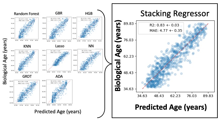
The figure reports an example of the results of the regression capability of the model built using 20 features in the eight regressors (left) and the final stacking layer (right). The coordinates of the blue points in the scatter plot represent the predicted age by the model vs the real one. The red line is a linear interpolation of the data. The performance of the final model had an MAE of 4.77 and the R^2 of 0.83.
GBR is gradient boost regressor, HGB is histogram gradient boost regressor, KNN is K neighbor regressor, NN is neural network regressor, ADA is ADA boost, GRDT is elastic net