0008
Accelerated 2D Cartesian MRI with an 8-channel local B0 coil array combined with parallel imaging1High-Field MR Center, Max Planck Institute for Biological Cybernetics, Tübingen, Germany, 2Institute of Biomedical Imaging, Graz University of Technology, Graz, Austria, 3Institute for Diagnostic and Interventional Radiology, University Medical Center Göttingen, Göttingen, Germany, 4German Centre for Cardiovascular Research (DZHK), Partner Site Göttingen, Göttingen, Germany, 5BioTechMed-Graz, Graz, Austria, 6A. A. Martinos Center for Biomedical Imaging, Department of Radiology, Massachusetts General Hospital, Charlestown, MA, United States, 7Harvard Medical School, Boston, MA, United States, 8Department of Health Technology, Technical University of Denmark, Kongens Lyngby, Denmark, 9Danish Research Centre for Magnetic Resonance, Centre for Functional and Diagnostic Imaging and Research, Copenhagen University Hospital Amager and Hvidovre, Hvidovre, Denmark, 10Department for Biomedical Magnetic Resonance, University of Tübingen, Tübingen, Germany
Synopsis
Keywords: YIA, Hybrid & Novel Systems Technology, Physics & Engineering, Acquisition & Reconstruction, New Trajectories & Spatial Encoding Methods, Acquisition Methods, nonlinear gradient
Motivation: The inherently slow MRI scans can be accelerated through rapid modulation of nonlinear gradient fields; however, its fundamental mechanisms and limits remain incompletely understood and validated.
Goal(s): We investigate accelerated MRI with flexible modulations of nonlinear B0 fields using a custom-built local B0 array.
Approach: The sampling theory is extended to rigorously compare nonlinear field modulation schemes in a quantitative k-space. A novel field calibration technique is proposed to enhance reconstruction. With safety evaluations, we perform in-vivo accelerated scans.
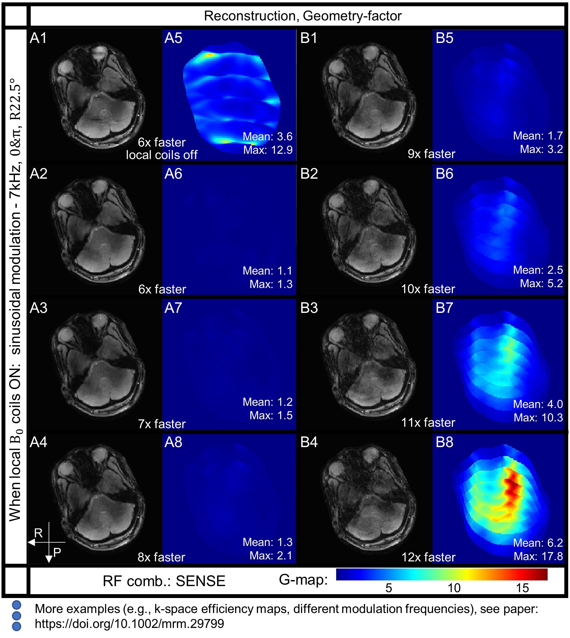
Results: Our in-vivo 2D FLASH scans make significant steps to speed up MRI with local B0 array, achieving eight-fold joint acceleration with parallel imaging.
Impact: For the first time, the sampling efficiency of nonlinear gradients in the entire k-space is quantitatively visualized, allowing rigorous comparison of distinct B0 modulations. Furthermore, the field estimation technique enables fast and robust in-vivo scans accelerated by flexible nonlinear fields.
Introduction
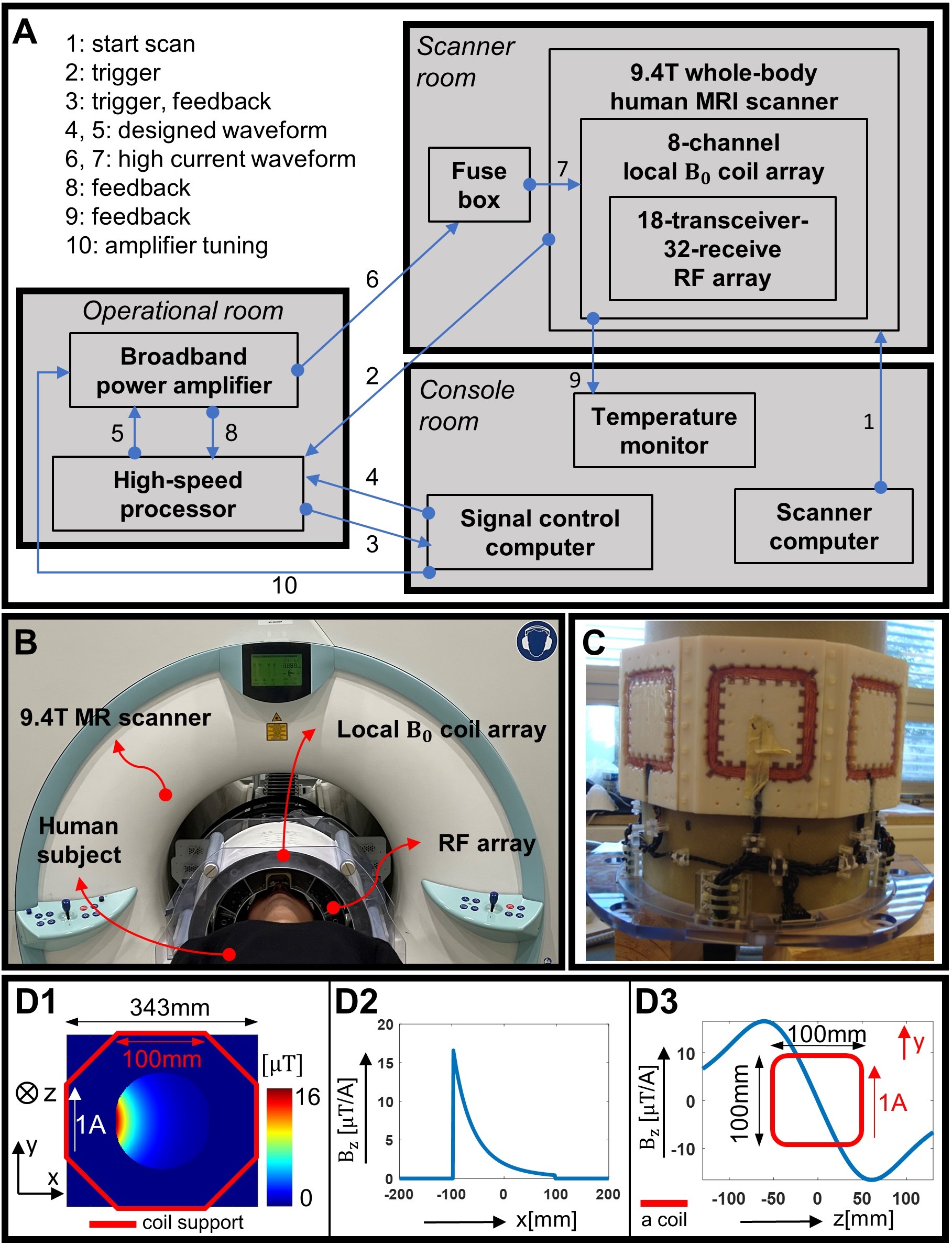
Despite significant advancements1–11 over recent decades, MRI remains inherently slow due to sequential data acquisitions. While methods using multiple RF receivers12,6–8 and rapid switching of linear gradient13,14 became well explained15, fast MRI techniques leveraging nonlinear gradient fields16–20 appeared much less investigated and understood. To unlock further acceleration potentials, we developed an 8-channel local B0 coil array, adapted from multi-coil shim design21 and accommodates a 16-transceive-32-receive RF array22, to explore rapid and flexible modulation of nonlinear B0 fields for fast 2D Cartesian MRI.As a more general technique23 than wave-CAIPI13 and FRONSAC19, we employ eight small B0 loops (i.e., 10cm x 10cm) driven by independent current waveforms (here, sinusoidal) during the signal readout of multi-slice FLASH1 scans (see Figure 1). This approach imposes additional localized spin phase evolution on objects, rather than global13,19, to disentangle mixed pixels in accelerated scans7. Moreover, it can generate various nonlinear gradient fields24 to identify the optimal modulation schemes.
To rigorously compare the k-space encoding efficiency across distinct modulation schemes in arbitrary acquisition time durations, we extend a mathematical framework for parallel imaging25 based on reproducing kernel Hilbert space (RKHS) to incorporate nonlinear B0 field encoding23. Additionally, a novel field calibration technique utilizing amplifier current monitors and the ESPIRiT algorithm15 is proposed to calibrate our setup and enhance reconstruction. Safety evaluations are performed26,27, and ex-vivo and in-vivo brain scans validate the feasibility of MRI acceleration using local B0 modulations28.
Methods
With RKHS, image reconstruction can be seen as interpolating the continuous k-space signal representations of the objects from discrete MRI samples acquired using arbitrary “spatial encoders”. The norm of interpolation weights (i.e., cardinal function) represents time-domain noise amplification of sampling operators. According to Equation (14-16)28, relative to fully sampled k-space, the time-domain encoding efficiency incorporating linear, nonlinear gradient fields and RF receivers’ sensitivity can be computed from the signal encoding matrix:$$E_{\bar{t},j}E_{t,i}^\ast U=E_{\bar{t},j}E_{\dot{t},k}^\ast$$
$$P^{\dot{t},k}\ =\ \sqrt{\left(\ E_{\dot{t},k}E_{\dot{t},k}^\ast\ -\ \ {U^\ast E}_{t,i}E_{\dot{t},k}^\ast\right)_{diag}}$$
where $$$E_{t,i},E_{\bar{t},j}$$$ are both the tested encoding matrix including all “spatial encoders”, and $$$E_{\dot{t},k}$$$ is a reference encoding matrix (e.g., DFT matrix), $$$U$$$ is the matrix to be solve for cardinal function, $$$P^{\dot{t},k}$$$ is the power function representing approximation errors. The noise amplification factor is obtained by vertically summing $$$U$$$. The asterisk $$$\ast$$$ denotes the conjugate transpose operation.
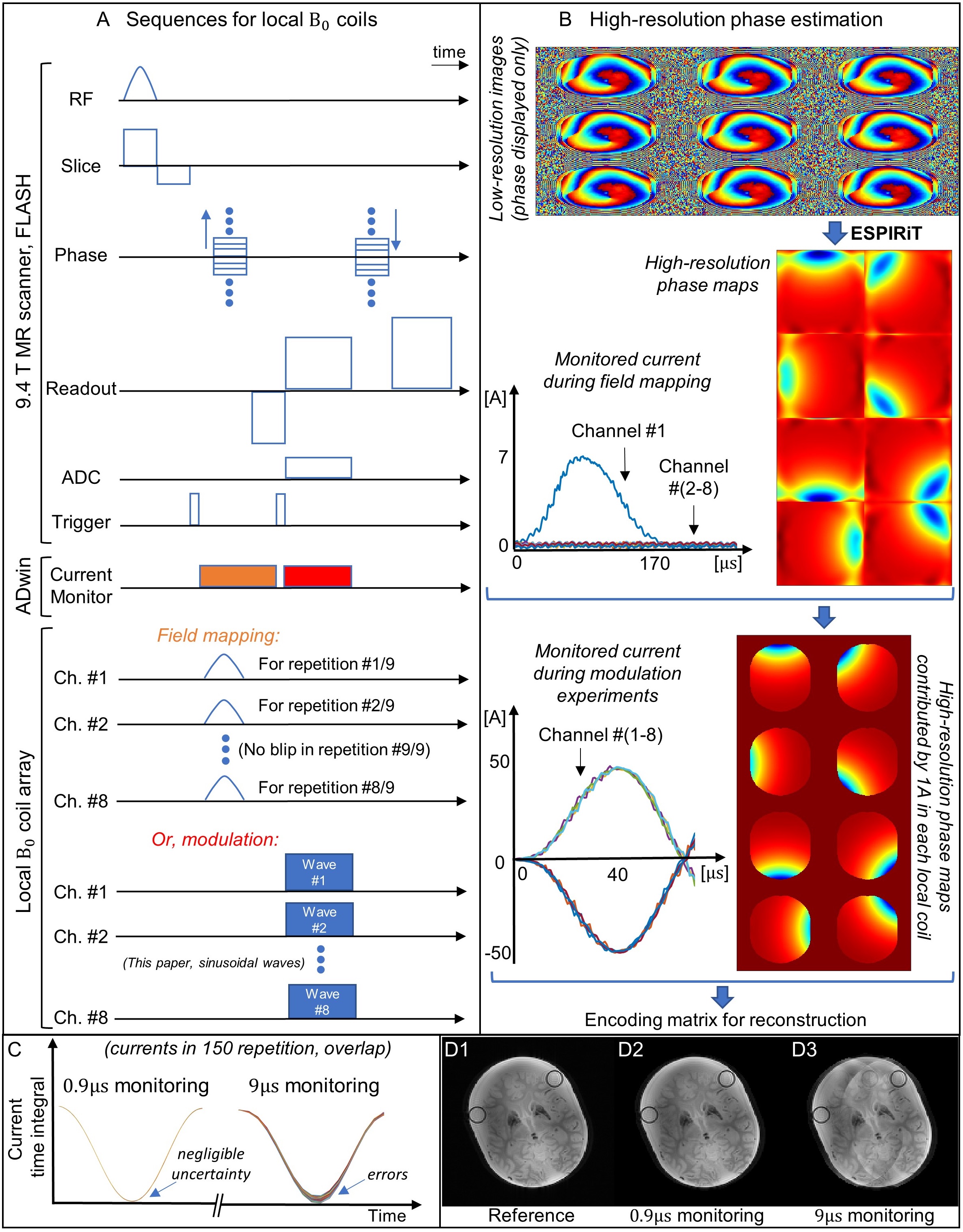
Only one field calibration scan is required for all distinct modulation experiments (see Figure 2). Essentially, low-resolution field mapping scans with a current blip in each local coil are input to the ESPIRiT algorithm15 to extrapolate phase maps in high-resolution grid with robustness. The current monitors are exploited to resolve phase offset maps by unit current in a local B0 coil, which are later combined with monitored modulation currents for forward model reconstruction13.
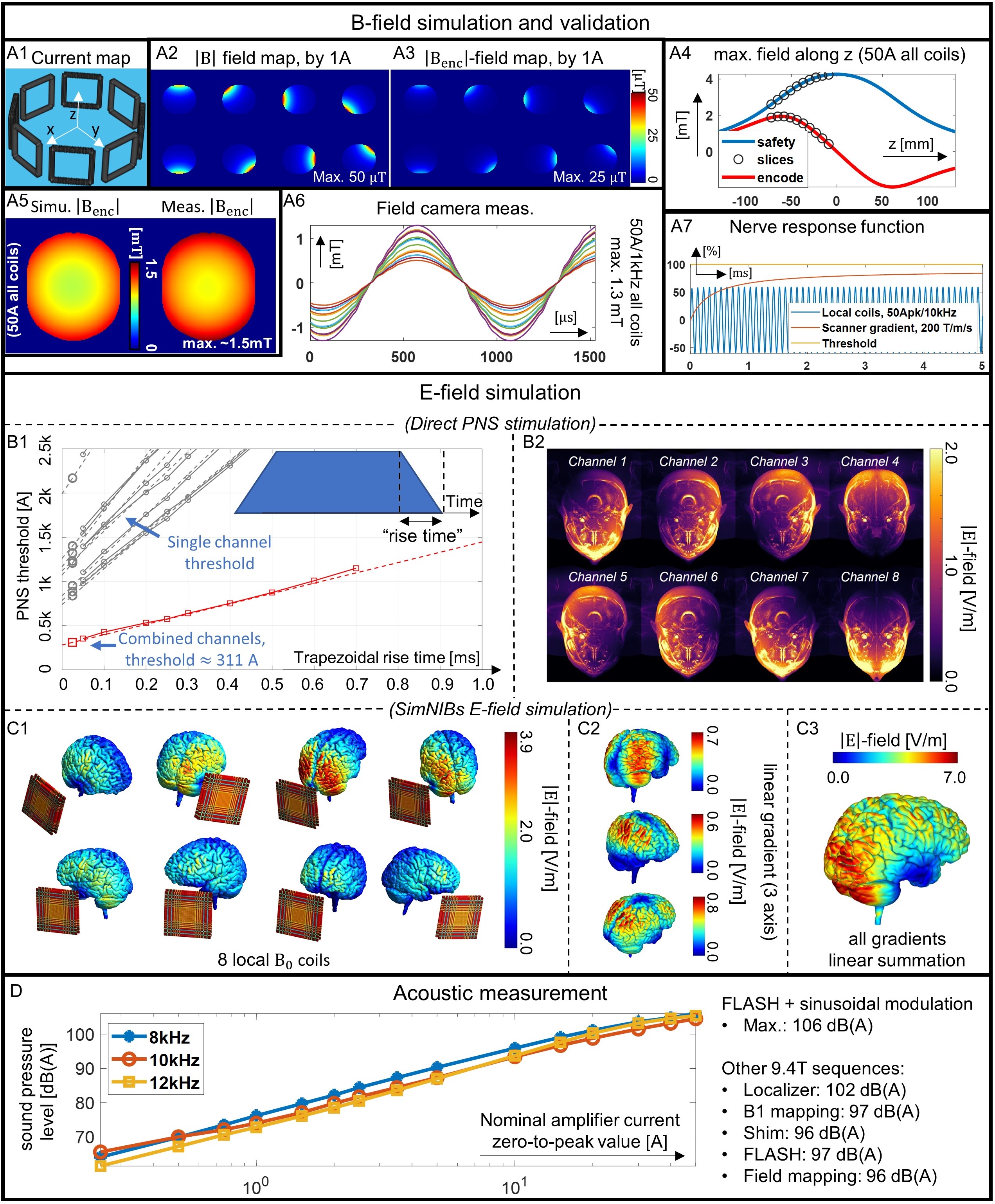
A comprehensive safety evaluation of our local B0 array is performed with respect to PNS stimulation and acoustic noise (see Figure 3). Ex-vivo and in-vivo brain scans in a 9.4T human scanner (Siemens Healthineers, Germany) are tested with various B0 modulation schemes, including distinct nonlinear field shapes, and time shifts of current waveforms between consecutive phase encoded steps similar to aliasing control9.
Results
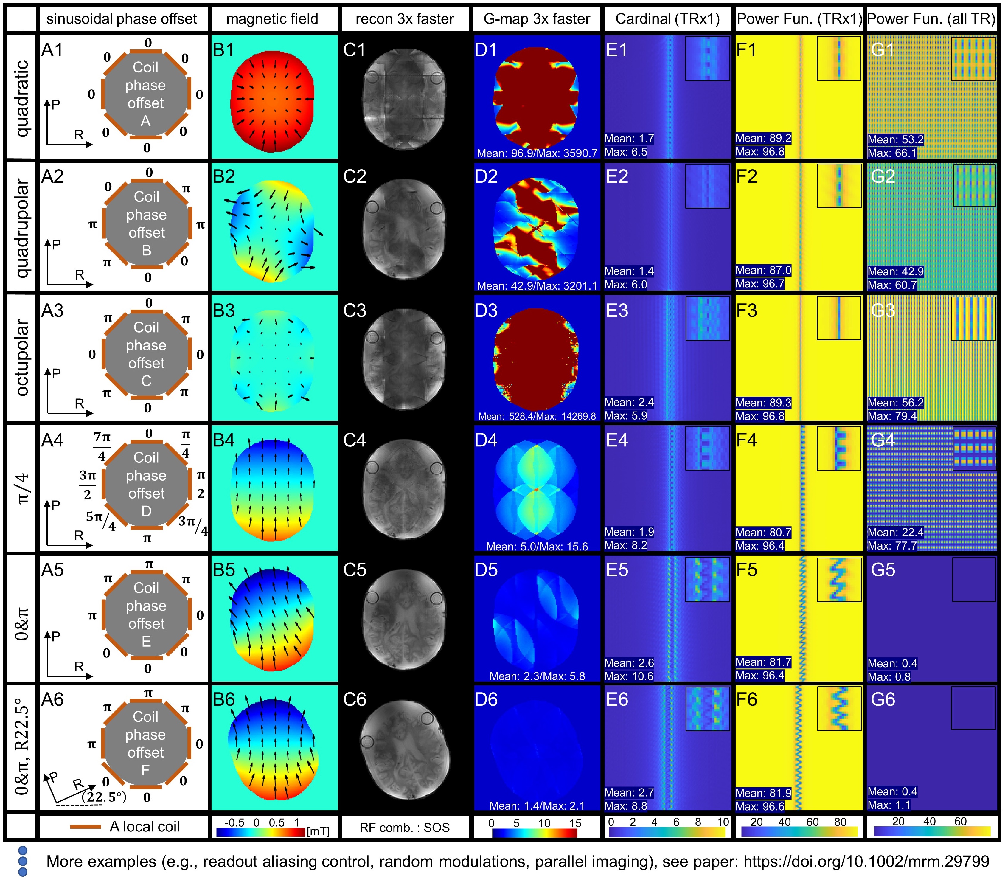
Utilizing the RKHS framework, we visualized the quantitative k-space sampling efficiency (see Figure 4), identifying a nearly linear gradient along the phase encoding dimension as the optimal modulation field for 2D Cartesian MRI, similar to bunched phase encoding10. The usefulness of readout aliasing control is demonstrated only in limited scenarios (e.g., 5kHz, scheme of “45 deg”). Facilitated by the field calibration technique, the brain scans with local B0 coils modulation are successfully reconstructed, reaching threefold and eightfold acceleration without and with SENSE7, respectively, and free of apparent artifacts (see Figure 5).Discussion & Conclusion
Our study highlights the extended RKHS framework as a novel mathematical foundation for pulse sequence design involving nonlinear gradient fields, potentially benefiting other nonconventional spatial encoding29–34 as well. Beyond the local k-space20 and the G-map7, this framework provides insights showing how the k-space efficiency maps computed based on a time segment (e.g., a single TR in this paper) can decouple the entire acquisition duration, and quantify individual contributions of nonlinear gradients encoding within smaller temporal periods to the full k-space.Eventually, our local B0 array substantially accelerate 2D Cartesian ex-vivo and in-vivo scans, validating the feasibility of this emerging technology in practice. Although the optimized local B0 coils converges to a linear gradient35 for accelerated 2D FLASH scans, our study doesn’t eliminate all possibility for potential advantages of more flexible nonlinear field modulation in spatial-temporal domain, especially in 3D Cartesian and other non-Cartesian acquisitions for future study.
Acknowledgements
This study is supported by the ERC Advanced Grant (No.834940). The ex vivo brain phantom was with courtesy of the Institute of Clinical Anatomy and Cell Analysis, Department of Anatomy, Eberhard Karls University of Tübingen. The first author thanks Dr. Thomas Shiozawa (Institute of Clinical Anatomy and Cell Analysis) for assistance with sample preparation, and Dr. Gisela Hagberg for assistance in scanning this phantom. M.D. receives research support from Siemens Healthineers. A.T. was supported by the Lundbeck foundation (grant R313-2019-622).References
1. Haase A, Frahm J, Matthaei D, Hanicke W, Merboldt K-D. FLASH Imaging. Rapid NMR Imaging Using Low Flip-Angle Pulses. J Magn Reson. 1969;67:258-266. doi:10.1016/0022-2364(86)90433-6
2. Mansfield P. Multi-planar image formation using NMR spin echoes. J Phys C Solid State Phys. 1977;10(3):L55-L58. doi:10.1088/0022-3719/10/3/004
3. Stehling MK, Turner R, Mansfield P. Echo-planar imaging: magnetic resonance imaging in a fraction of a second. Science. 1991;254:43-50. doi:10.1126/science.1925560
4. Ahn CB, Kim JH, Cho ZH. High-Speed Spiral-Scan Echo Planar NMR Imaging-I. IEEE Trans Med Imaging. 1986;5(1):2-7. doi:10.1109/TMI.1986.4307732
5. Madore B, Glover GH, Pelc NJ. Unaliasing by fourier-encoding the overlaps using the temporal dimension (UNFOLD), applied to cardiac imaging and fMRI. Magn Reson Med. 1999;42: 813-828.
6. Sodickson DK, Manning WJ. Simultaneous acquisition of spatial harmonics (SMASH): Fast imaging with radiofrequency coil arrays. Magn Reson Med. 1997;38(4):591-603. doi:10.1002/mrm.1910380414
7. Pruessmann KP, Weiger M, Scheidegger MB, Boesiger P. SENSE: sensitivity encoding for fast MRI. Magn Reson Med. 1999;42:952-962.
8. Griswold MA, Jakob PM, Heidemann RM, et al. Generalized autocalibrating partially parallel acquisitions (GRAPPA). Magn Reson Med. 2002;47(6):1202-1210. doi:10.1002/mrm.10171
9. Breuer FA, Blaimer M, Mueller MF, et al. Controlled aliasing in volumetric parallel imaging (2D CAIPIRINHA). Magn Reson Med. 2006;55(3):549-556. doi:10.1002/mrm.20787
10. Moriguchi H, Duerk JL. Bunched phase encoding (BPE): A new fast data acquisition method in MRI. Magn Reson Med. 2006;55(3):633-648. doi:10.1002/mrm.20819
11. Lustig M, Donoho D, Pauly JM. Sparse MRI: The application of compressed sensing for rapid MR imaging. Magn Reson Med. 2007;58(6):1182-1195. doi:10.1002/mrm.21391
12. Roemer PB, Edelstein WA, Hayes CE, Souza SP, Mueller OM. The NMR phased array. Magn Reson Med. 1990;16(2):192-225. doi:10.1002/mrm.1910160203
13. Bilgic B, Gagoski BA, Cauley SF, et al. Wave-CAIPI for highly accelerated 3D imaging: Wave-CAIPI for Highly Accelerated 3D Imaging. Magn Reson Med. 2015;73(6):2152-2162. doi:10.1002/mrm.25347
14. Cauley SF, Setsompop K, Bilgic B, Bhat H, Gagoski B, Wald LL. Autocalibrated wave-CAIPI reconstruction; Joint optimization of k-space trajectory and parallel imaging reconstruction. Magn Reson Med. 2017;78(3):1093-1099. doi:10.1002/mrm.26499
15. Uecker M, Lai P, Murphy MJ, et al. ESPIRiT-an eigenvalue approach to autocalibrating parallel MRI: Where SENSE meets GRAPPA. Magn Reson Med. 2014;71(3):990-1001. doi:10.1002/mrm.24751
15. Patz S, Hrovat MI, Pulyer YM, Rybicki FJ. Novel encoding technology for ultrafast MRI in a limited spatial region. Int J Imaging Syst Technol. 1999;10:216-224.
17. Hennig J, Welz AM, Schultz G, et al. Parallel imaging in non-bijective, curvilinear magnetic field gradients: a concept study. Magn Reson Mater Phys Biol Med. 2008;21(1-2):5. doi:10.1007/s10334-008-0105-7
18. Stockmann JP, Ciris PA, Galiana G, Tam L, Constable RT. O-space imaging: Highly efficient parallel imaging using second-order nonlinear fields as encoding gradients with no phase encoding. Magn Reson Med. 2010;64(2):447-456. doi:10.1002/mrm.22425
19. Wang H, Tam LK, Constable RT, Galiana G. Fast rotary nonlinear spatial acquisition (FRONSAC) imaging. Magn Reson Med. 2016;75(3):1154-1165. doi:10.1002/mrm.25703
20. Gallichan D, Cocosco ChrisA, Dewdney A, et al. Simultaneously driven linear and nonlinear spatial encoding fields in MRI. Magn Reson Med. 2011;65(3):702-714. doi:10.1002/mrm.22672
21. Juchem C, Nixon TW, McIntyre S, Boer VO, Rothman DL, de Graaf RA. Dynamic multi-coil shimming of the human brain at 7T. J Magn Reson. 2011;212(2):280-288. doi:10.1016/j.jmr.2011.07.005
22. Avdievich NI, Giapitzakis IA, Bause J, Shajan G, Scheffler K, Henning A. Double-row 18-loop transceive-32-loop receive tight-fit array provides for whole-brain coverage, high transmit performance, and SNR improvement near the brain center at 9.4T. Magn Reson Med. 2019;81(5):3392-3405. doi:10.1002/mrm.27602
22. Scheffler K, Loktyushin A, Bause J, Aghaeifar A, Steffen T, Schölkopf B. Spread-spectrum magnetic resonance imaging. Magn Reson Med. 2019;82:877-885. doi:10.1002/mrm.27766
24. Littin S, Jia F, Layton KJ, et al. Development and implementation of an 84-channel matrix gradient coil. Magn Reson Med. 2018;79(2):1181-1191. doi:10.1002/mrm.26700
25. Athalye V, Lustig M, Martin Uecker. Parallel magnetic resonance imaging as approximation in a reproducing kernel Hilbert space. Inverse Probl. 2015;31(4):045008. doi:10.1088/0266-5611/31/4/045008
26. IEC-60601-2-33-2022. Edition 4.0. International Electrotechnical Commission. Accessed February 2023. https://webstore.iec. ch/publication/67211
27. Tian R, Davids M, Thielscher A, Buckenmaier K, Steffen T, Scheffler K. Safety evaluation of an 8-channel local B0 coil array with sinusoidal modulation in a 9.4T human MR scanner. ISMRM Workshop on MR Safety: from Physics & Physiology to Policies & Practice. New York, USA: ISMRM; 2022.
28. Tian R, Uecker M, Davids M, et al. Accelerated 2D Cartesian MRI with an 8-channel local B0 coil array combined with parallel imaging. Magn Reson Med. 2024;91(2):443-465. doi:10.1002/mrm.29799
29. Katscher U, Lisinski J, Börnert P. RF encoding using a multielement parallel transmit system. Magn Reson Med. 2010;63(6):1463-1470. doi:10.1002/mrm.22439
30. Sharp JC, King SB. MRI using radiofrequency magnetic field phase gradients. Magn Reson Med. 2010;63(1):151-161. doi:10.1002/mrm.22188
31. Kartäusch R, Driessle T, Kampf T, et al. Spatial phase encoding exploiting the Bloch–Siegert shift effect. Magn Reson Mater Phys Biol Med. 2014;27(5):363-371. doi:10.1007/s10334-013-0417-0
32. Glang F, Nikulin AV, Bause J, et al. Accelerated MRI at 9.4 T with electronically modulated time-varying receive sensitivities. Magn Reson Med. 2022;88(2):742-756. doi:10.1002/mrm.29245
33. Tian R, Hennel F, Bianchi S, Pruessmann KP. Superresolution MRI with a Structured-Illumination Approach. Phys Rev Appl. 2023;19(3):034074. doi:10.1103/PhysRevApplied.19.034074
34. Setsompop K, Fan Q, Stockmann J, et al. High-resolution in vivo diffusion imaging of the human brain with generalized slice dithered enhanced resolution: Simultaneous multislice (gSlider-SMS). Magn Reson Med. 2018;79(1):141-151. doi:10.1002/mrm.26653
35. Versteeg E, Klomp DWJ, Siero JCW. A silent gradient axis for soundless spatial encoding to enable fast and quiet brain imaging. Magn Reson Med. 2022;87(2):1062-1073. doi:10.1002/mrm.29010Figures