0005
Diffusion-Weighted SPECIAL Improves the Detection of J-Coupled Metabolites at Ultrahigh Magnetic Field1LIFMET, EPFL, Lausanne, Switzerland, 2CIBM Center for Biomedical Imaging, Lausanne, Switzerland, 3Animal Imaging and Technology, EPFL, Lausanne, Switzerland
Synopsis
Keywords: YIA, Diffusion/other diffusion imaging techniques, Diffusion-weighted MR spectroscopy, rodent brain, high field, SPECIAL, glutamine, J-coupled metabolites
Motivation: Diffusion-weighted MR spectroscopy (dMRS) uniquely probes cell-specific tissue microstructure in vivo but most sequences suffer from long TE leading to signal loss by J-evolution and T2 relaxation.
Goal(s): To propose an alternative dMRS sequence (DW-SPECIAL) with a shorter TE while preserving the benefits of the current gold-standard rodent sequence at high field (STE-LASER).
Approach: DW-SPECIAL was tested in vivo in the rat brain and compared to STE-LASER.
Results: DW-SPECIAL halved the minimum TE while reducing specific absorption rate compared to STE-LASER, thereby 1) improving the J-coupled metabolites’ diffusion properties estimation and 2) offering a new candidate sequence for human dMRS.
Impact: With its shorter TE, our newly proposed DW-SPECIAL can serve as an alternative to STE-LASER when strongly J-coupled metabolites like glutamine are investigated, thereby extending the range of accessible metabolites in the context of diffusion-weighted MRS acquisitions at high field.
Introduction
In vivo diffusion-weighted magnetic resonance spectroscopy (dMRS) has emerged as a powerful technique to probe tissue morphology at the micrometre scale via the non-invasive assessment of metabolite diffusion properties1,2. When diffusion-sensitizing gradients are added to single-voxel MR spectroscopy (MRS) sequences, cell-specific microstructure can be inferred by assessing the diffusion properties of the major brain metabolites3–6, which are differentially concentrated in specific brain cells. The STE-LASER sequence7, consisting of a stimulated echo (STE) diffusion module followed by a LASER localization, has become the gold-standard for rodent dMRS studies at high field. In addition to preserving the advantages of diffusion-weighted STEAM, its block-design yields a sharp voxel selection with the absence of cross-terms in the b-value. However, STE-LASER suffers from a long minimum echo time which hampers the detection of J-coupled metabolites. In the present work, we aimed at improving the detection and subsequent estimation of the diffusion properties of J-coupled and low-concentrated metabolites in dMRS acquisitions at high field. To do so, we developed a new dMRS sequence, named DW-SPECIAL, based on the semi-adiabatic SPECIAL sequence8 used for single-voxel spectroscopy, which benefits from the same advantages as STE-LASER with half the minimum echo time.Methods
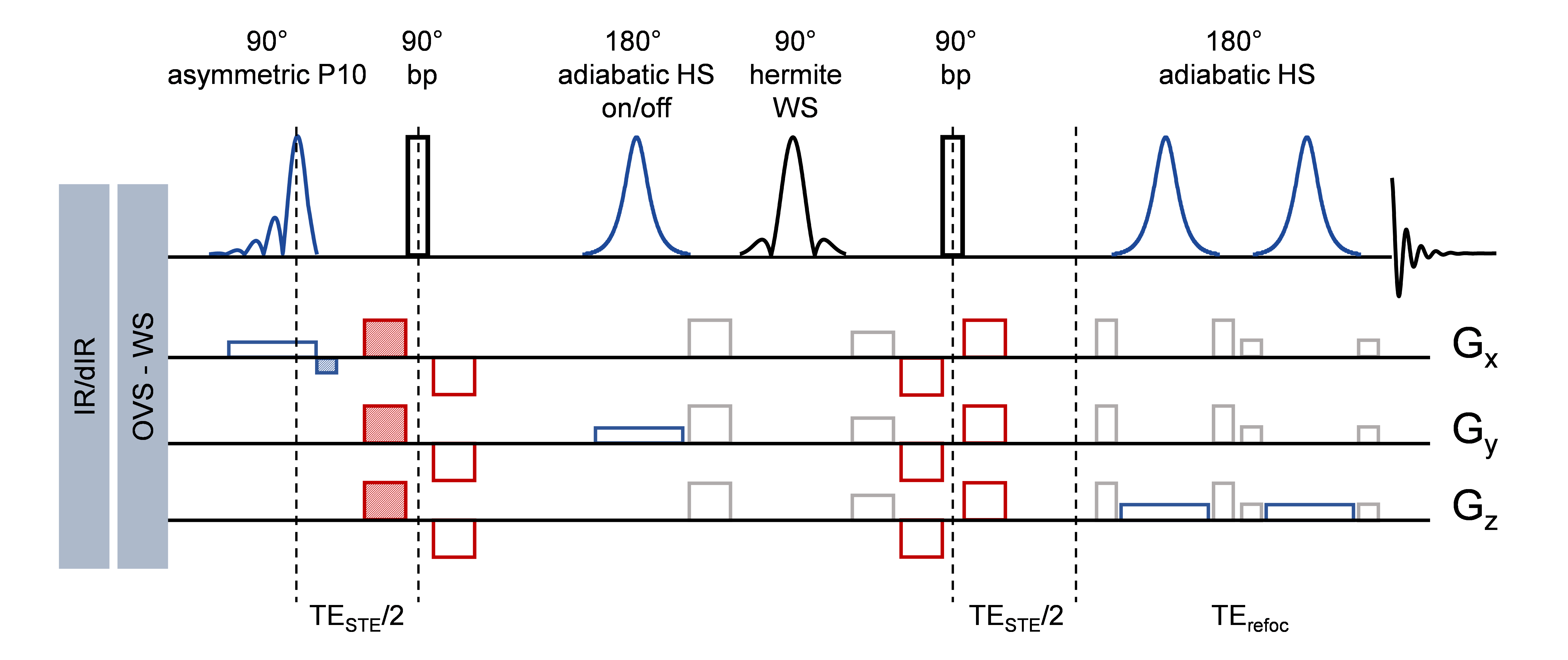
DW-SPECIAL (Fig 1) combines an STE diffusion block with a semi-adiabatic SPECIAL localization: the first 90° pulse of the STE is rendered slice-selective, an on/off slice-selective adiabatic inversion is inserted in the mixing time and two 180° slice-selective adiabatic pulses after the STE block. Diffusion gradients are inserted in the STE block in a bipolar fashion. The on/off ISIS scheme performs a slice-selective adiabatic inversion (HS, 2 ms, 10 kHz) on the inhomogeneous direction (y), here perpendicular to the transmit/receive quadrature surface coil, and requires a two-step phase cycling. Acquisitions with DW-SPECIAL and STE-LASER7 (used as reference) were performed at 14.1T (with the following parameters: TR/Δ/δ=3000/43/3 ms, Δ: diffusion time, δ: duration of diffusion gradient in the direction (1,1,1), 5 b-values between 0.05 and 10 ms/µm2) in vivo on the rat brain (N=5, voxel size: 7x5x5 mm3), using the shortest achievable TE for each sequence. Eddy currents, B0 drift and phase corrections were performed on individual shots using spectral registration in FID-A9 and outlier shots removed as detailed in our recent consensus10. A metabolite basis-set was simulated with NMRScope-B11 (jMRUI) and a macromolecule (MM) spectrum was acquired in vivo with each sequence using double inversion recovery (TI=2200/800ms) with b=5ms/µm2, where residual metabolites were removed using AMARES (jMRUI)12,13. Concentrations were quantified with LCModel14 and the apparent diffusion coefficient and intra-stick diffusivity (Callaghan’s model15) were fitted and compared between the sequences for glutamate (Glu), glutamine (Gln), myo-inositol (mIns), taurine (Tau), total N-acetylaspartate (tNAA), total choline (tCho), total creatine (tCr) and the mobile macromolecules (MM).Results and discussion
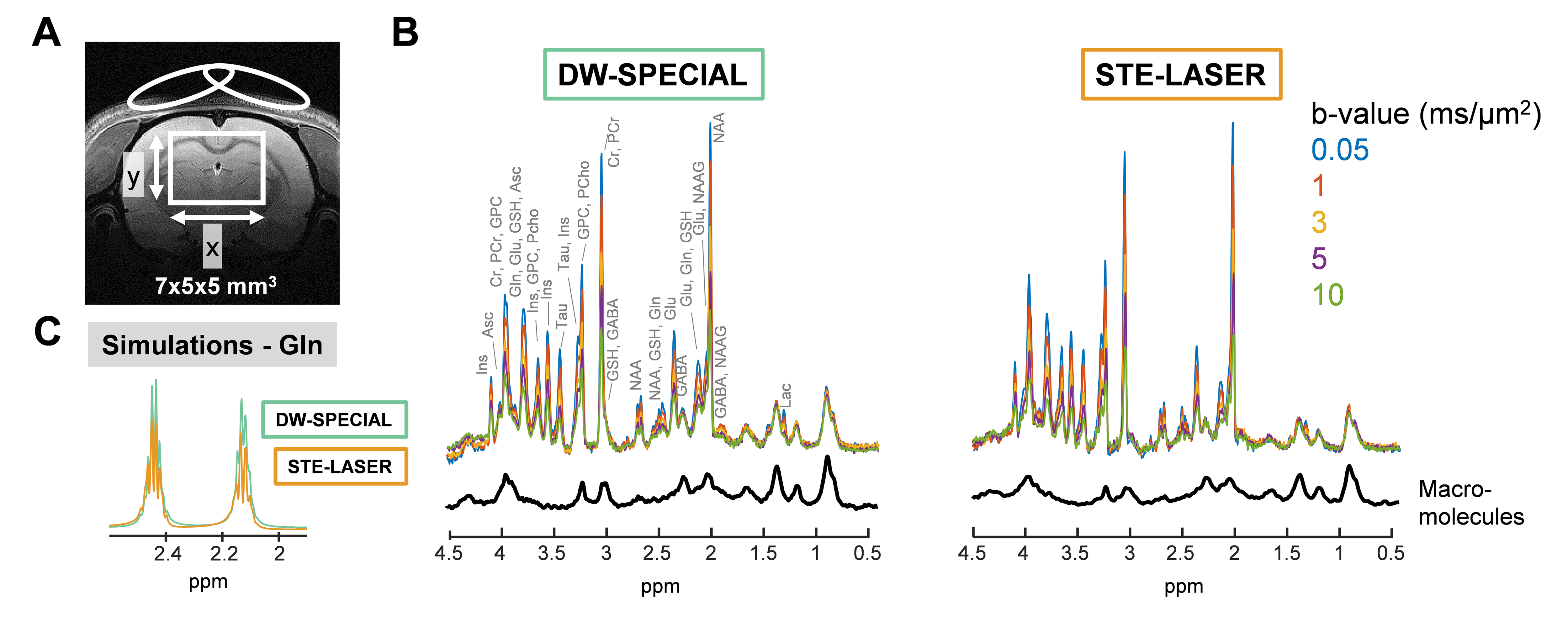
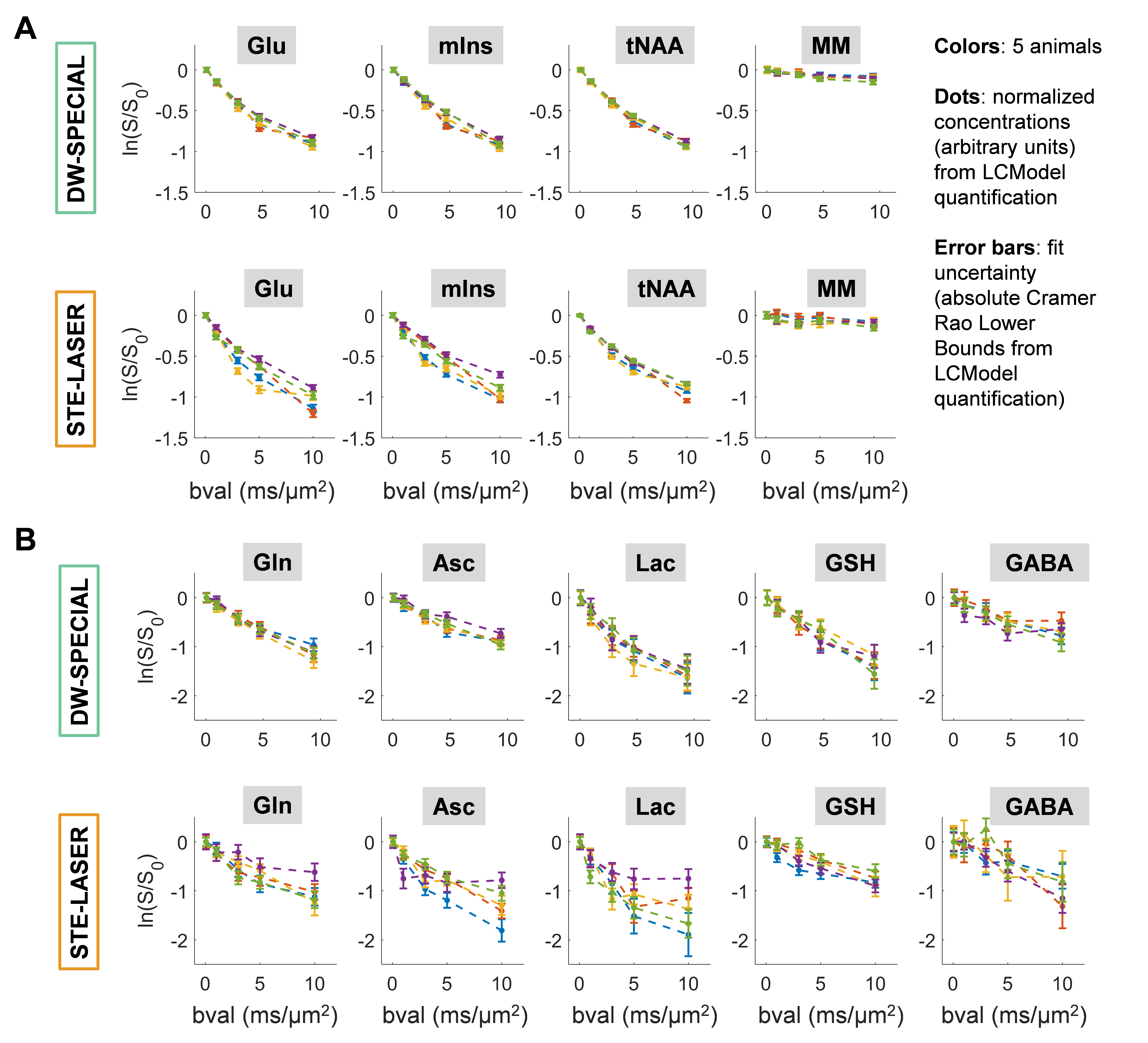
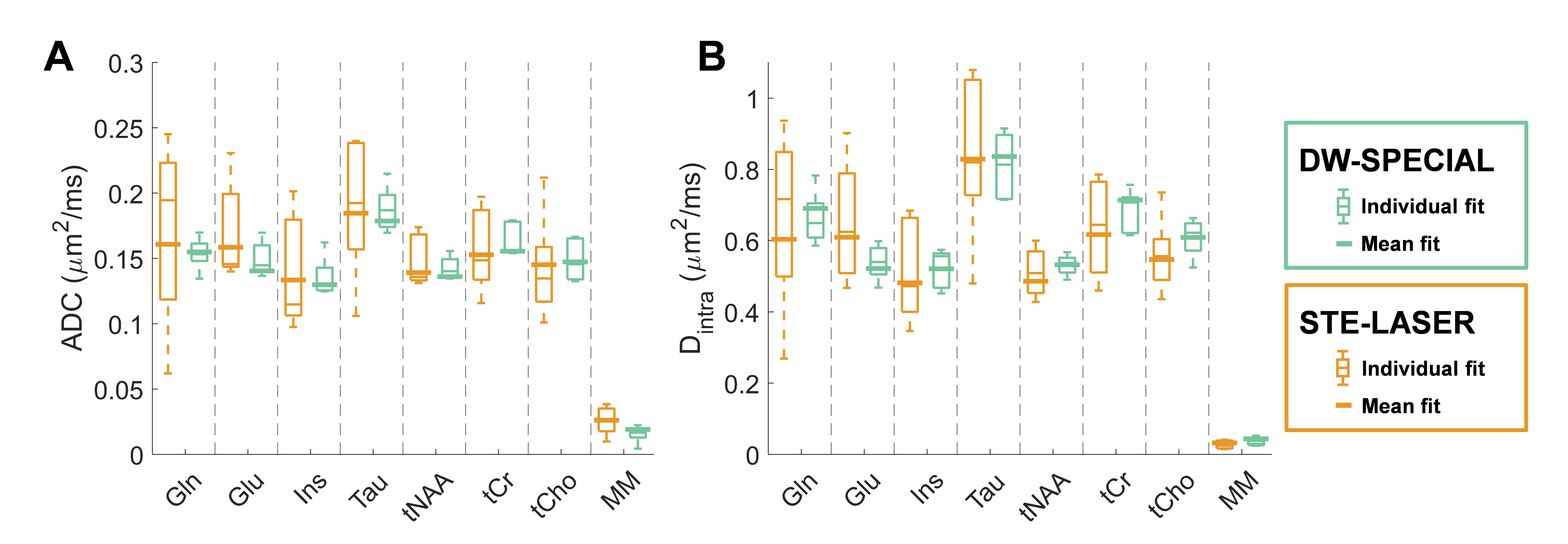
The shorter echo time achieved with DW-SPECIAL (18 ms against 33 ms with STE-LASER) limited the metabolites’ signal loss caused by J-evolution, as predicted by simulations (e.g., for Gln (Fig 2C)). In addition, DW-SPECIAL preserved the main advantages of STE-LASER7: diffusion time during a STE, a sharp volume selection and limited sensitivity to B1 inhomogeneities with the use of adiabatic pulses. Interestingly, although DW-SPECIAL does not retain the block-design of STE-LASER, the absence of cross terms in the b-value is ensured as long as the two shaded gradients on Fig 1 do not overlap. In vivo, compared to STE-LASER, DW-SPECIAL yielded the same spectral quality, benefitting from the good spectral separation and high SNR at 14.1T (Fig 2A-B). DW-SPECIAL reduced the group variability of diffusion decays for most quantified metabolites, most strikingly for low-concentrated ones which were hardly reported before (Fig 3). Metabolites’ diffusion estimates were in good agreement between the two sequences (p>0.05, 2-way ANOVA) and DW-SPECIAL reduced the standard deviation of the diffusion estimates based on individual animal fitting (e.g., ADCgln F-test p-value: 0.0054) without loss of accuracy compared to the fit on the diffusion decay averaged across animals (Fig 4). Finally, DW-SPECIAL led to a smaller average power deposition in the RF coil (proportional to the SAR) than STE-LASER, rendering its implementation on human scanners feasible (Table 1). We have recently extended DW-SPECIAL to the study of the diffusion properties of the 1H MRS downfield region using a metabolite cycling approach16, benefitting from the ultra-high field to separate the contributions of slow-diffusing MM and of fast-diffusing metabolites.Conclusion
With its shorter echo time, DW-SPECIAL can serve as an alternative to STE-LASER and holds the promise of extending the range of accessible metabolites in the context of dMRS acquisitions while providing, with its reduced SAR, a new candidate sequence for human dMRS studies.Acknowledgements
Supported by the European Union's Horizon 2020 research and innovation program under the Marie Sklodowska-Curie grant agreement No 813120 (INSPiRE-MED), the SNSF projects no 310030_173222, 310030_201218 and the Leenaards and Jeantet Foundations. We acknowledge access to the facilities and expertise of the CIBM Center for Biomedical Imaging, founded and supported by Lausanne University Hospital (CHUV), University of Lausanne (UNIL), Ecole polytechnique fédérale de Lausanne (EPFL), University of Geneva (UNIGE) and Geneva University Hospitals (HUG). We thank Stefanita Mitrea and Dario Sessa, Analina Da Silva and Mario Lepore for veterinary support, Thanh Phong Lê for technical support, Vladimir Mlynarik for experimental advice and Katarzyna Pierzchala for her help with phantom preparation.References
1. Ronen, I. & Valette, J. Diffusion-Weighted Magnetic Resonance Spectroscopy. in eMagRes 733–750 (American Cancer Society, 2015). doi:10.1002/9780470034590.emrstm1471.
2. Palombo, M., Shemesh, N., Ronen, I. & Valette, J. Insights into brain microstructure from in vivo DW-MRS. NeuroImage 182, 97–116 (2018).
3. Ronen, I., Ercan, E. & Webb, A. Axonal and glial microstructural information obtained with diffusion-weighted magnetic resonance spectroscopy at 7T. Frontiers in Integrative Neuroscience 7, 13 (2013).
4. Palombo, M., Ligneul, C., Hernandez-Garzon, E. & Valette, J. Can we detect the effect of spines and leaflets on the diffusion of brain intracellular metabolites? NeuroImage 182, 283–293 (2018).
5. Valette, J., Ligneul, C., Marchadour, C., Najac, C. & Palombo, M. Brain Metabolite Diffusion from Ultra-Short to Ultra-Long Time Scales: What Do We Learn, Where Should We Go? Front Neurosci 12, 2 (2018).
6. Najac, C., Branzoli, F., Ronen, I. & Valette, J. Brain intracellular metabolites are freely diffusing along cell fibers in grey and white matter, as measured by diffusion-weighted MR spectroscopy in the human brain at 7 T. Brain Struct Funct 221, 1245–1254 (2016).
7. Ligneul, C., Palombo, M. & Valette, J. Metabolite diffusion up to very high b in the mouse brain in vivo: Revisiting the potential correlation between relaxation and diffusion properties. Magnetic Resonance in Medicine 77, 1390–1398 (2017).
8. Mlynárik, V., Gambarota, G., Frenkel, H. & Gruetter, R. Localized short-echo-time proton MR spectroscopy with full signal-intensity acquisition. Magnetic Resonance in Medicine 56, 965–970 (2006).
9. Near, J. et al. Frequency and phase drift correction of magnetic resonance spectroscopy data by spectral registration in the time domain. Magn Reson Med 73, 44–50 (2015).
10. Ligneul, C. et al. Diffusion-weighted MR spectroscopy: Consensus, recommendations, and resources from acquisition to modeling. Magnetic Resonance in Medicine 91, 860–885 (2024).
11. Starčuk, Z. & Starčuková, J. Quantum-mechanical simulations for in vivo MR spectroscopy: Principles and possibilities demonstrated with the program NMRScopeB. Anal Biochem 529, 79–97 (2017).
12. Kunz, N. et al. Diffusion-weighted spectroscopy: a novel approach to determine macromolecule resonances in short-echo time 1H-MRS. Magnetic Resonance in Medicine 64, 939–946 (2010).
13. Simicic, D. et al. In vivo macromolecule signals in rat brain 1H-MR spectra at 9.4T: Parametrization, spline baseline estimation, and T2 relaxation times. Magnetic Resonance in Medicine 86, 2384–2401 (2021).
14. Provencher, S. W. Estimation of metabolite concentrations from localized in vivo proton NMR spectra. Magn Reson Med 30, 672–679 (1993).
15. Callaghan, P. T., Jolley, K. W. & Lelievre, J. Diffusion of water in the endosperm tissue of wheat grains as studied by pulsed field gradient nuclear magnetic resonance. Biophys J 28, 133–141 (1979).
16. Mosso, J., Döring, A., Kreis, R., Cudalbu, C. & Lanz, B. Metabolite-cycling at 14.1T: sequence implementation and initial explorations using SPECIAL and diffusion-weighted SPECIAL. in 2024 ISMRM & ISMRT Annual Meeting & Exhibition, accepted as an oral presentation.
17. Tkác, I., Starcuk, Z., Choi, I. Y. & Gruetter, R. In vivo 1H NMR spectroscopy of rat brain at 1 ms echo time. Magn Reson Med 41, 649–656 (1999).Figures