0001
MRI Assessment of Cerebral White Matter Microvascular Hemodynamics Across the Adult Lifespan1Athinoula A. Martinos Center for Biomedical Imaging, Massachusetts General Hospital, Boston, MA, United States, 2Department of Radiology, Harvard Medical School, Boston, MA, United States, 3Department of Neurology, Harvard Medical School, Boston, MA, United States, 4Neuroimaging Research for Veterans Center, VA Boston Healthcare System, Boston, MA, United States
Synopsis
Keywords: YIA, White Matter
Motivation: The mechanisms underlying age-related structural neurodegeneration are not well understood, limiting our knowledge of atypical versus typical aging.
Goal(s): Our goal was to use data involving advanced hemodynamic and structural MRI techniques tailored to white matter to characterize the aging process.
Approach: We used data from a large cohort of the Human Connectome Project in Aging to investigate the relationship between brain blood flow and white matter tract microstructural integrity.
Results: We found strong relationships between white matter hemodynamics and tract integrity that were affected by both age and sex.
Impact: These findings could reveal potential underlying physiological mechanisms behind structural changes during typical aging. This could help us understand healthy brain aging and encourage future research to target hemodynamic biomarkers to understand neurodegeneration.
Introduction
It is well established that cerebral white matter microstructure deteriorates with advancing age, though the mechanisms behind this effect are little understood. Previous studies have indicated that these structural changes may be tract-dependent (1), and that there is an apparent relationship between white matter lesion development and elevated vascular risk (2). Furthermore, there is a strong negative correlation between global gray matter cerebral blood flow (CBF) and increasing age (3). Therefore, imaging of white matter hemodynamics could provide insights into age-related neurodegeneration. However, white matter exhibits lower vascular density and longer arterial arrival times compared to gray matter, requiring specialized imaging approaches. The Human Connectome Project in Aging (HCP-A) collected cross-sectional data in typically aging adults using innovative approaches for understanding white matter hemodynamic physiology (4). In this study, we investigated the impact of age and sex on white matter hemodynamics in the HCP-A cohort using tract-based spatial statistics (TBSS) (5).Methods
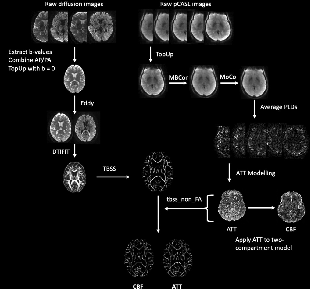
Participants: 678 typically aging individuals (381 female; aged 36–100 years) were extracted from publicly available HCP-A data. Inclusion criteria consisted of the presence of both diffusion tensor imaging (DTI) and multi-delay pseudo-continuous arterial spin labeling (ASL) data.Data Acquisition and Processing: Imaging data were acquired at four sites using a 32-channel head coil and 3T Siemens Prisma MRI scanners. ASL data were collected with labeling duration=1500 ms and five post-labeling delays=200 ms (control/label pairs=6), 700 ms (pairs=6), 1200 ms (pairs=6), 1700 ms (pairs=10), and 220 ms (pairs=15). DTI data were acquired with b-values=0, 1.5, 3 s/mm2, TR=3230 ms, TE=89.2 ms. More details of the acquisition parameters have been described previously (6). The full processing pipeline is demonstrated in Figure 1.
Image Analysis: To investigate the white matter microstructure, a skeleton of mean fractional anisotropy (FA) was produced using the TBSS skeletonization procedure (5). This skeleton was used to project ASL-derived CBF and arterial transit time (ATT) measures onto white matter tracts. General linear models were then applied to white matter FA, CBF, and ATT maps, while covarying for age and sex. Threshold-free cluster enhancement multiple comparisons correction was performed for the effects of age and sex, thresholded at pFWE<0.05. CBF, ATT, and FA were then compared between sex for each tract using analysis of covariance, with multiple comparisons correction for the number of tracts at pFDR<0.05. Pearson’s correlations were also performed between FA and each perfusion measure, followed by a paired samples t-test between correlation coefficients controlled for multiple comparisons using Bonferonni-adjusted padj<0.05 across tracts.
Results
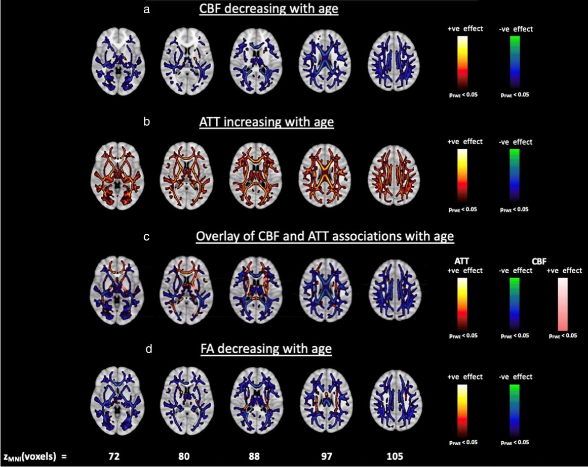
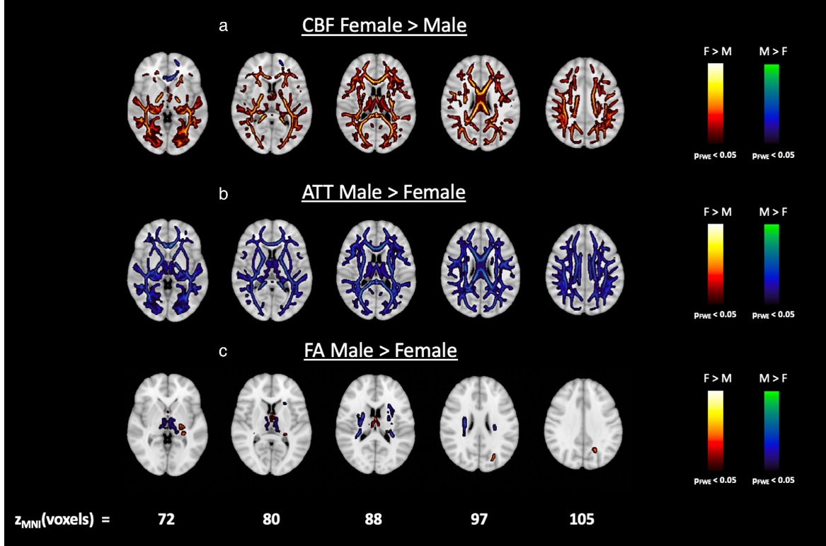
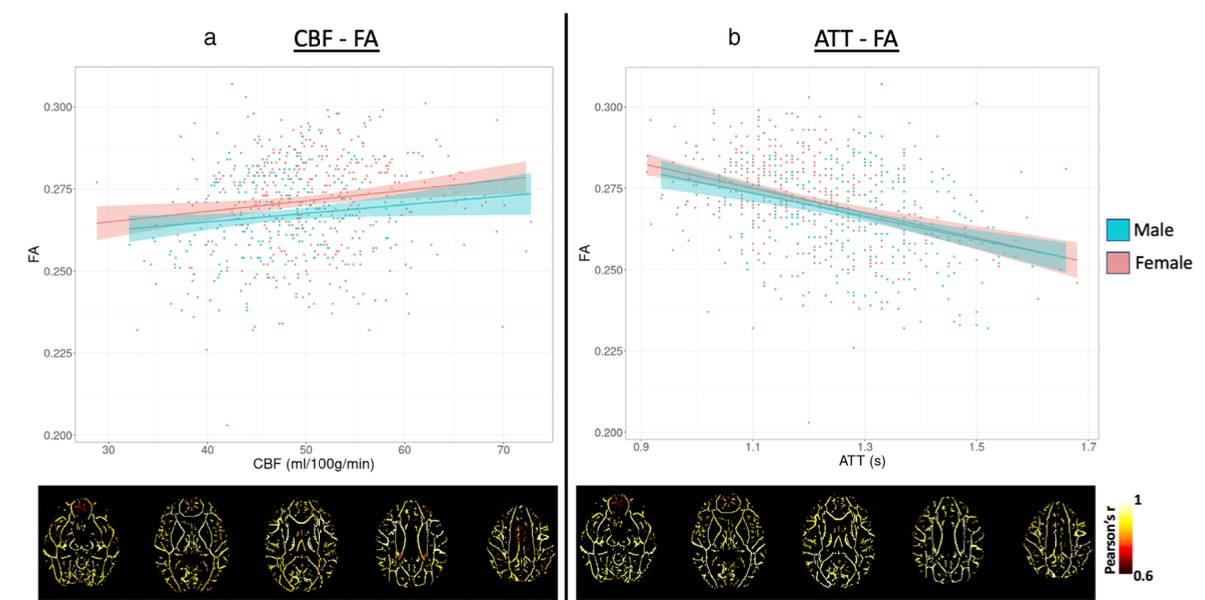
Figure 2 demonstrates that lower white matter CBF and longer white matter ATTs were significantly associated with older age. These effects were widespread across tracts for ATT. Figure 3 shows that significant (pFDR < 0.05) sex differences in ATT were observed across all tracts, and significant sex differences in CBF were observed in all tracts except the bilateral uncinate fasciculus. Females demonstrated significantly higher CBF compared to males across the lifespan, but only a few tracts demonstrated significant sex differences in FA. Figure 4 highlights that stronger relationships were observed between FA and ATT compared to FA and CBF.Discussion
This study investigated the relationship between white matter hemodynamics and microstructural integrity. Older age was associated with lower CBF, lower FA, and longer ATT across most white matter tracts, in agreement with existing literature (7, 8). Lower white matter ATT was observed globally in females in comparison to males, whereas higher CBF in females was relatively more localized. These findings could be related to sex-specific mechanisms of arterial stiffening (9), but further work is required to investigate this hypothesis. The strong relationship between FA and ATT could suggest that ATT is a more sensitive measure of hemodynamic compromise compared to CBF. Longitudinal research using advanced white matter imaging techniques would benefit the validation of our findings, aiding in the characterization of the normative aging process.Acknowledgements
This research was made possible in part by the computational hardware generously provided by the Massachusetts Life Sciences Center (https:// www.masslifesciences.com/). Data and/or research tools used in the preparation of this manuscript were obtained from the National Institute of Mental Health (NIMH) Data Archive (NDA). NDA is a collaborative informatics system created by the National Institutes of Health to provide a national resource to support and accelerate research in mental health. Dataset identifier(s): 10.15154/876w-3a49. This manuscript reflects the views of the authors and may not reflect the opinions or views of the NIH or of the Submitters submitting original data to NDA. This work was supported by the National Institutes of Health/National Institute on Aging (R21AG072068, U01AG052564, and U01AG052564-S1) and the American Heart Association (19CDA34790002 and 23POST1030607).References
1. Grieve SM, Williams LM, Paul RH, Clark CR, Gordon E. Cognitive aging, executive function, and fractional anisotropy: A diffusion tensor MR imaging study. Am J Neuroradiol 2007;28(2):226-235.
2. Williams OA, An Y, Beason-Held L, et al. Vascular burden and APOE ε4 are associated with white matter microstructural decline in cognitively normal older adults. Neuroimage 2019;188:572-583.
3. Juttukonda MR, Li B, Almaktoum R, et al. Characterizing cerebral hemodynamics across the adult lifespan with arterial spin labeling MRI data from the Human Connectome Project-Aging. Neuroimage 2021; 230:117807.
4. Bookheimer SY, Salat DH, Terpstra M, et al. The lifespan human connectome project in aging: An overview. Neuroimage 2019;185: 335-348.
5. Smith SM, Jenkinson M, Johansen-Berg H, et al. Tract-based spatial statistics: Voxelwise analysis of multi-subject diffusion data. Neuroimage 2006; 31(4):1487-1505.
6. Damestani, N.L., Jacoby, J., Michel, C.B., et al. MRI assessment of cerebral white matter microvascular hemodynamics across the adult lifespan. Journal of Magnetic Resonance Imaging 2024.
7. Alisch JS, Khattar N, Kim RW, et al. Sex and age-related differences in cerebral blood flow investigated using pseudo-continuous arterial spin labeling magnetic resonance imaging. Aging (Albany NY) 2021;13(4): 4911-4925.
8. Aslan S, Huang H, Uh J, et al. White matter cerebral blood flow is inversely correlated with structural and functional connectivity in the human brain. Neuroimage 2011;56(3):1145-1153.
9. DuPont JJ, Kenney RM, Patel AR, Jaffe IZ. Sex differences in mechanisms of arterial stiffness. Br J Pharmacol 2019;176(21):4208-4225.
Figures