TIPS & TRICKS TO OPTIMIZE YOUR INFLAMMATORY BRAIN PROTOCOLS
1Oslo University Hospital, Norway
Synopsis
Keywords: Neuro: Brain
For neuro imaging there are many sequences to choose depending on the cases. Thereby, it is important to have a better understanding of which sequences are the most important when it comes to inflammatory brain protocols. To minimize errors at a big department with many technicians, it is mandatory to setup the protocol with an easy workflow. Today, we have artificial intelligence in our hands as an important tool not only for enhancing the quality and/or speeding our sequences, but also for the protocol setup. With that said, we are not only minimizing errors, but also increasing efficiency.
Acknowledgements
Special thanks to my colleagues Vanja Cengija and Rolf SvendsmarkReferences
1. David J., et.al. Neuro MR: Protocols. JOURNAL OF MAGNETIC RESONANCE IMAGING 26:838–847 (2007)
2. Benjamin V. Ineichen., et.al. Leptomeningeal enhancement in multiple sclerosis and other neurological diseases: A systematic review and Meta-Analysis. NeuroImage: Clinical 33 (2022) 102939
3. Kalaivani J., et.al. Magnetic Resonance Imaging Findings in Viral Encephalitis: A Pictorial Essay. Journal of Neurosciences in Rural Practice, Volume 9, Issue 4 (2018)
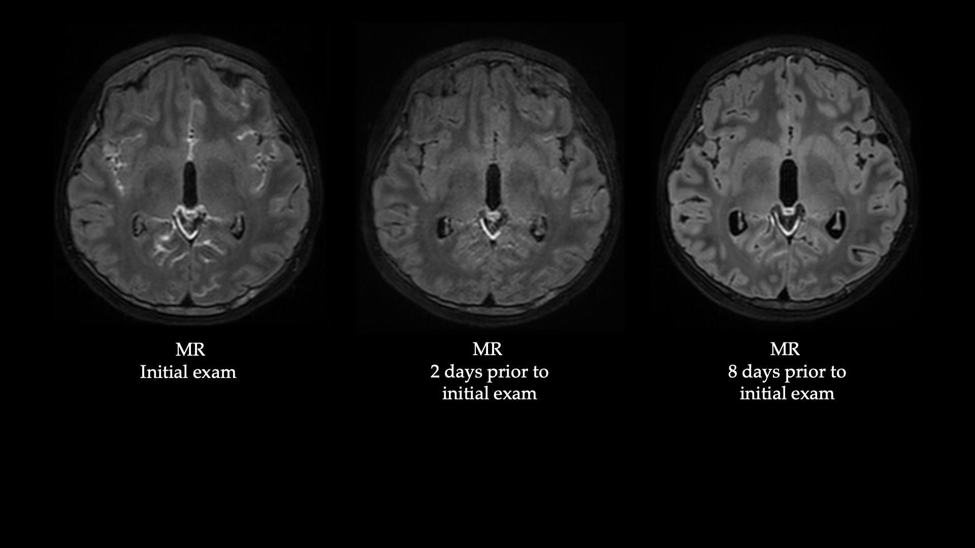
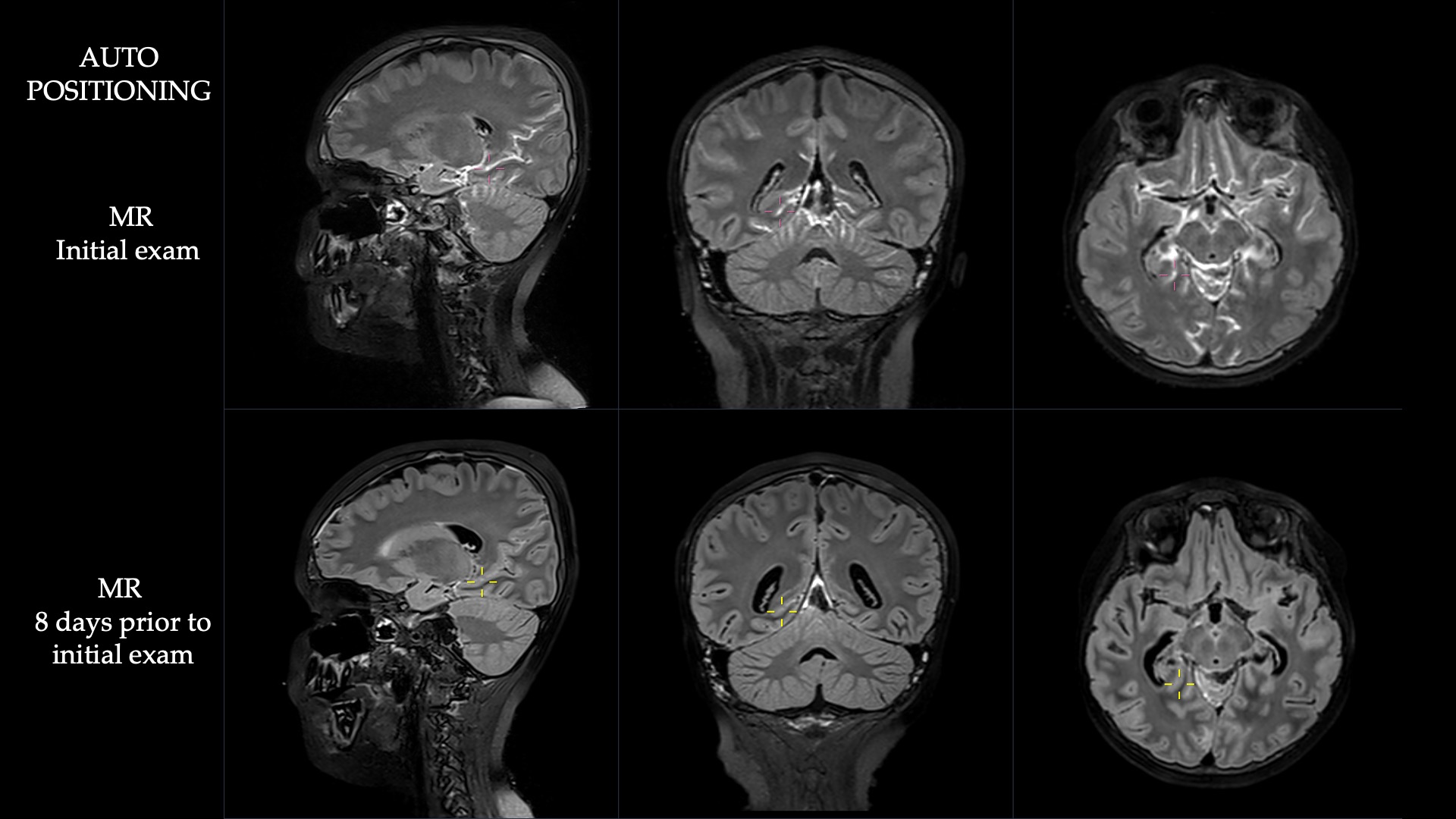
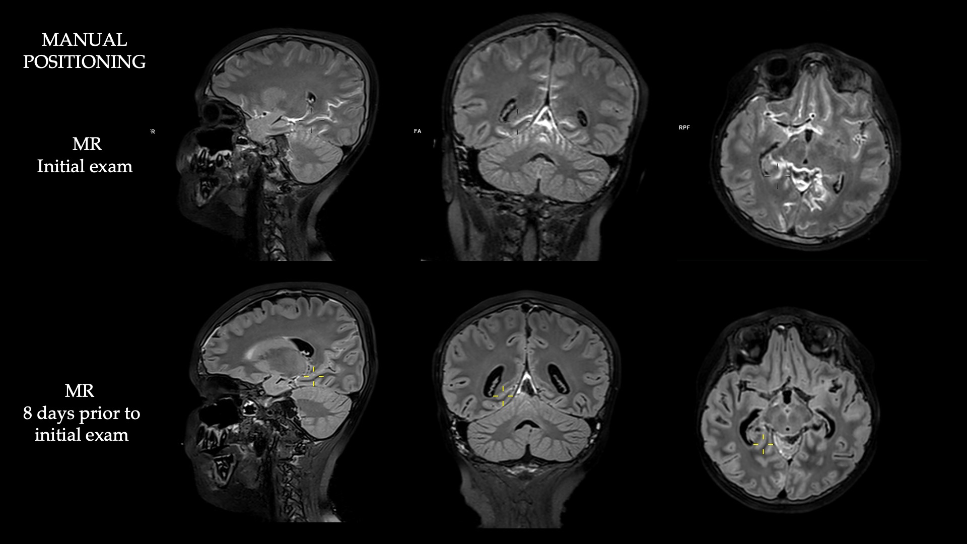
Figures