Creating Chef's Signature Dishes: MR in Clinical Practice
Alessandro André Mazzola1
1MRIONLINE, Brazil
1MRIONLINE, Brazil
Synopsis
Keywords: Education Committee: Clinical MRI, Physics & Engineering: Physics
MRI has always had a connection with food. The idea of using a magnetic field gradient came up while professor Paul Lauterbur was having dinner and scribbled the idea on a napkin. Choosing a good dish in a restaurant starts with a menu. Protocols are our menu and, like dishes in a restaurant, we need standardization and quality. The master chef in MRI will be the person who can deliver the best dish, with the highest quality at the shortest time. Artificial Intelligence will help us to perform MRI scans even more but we should pay more attention to safety.Introduction
I could say that “MRI has always had a connection with food!” Could we say that MRI started in a restaurant?Early Days until Now: MRI & Food
Could we say that MRI started in a restaurant? At least the idea of using a magnetic field gradient came up while Professor Paul Lauterbur was having dinner with his friend Donald Vickers and scribbled the idea on a napkin at the Big Boy Restaurant in Pittsburg. Later that day, he jotted down the ideas in his notebook and asked Donald Vickers to sign at the bottom of the page (figure 1).Lauterbur himself placed different objects such as small crabs and shells inside his spectrometer while applying the zeugmatography technique. Professor Waldo Hinshaw in 1974 made an image of a spring onion while working at Professor Edward Raymond Andrew's Cavendish laboratory.Its not food but it a nice reference of Ratatouille Film from Disney. The image is a relaxation map of a Mouse made by the Abeerden Group and presented in 1974 in Amper Congress in Nottingham. The most intersting part of this picture resides in the fact that the colors were painted manually by professor Jim Hutichinson´s wife Meg Foster just after the conference. In 1976 Peter Mansfield and Andrew Maudsley showed an image of a Turkey Leg immersed in oil obtained by the Line Scan technique. A year later, in 1977, Professor Andrew had a very vivid image of a lemon published on the cover of Nature. According to Professor Andrew, the image convinced many people that geometrically reliable images could indeed be obtained by n.m.r.. The matrix size here was 128 x 128 interpolated to 256 x 256; the resolution was about 0.5 mm (figure 2).A nectarine was used in 1980 by Paul Lauterbur to demonstrate the 16 slices from a 3D acquisition with a resolution of 3.5 mm. With this he showed that 3D reconstruction was a practical and efficient method of obtaining MR images.More recently, in September 2021, researchers from the ISEULT project in France showed images of a pumpkin as the first images taken by the 11.7T superconducting magnet.Menu, Kitchen, Ingredients & Flavours
Choosing a good dish in a restaurant starts with a menu. In clinical MRI, the protocols saved in the equipment will have to be correlated with the medical request. Protocols, like dishes in a restaurant, need standardization and quality. In the past we had more restricted menus, but currently the number of protocols saved in a typical 1.5 or 3.0T equipment is around 150 to 200 protocols.Take a look at the example of the head or brain protocols. We can have more than 20 different protocols to investigate brain tissue.Can kitchens with different features and appliances produce the same dish?In cooking perhaps yes, but it is difficult to achieve this on a large scale or maintaining the same quality. Kitchen size, structure and appliances are essential to produce faster and with high quality. In the same way we can look at the clinical acquisitions on MRI.A few years ago it seemed that Clinical MRI would focus on exams performed on 1.5T or 3T equipment. However in the last few years we have seen a large number of options, both in fields larger than 3T as the 7T, such as fields as low as 60 or 80 mT, point-of-care equipment for specific applications. It is important to highlight that each “dish” will require good appliances in our kitchen and also the correct understanding of the limitations of each kitchen!And each clinical protocol will have its pulse sequences and parameters. I believe that just like a good dish, following a correct and standardized recipe is the guarantee that the minimum quality will be reached to investigate a certain pathology. PIRADS may be an interesting example of how this standardization has benefited not only the search of better image quality but also the medical report made by the radiologist.All these different ingredients and flavors are to produce contrast in the images. Clinical MRI has increasingly been using different flavors in each protocol. Many people call this “multiparametric acquisitions”, however, I think this term should not be used, because, since its origin, MRI has always been multiparametric. We never serve a single dish with a single flavor in a clinical protocol. It is the combination of different contrasts and techniques that allows us to see anatomical, physiological or functional characteristics with greater properties.Master Chef, Fast Food and Slow Food
The Master Chef in MRI will be the person who can deliver the best dish, with the highest quality at the shortest time. I have developed a method for adjusting pediatric protocols that I call the “orange method”. An orange or a lemon or even other fruits are similar in size to a newborn's head or body. In this way, it is possible to adjust parameters such as field of view and slice thickness so as not to impair the resolution of the images. I recently adjusted an MRA protocol with GE's TRICKS technique using an orange and a pineapple as the head and body of a 3 to 4 months old child. Making the previous adjustments to the protocol in this way already allowed a good spatial and temporal resolution to perform an angiography of the head and neck vessels using 0.3 milliliters of gadobutrol (Figure 3).The pressure for greater productivity means that we have to choose between Fast Food or Slow Food. It is important to highlight that reducing acquisition time is only part of the process. Currently, it is also necessary to optimize the preparation and removal time of patients. Techniques such as Siemens Healthineers' CAIPIRINHA have been helping a lot in this task. I keep imagining the face of the German researcher when he managed to create the acronym CAIPIRINHA to describe this technique. And what do you prefer? Fast Food or Slow Food?Artificial Inteligence and Future of MRI
And the subject of the moment seems to be Artificial Intelligence!Will we soon have an AI Master Chef in MRI?Will our AI Master Chef help us perform MRI scans even more?Positioning of Slices, better Combination of Coils and noise removal is already a reality in many MRI scanners. But, may we, very soon, have algorithms for automatic decision making in case of artifacts, parameter adjustments for image quality or, even, the complete operation of a MRI exam? Or even deciding the information that counts in the medical prescription and clinical data without human interaction?I believe that ethical and safety discussions will continue to grow as it is the case with the remote operation of MRI. I imagine that there will be a lot of controversy when the possibility arises of performing exams without the presence of an MRI technologist.Well, my personal vision of the future of clinical MR imaging goes through the following topics:Greater patient accessibility to MRI scans, especially in poorer countriesExpansion of clinical applicationsAdoption of various Artificial Intelligence techniques, not only to remove noise from imagesShorter acquisition and exam timesSuperconducting magnets with less helium and MR system with less power consumptionSpecific clinical applications with low-field, low-cost equipmentAnd like all of this, we will have to face greater challenges to improve MR safety!Acknowledgements
I would like to thank my colleague Liana Guerra Sanches for inviting me and organizing this session. My thanks to my colleagues at MRIONLINE, and in this session.References
No reference found.Figures
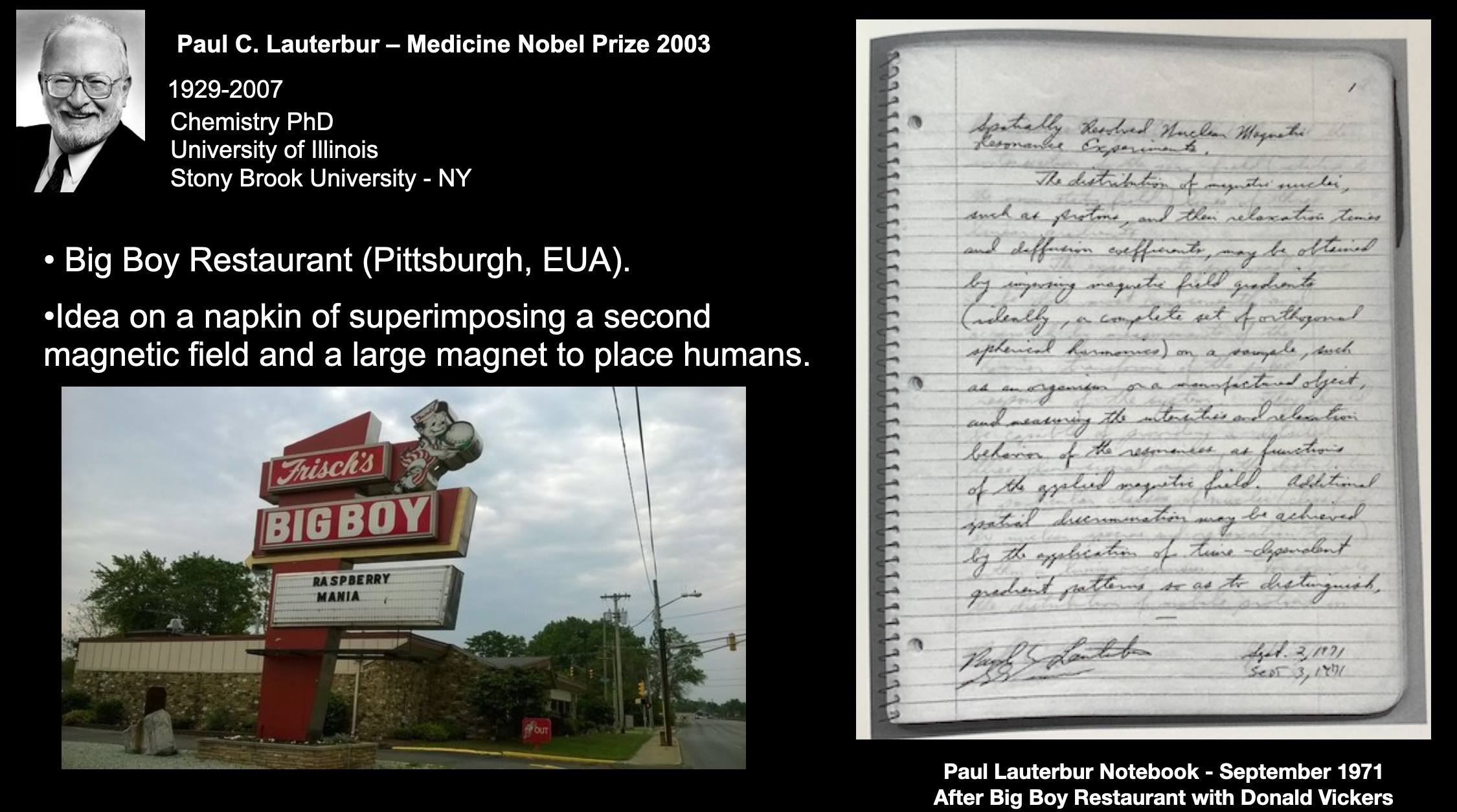
Figure 01
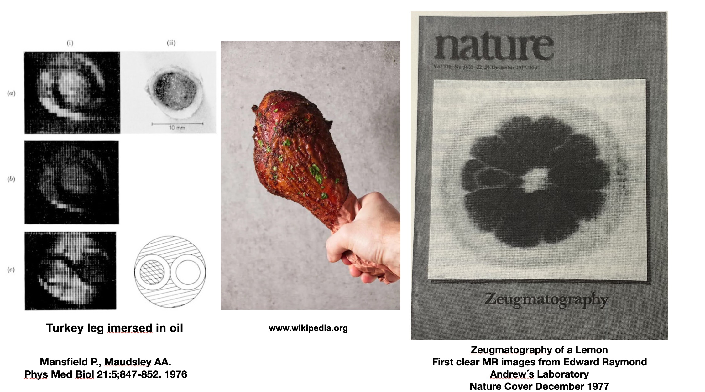
Figure 02
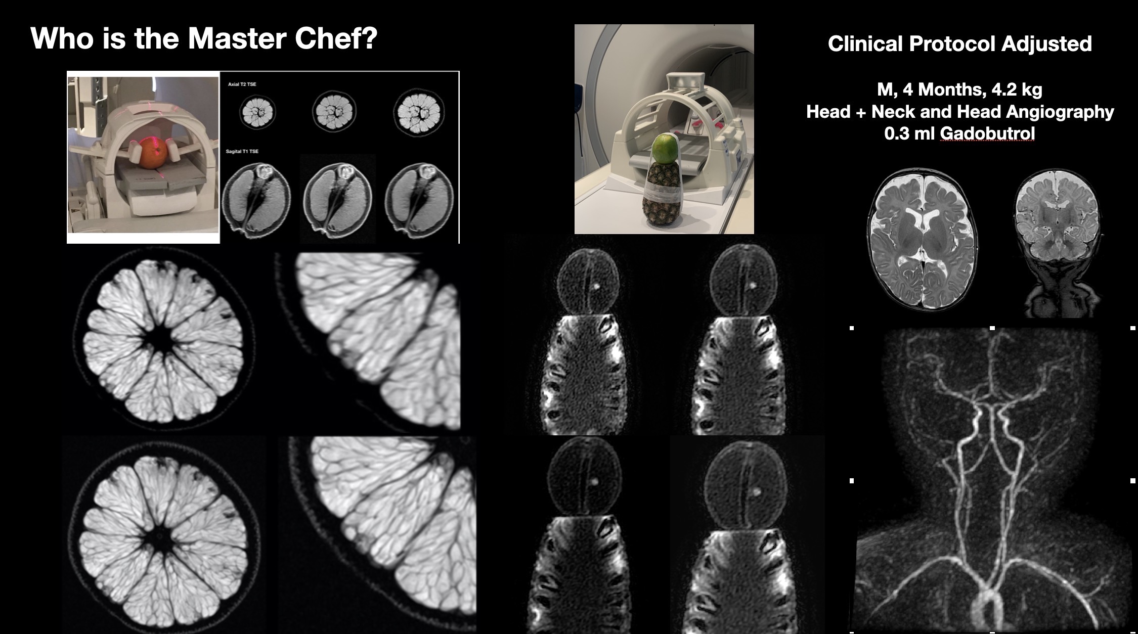
Figure 03