Preclinical & Histological Validation of Body/Tumor Microstructure Mapping
Yuko Someya1,2
1Dep. of diagnostic imaging and nuclear medicine, Kyoto University Graduate School of Medicine, Japan, 2Dep. of Radiology, Kobe City Medical Center General Hospital, Kobe, Japan
1Dep. of diagnostic imaging and nuclear medicine, Kyoto University Graduate School of Medicine, Japan, 2Dep. of Radiology, Kobe City Medical Center General Hospital, Kobe, Japan
Synopsis
Keywords: : Preclinical/Animal, Cross-organ: Cancer
Diffusion MRI, which visualizes the non-Gaussian movement of water molecules in a heterogeneous tumor microenvironment, has the potential to add new information about tissue microstructure non-invasively. This talks Introduce diffusion MRI methods for studying tumor microstructure, and then describe their sensitivity to tumor microstructural differences (cell size, shapes and configurations, cellularity, amount of extracellular space, intra-tumor heterogeneity, etc.), considering how the submicron resolution of tissue microscopy and the millimeter resolution of diffusion MRI can be linked to. Since the assessment of tumor angiogenesis is essential in oncology, the assessment of tissue microvasculature through the IVIM approach will also be discussed.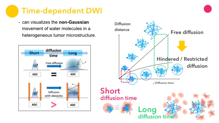 Slide #1