How Are MRI & PET/MRI Used in the Diagnosis & Management of Medium & Large Vessel Vasculitis?
Thorsten Bley1
1University of Wurzburg, Germany
1University of Wurzburg, Germany
Synopsis
Keywords: Cardiovascular: Vascular, Cross-organ: Inflammation
Next to CCDS, MRI and PET are recommended imaging modalities in large vessel vasculitides. With an in-plane spatial resolution of 200x250µm2 MRI displays mural inflammatory changes (circumferential thickening, contrast enhancement) of the superficial temporal arteries in giant cell arteritis. Combined with a first pass angiography of the thoracic aorta and its branch vessels the inflammatory disease burden and vascular damage (dilatation, stenosis) can be assessed within one examination. PET is considered the most sensitive imaging modality for assessing large vessel vasculitis. Combining sensitivity of PET with anatomic information of MRI in one hybrid scan offers promising perspectives for vasculitis imaging. Slide #1
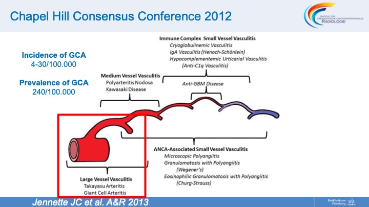 Slide #2
 Slide #3
 Slide #4
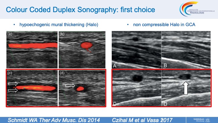 Slide #5
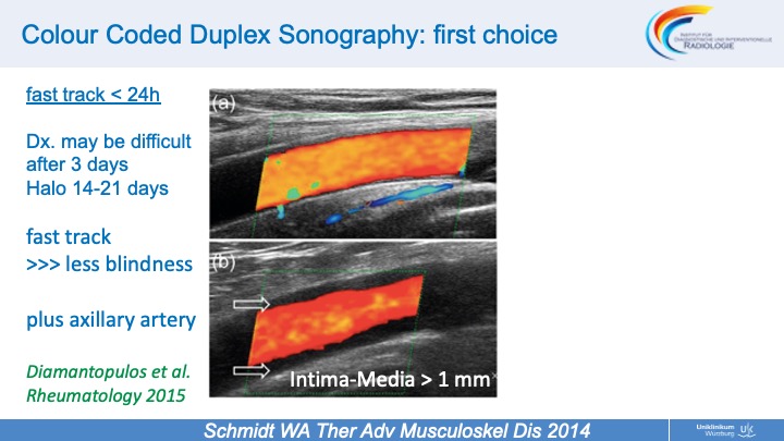 Slide #6
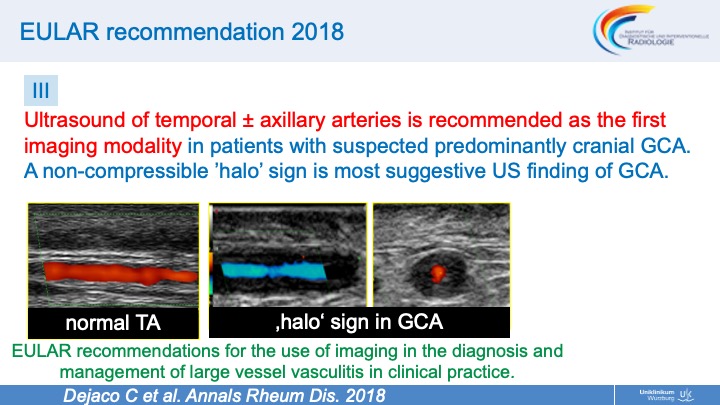 Slide #7
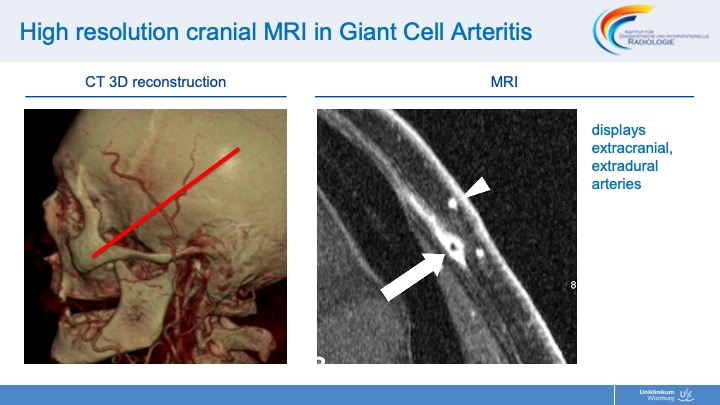 Slide #8
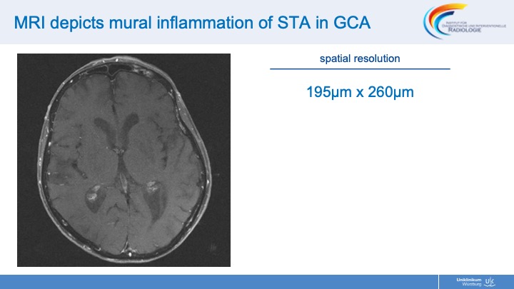 Slide #9
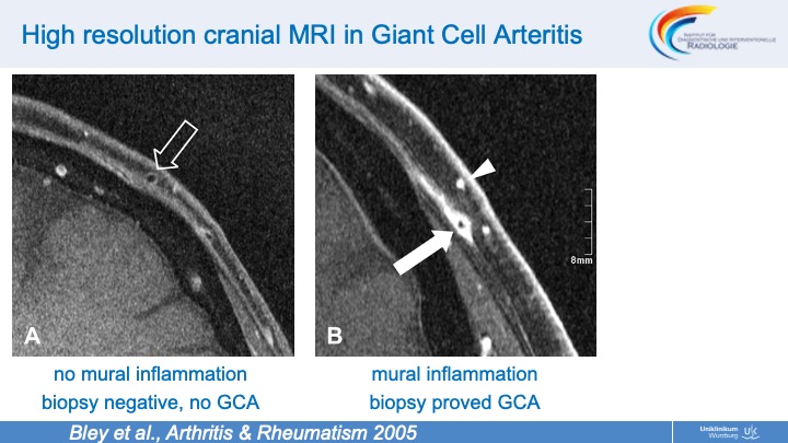 Slide #10
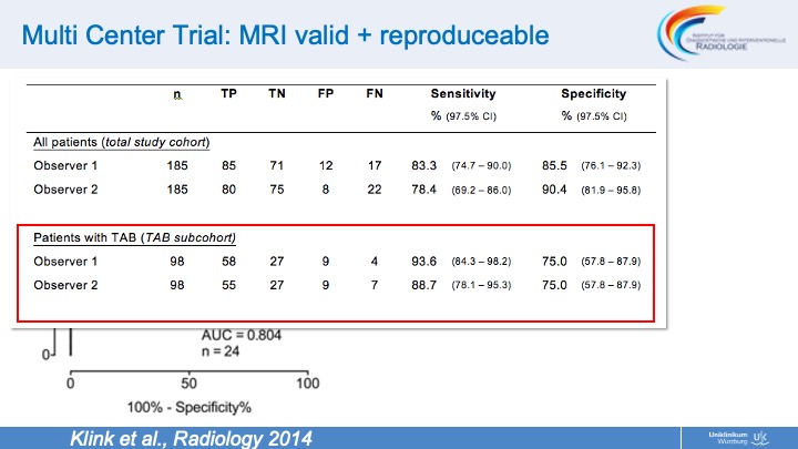 Slide #11
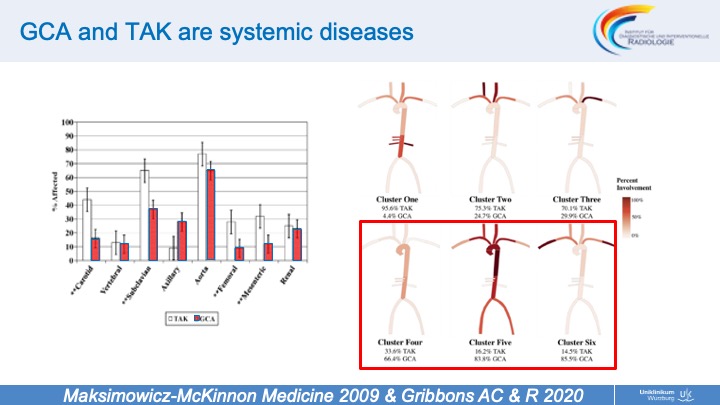 Slide #12
 Slide #13
 Slide #14
 Slide #15
 Slide #16
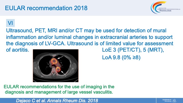 Slide #17
 Slide #18
 Slide #19
 Slide #20
 Slide #21
 Slide #22
 Slide #23
 Slide #24
 Slide #25
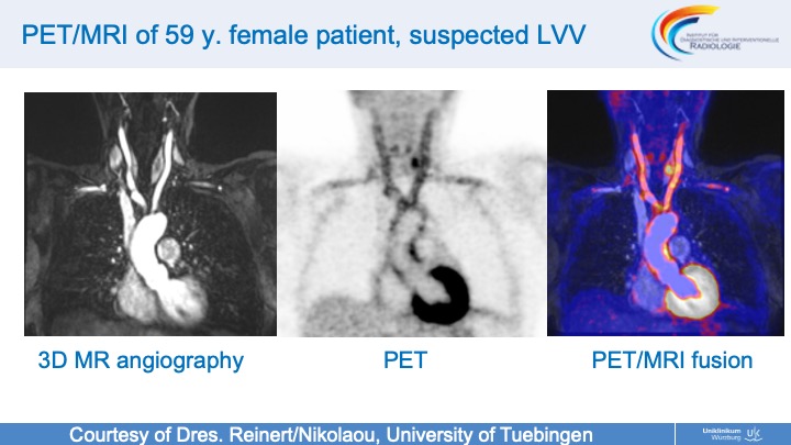 Slide #26
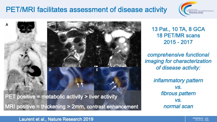 Slide #27
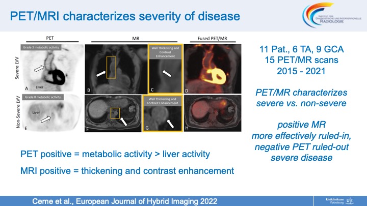 Slide #28
 Slide #29
 Slide #30