With PET/MRI & with PET Tracers
Bruno Stankoff1
1Sorbonne University, Paris, France, Paris, France
1Sorbonne University, Paris, France, Paris, France
Synopsis
Keywords: Cross-organ: Inflammation, Neuro: Neurodegeneration, Contrast mechanisms: Molecular imaging
Whereas the iron component sometimes associated with inflammation is accessible to MRI, the molecular specificity of PET is required to quantify neuroinflammation linked to innate immune cells. Tracers targeting TSPO are becoming largely available, with second generation tracers allowing an optimized sensitivity and specificity. Developing a TSPO PET imaging protocol implies the selection of an appropriate tracer, and the application of a robust and reproducible quantification model for data analysis. Following these requisites, promising results have been recently obtained in multiple sclerosis unravelling that an unexpectedly high proportion of lesions have a persistent neuroinflammatory content that drives progression.Imaging inflammation in the brain with PET/MRI and with PET Tracers
Whereas the iron component sometimes associated with inflammation is accessible to MRI, the molecular specificity of PET is required to quantify neuroinflammation linked to innate immune cells. Tracers targeting TSPO are becoming largely available, with second generation tracers allowing an optimized sensitivity and specificity. Developing a TSPO PET imaging protocol implies the selection of an appropriate tracer, and the application of a robust and reproducible quantification model for data analysis. Following these requisites, promising results have been recently obtained in multiple sclerosis unravelling that an unexpectedly high proportion of lesions have a persistent neuroinflammatory content that drives progression.Acknowledgements
We thanks ANR (Agence Nationale de la Recherche) and fondation ARSEP for funding support.References
Hamzaoui M *, Garcia J *, Boffa G, Lazzarotto A, Absinta M, Ricigliano VAG, Soulier T, Tonietto M, Gervais P, Bissery A, Louapre C, Bottlaender M, Bodini B, Stankoff B. Positron emission tomography with [18F]-DPA-714-PET unveils a smoldering component in most multiple sclerosis lesions which drives disease progression. Annals of neurology 2023 10.1002/ana.26657. Online ahead of print
Bodini B, Tonietto M, Airas L, Stankoff B. Positron emission tomography in multiple sclerosis - straight to the target. Nat Rev Neurol. 2021 Nov;17(11):663-675
Bodini B, Poirion E, Tonietto M, Benoit C, Palladino R, Maillart E, Portera E, Battaglini M, Bera G, Kuhnast B, Louapre C, Bottlaender M, Stankoff B. Individual Mapping of Innate Immune Cell Activation Is a Candidate Marker of Patient-Specific Trajectories of Worsening Disability in Multiple Sclerosis. J Nucl Med. 2020 Jul;61(7):1043-1049Figures
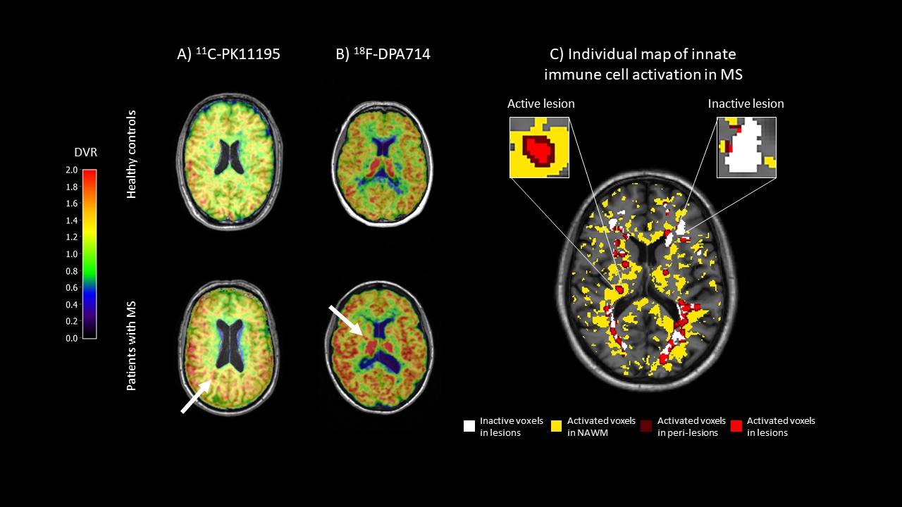
A, B, 11C-PK11195 (A) and 18F-DPA714 (B) DVR maps of controls and patients with multiple sclerosis (MS). Arrows points areas of increased DVR reflecting the presence of active innate immune cells. C, an individual map of innate immune cells obtained after thresholding a 18F-DPA714 DVR map in a patient with MS. Voxels characterised by a significant activation of innate immune cells are depicted in yellow in the normal-appearing white matter, dark red in perilesional areas and light red inside lesions visible on T2-weighted images Bodini B, et al. Nat Rev Neurol. 2021 Nov;17(11):663-675.