Imaging Immune-Mediated Lesions in Neuroinflammatory Diseases
Cornelius Faber1
1University of Muenster, Germany
1University of Muenster, Germany
Synopsis
Keywords: Neuro: Nervous System
Auto-immune neuroinflammatory disorders may be classified as either predominantly demyelinating, parenchymal non-demyelinating, meningeal-involving or vascular type. Similar to any type of neuroinflammation, underlying mechanisms that result in detectable lesions involve activation of resident immunocompetent cells, disruption of BBB and infiltration of peripheral immune cells. Lesions may display demyelination, edema formation, iron accumulation and atrophy, which all can be best detected using a variety of MRI sequences. Altered metabolic profiles are best observed by MR spectroscopy methods, while apoptosis/cell loss and upregulation of receptors is best detected by radioisotope-based methods, for which a wealth of tracers is available. Slide #1
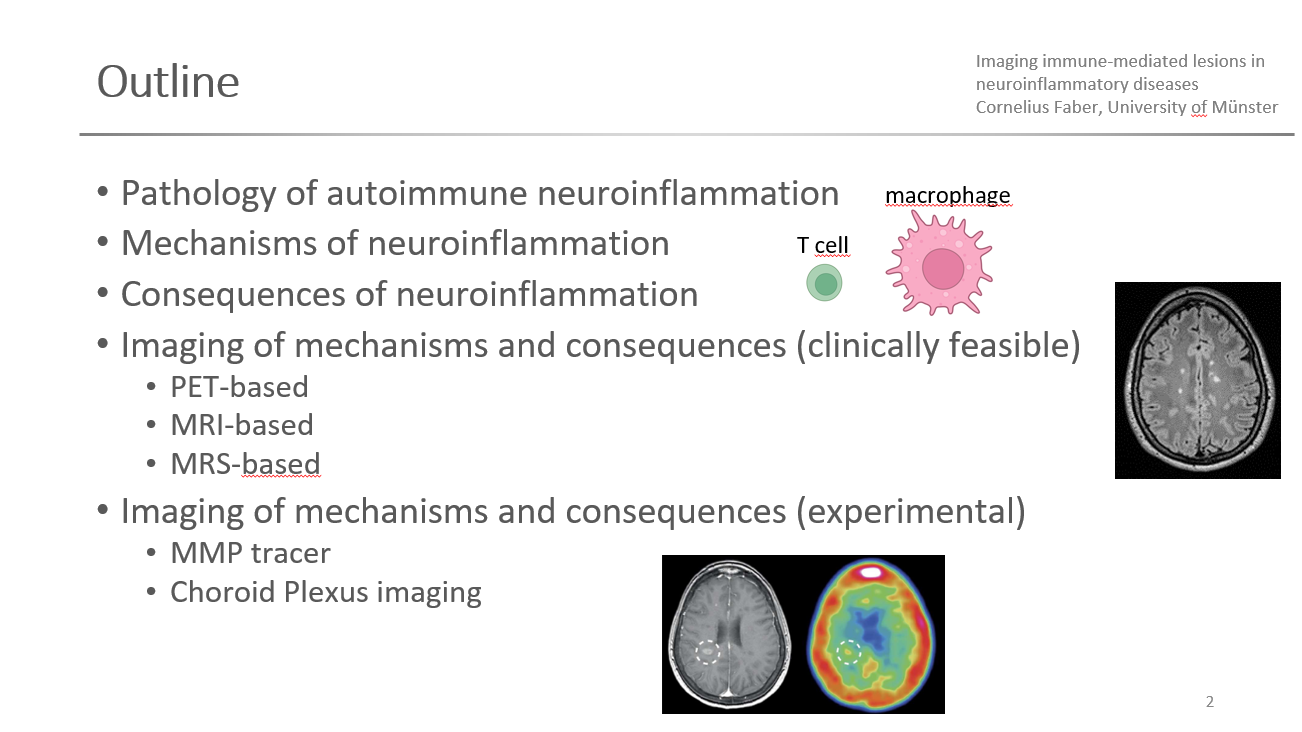 Slide #2
 Slide #3
 Slide #4
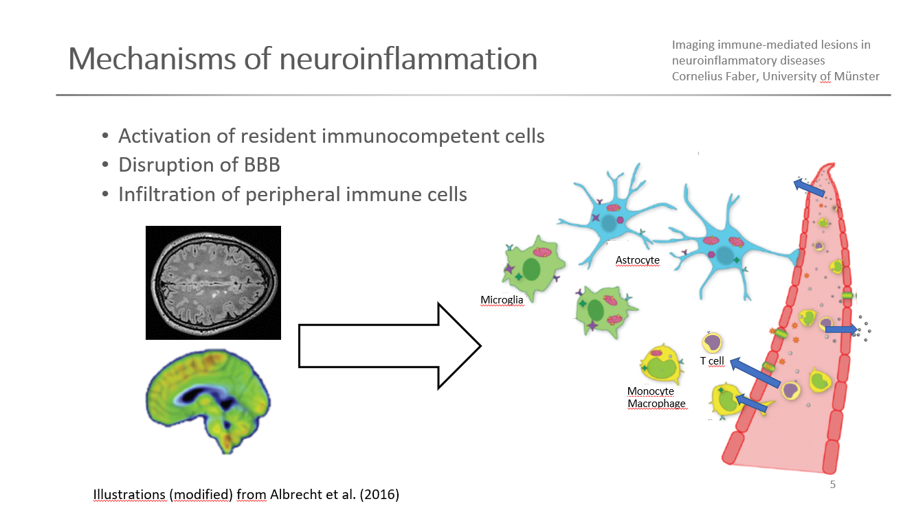 Slide #5
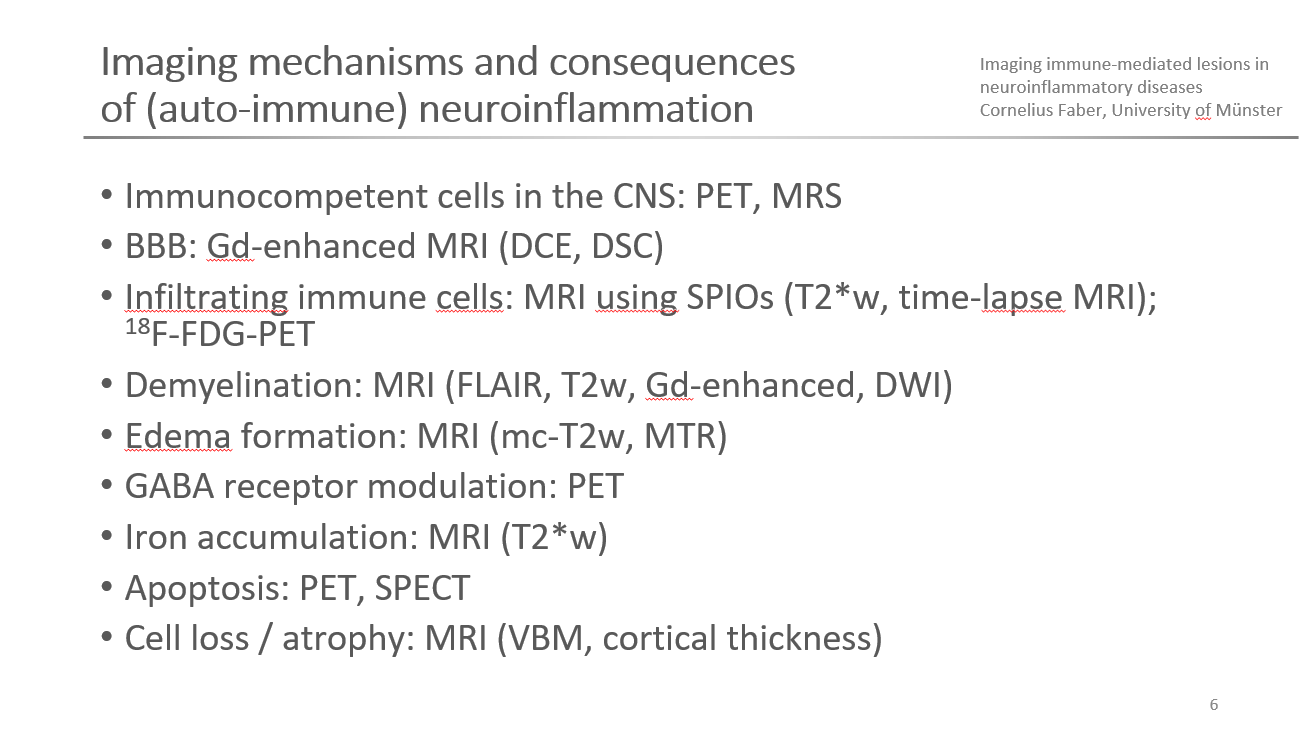 Slide #6
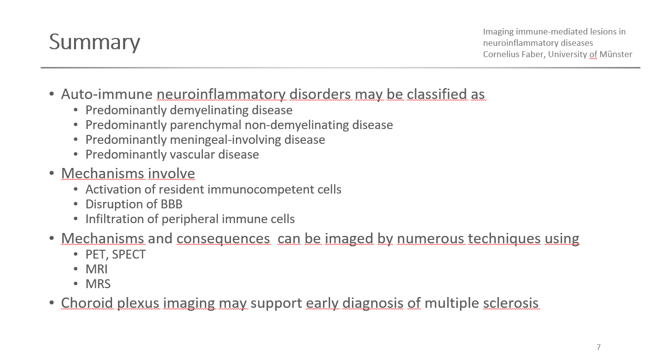 Slide #7
 Slide #8