MRI in Small & Large Bowel Tumours: One-Stop Shop
Andrea Laghi1
1Sapienza - University of Rome, Rome, Italy
1Sapienza - University of Rome, Rome, Italy
Synopsis
Keywords: Body: Digestive
In current clinical practice, MR imaging plays a limited role in the study of small and large bowel tumors, if we exclude rectal cancer. In fact, most patients with either a suspected small bowel neoplasm or with a need of colon cancer staging is referred to a MDCT evaluation. However, high soft tissue contrast, multiplanar imaging acquisition, as well as the possibility of performing functional studies make MR a promising technique to study these diseases. The possibility of using MRI in assessing small and large bowel tumors depends on the unmet clinical needs which cannot be fully satisfied by MDCT.In current clinical practice, MR imaging plays a limited role in the study of small and large bowel tumors, if we exclude rectal cancer. In fact, now most patients with either a suspected small bowel neoplasm or with a need of colon cancer staging is referred to a MDCT evaluation. Reasons are variable: CT availability; exam robustness (especially in elderly and fragile patients); patient clinical presentation (in the case of bowel obstruction CT is performed in an emergency setting).However, high soft tissue contrast, multiplanar imaging acquisition, multiparametric analysis, including “functional” studies (i.e., diffusion and perfusion) make MR a promising technique to study these diseases.Small and large bowel tumors are two very different entities: rare, heterogenous in histology (the most common tumors are adenocarcinoma, neuroendocrine neoplasms, gastrointestinal stromal tumors, and lymphoma), growth pattern and aggressiveness the small bowel tumors; very common and homogeneous in histology the colon cancers.The future success of a routinary use of MRI in assessing small and large bowel tumors depends on the unmet clinical needs which cannot be fully satisfied by a MDCT study.In the case of small bowel tumors, advantages of using MRI are the more accurate assessment of liver metastatic disease, especially in NET, the evaluation of distant spread outside liver, especially if a whole-body MR approach is used, the precise characterization of a primary lesion, based on a multiparametric analysis. In the case of colon cancer, there is a clear demand of a more accurate local staging, especially in locally advanced disease. This is due to the widespread use of neoadjuvant chemotherapy in colon cancer which improves patient outcome. Thus, local staging of colon cancer needs a detailed study of several factors affecting patient outcome, similarly to rectal cancer: extra-mural spread, extramural vascular invasion (EMVI), tumor deposits and lymphnodes. Although a limited number of studies is available, it can be argued that MR may be beneficial compared with CT in differentiating locally aggressive (T3cd/T4) vs less aggressive (T2/T3ab) tumors; in assessing venous invasion and in characterizing tumor deposits and lymphnodes.
Acknowledgements
No acknowledgement found.References
- Masselli G, Guida M, Laghi F, Polettini E, Gualdi G. Magnetic Resonance of Small Bowel Tumors. Magn Reson Imaging Clin N Am. 2020 Feb;28(1):75-88.
- Jasti R, Carucci LR. Small Bowel Neoplasms: A Pictorial Review. Radiographics. 2020 Jul-Aug;40(4):1020-1038.
- Schmidt SA, Beer M, Vogele D. Update: Dünndarmerkrankungen in Computertomographie und Magnetresonanztomographie [Update: Small bowel diseases in computed tomography and magnetic resonance imaging]. Radiologie (Heidelb). 2023 Apr 4. German.
- Rafaelsen SR, Dam C, Vagn-Hansen C, Møller J, Rahr HB, Sjöström M, Lindebjerg J, Hansen TF, Pedersen MRV. CT and 3 Tesla MRI in the TN Staging of Colon Cancer: A Prospective, Blind Study. Curr Oncol. 2022 Feb 13;29(2):1069-1079.
- Zhao Z, Zhou Y, Jiang M, Dang L. Application Value of MRI Combined with MSCT in Diagnosis and Staging of Colon Carcinoma. Comput Math Methods Med. 2022 May 23;2022:2593844.
- Liu LH, Lv H, Wang ZC, Rao SX, Zeng MS. Performance comparison between MRI and CT for local staging of sigmoid and descending colon cancer. Eur J Radiol. 2019 Dec;121:108741.
- Taylor SA, Mallett S, Miles A, Morris S, Quinn L, Clarke CS, Beare S, Bridgewater J, Goh V, Janes S, Koh DM, Morton A, Navani N, Oliver A, Padhani A, Punwani S, Rockall A, Halligan S. Whole-body MRI compared with standard pathways for staging metastatic disease in lung and colorectal cancer: the Streamline diagnostic accuracy studies. Health Technol Assess. 2019 Dec;23(66):1-270
- Dohan A, Hoeffel C, Soyer P, Jannot AS, Valette PJ, Thivolet A, Passot G, Glehen O, Rousset P. Evaluation of the peritoneal carcinomatosis index with CT and MRI. Br J Surg. 2017 Aug;104(9):1244-1249.
Figures
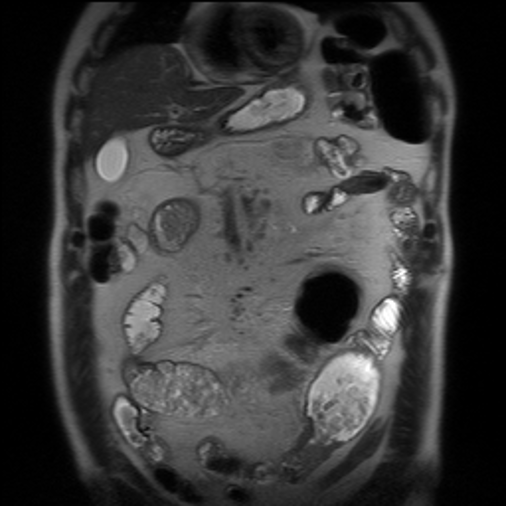
Ileal adenocarcinoma: T2w ss-FSE
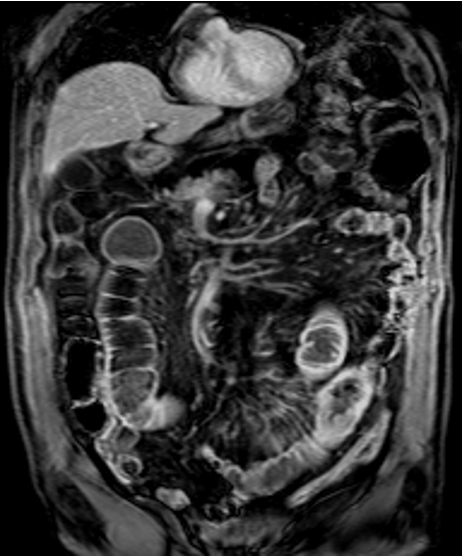
Ileal adenocarcinoma: CE-FS GRE T1w