fMRI vs. Intra-Cranial Measurements in Animals
Russell Chan1
1Gense Technologies Ltd., Hong Kong
1Gense Technologies Ltd., Hong Kong
Synopsis
Keywords: Contrast mechanisms: fMRI, Neuro: Brain function
Understanding how individual cells and complex brain networks interact in both time and space is a grant challenge. Recent fMRI advancements provide opportunities to measure layer-specific cortical responses to potentially address key neuroscience questions. optogenetic fMRI (ofMRI) can map effects of controlling cell-type specific neuronal population. In this session, technical considerations in applying ofMRI and intra-cranial electrophysiological recordings will be examined. Subsequently, distinct local and brain-wide networks activated by optogenetic stimulation of neurons specific to different cortical layers will be reviewed, and layer-specific fMRI responses and their neuronal origins will be explored. Lastly, the opportunities and challenges will be discussed.Target audience
Neuroimaging clinicians, scientists and engineers who are interested in multiscale and multimodal neuroimaging, neuromodulation, optogenetic, brain networks, brain functions and pre-clinical imaging.Highlights
- Technical considerations in optogenetic fMRI and intra-cranial measurements in animals
- Dissecting brain networks of neurons specific to each layer using optogenetic fMRI
- Investigating layer-specific fMRI responses and their neuronal origins
Objective
- Identify the practical issues and technical challenges of optogenetic fMRI and intra-cranial measurements in animals.
- Recognize the importance of layer-specific fMRI responses and their neuronal origins in dissecting brain networks and properties.
Purpose
The brain is a highly interconnected structure with parallel and hierarchical networks distributed within and between neural systems. This integrative architecture dictates the underlying principles of how brain-wide neural connectivity supports and organizes sensory, behavioral, and cognitive processes. Hence, it is imperative to investigate the spatiotemporal properties of brain-wide large-scale neural interactions. Normal operation of brain-wide networks requires precise and spatiotemporally structured activity propagation and interaction patterns to support the functional properties of these networks. Electrical stimulation cannot target cell-type specific neurons as it excites circuit components in the stimulation region that may not be involved in the particular behavior. Hence, this technique will not likely permit a reliable probe of the spatiotemporal activity propagation dynamics and their functional output. Furthermore, electrode recording site density and spatial coverage limit the characterization of properties in brain-wide large-scale brain functional networks. Recent fMRI advancements provide opportunities to measure layer-specific cortical responses to potentially address key neuroscience questions by distinguishing bottom-up from top-down cortical responses and investigating the interactions between the two, with limited studies on the neuronal origins of fMRI layer-specific cortical responses. Optogenetic fMRI (ofMRI) within the living mammalian brain reveals BOLD signals in downstream targets distant from the stimulation site, indicating that this approach can be used to map the global and local effects of controlling a local cell-type specific neuronal population. This method can overcome the limitations of electrical stimulation non-specificity and electrode recording spatial coverage in characterizing large-scale brain-wide neural activity propagation and interaction dynamics. In this session, technical considerations in applying ofMRI and intra-cranial electrophysiological recordings will be examined. Subsequently, distinct local and brain-wide networks activated by optogenetic stimulation of neurons specific to different cortical layers will be reviewed, and layer-specific fMRI responses and their neuronal origins will be explored. Lastly, the opportunities and challenges will be discussed.Acknowledgements
No acknowledgement found.References
No reference found.Figures
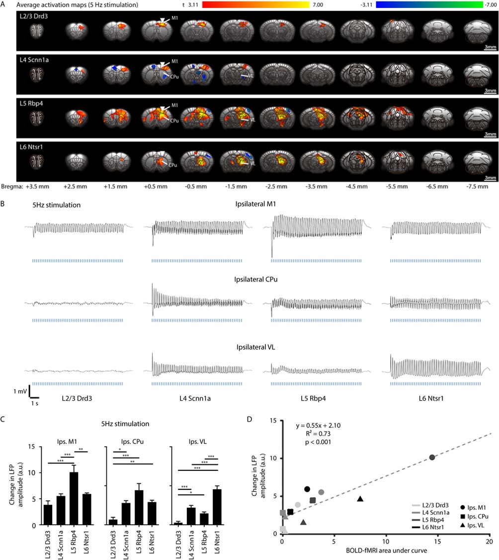
Stimulations of neurons specific to M1 layers evoke distinct brain-wide ofMRI responses (A) while LFP recordings reveal activations in M1 for all stimulations of neurons specific to M1 layers, and in CPu and VL for stimulations within L4, L5, and L6 only (B-C), which correlates with fMRI activations (D).