Dynamic Imaging: Clinical Importance & Challenges
1NYU Langone Medical Center, United States
Synopsis
Keywords: Musculoskeletal: Joints, Musculoskeletal: Knee, Neuro: Spinal Cord
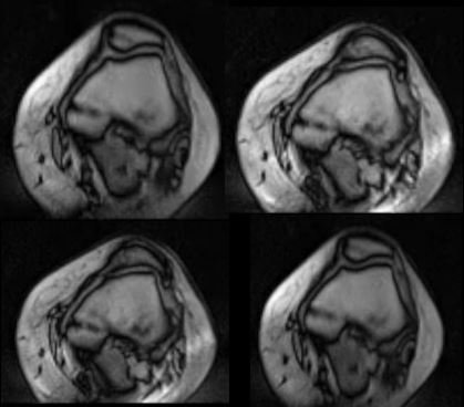
The importance of kinematic imaging will be explained in three promising clinical contexts. In the knee, where direct real-time visualization and quantification of patellofemoral maltracking may support findings on standard static imaging and guide surgical management. In the hip, where femoroacetablular impingement identified on examination can be confirmed directly with dynamic imaging alongside predisposing morphological features potentially guiding surgical decision-making. In the spine, where dynamic listhesis and cord impingement can have implications for the development of myelomalacia and necessity for decompression and fusion. The technical challenges will be addressed alongside future potential work to address these hurdles.Dynamic Imaging: Clinical Importance & Challenges
Acknowledgements
No acknowledgement found.References
Burke CJ, Kaplan D, Block T, Chang G, Jazrawi L, Campbell K, Alaia M. Clinical Utility of Continuous Radial Magnetic Resonance Imaging Acquisition at 3 T in Real-time Patellofemoral Kinematic Assessment: A Feasibility Study. Arthroscopy. 2018 Mar;34(3):726-733. doi: 10.1016/j.arthro.2017.09.020. Epub 2017 Dec 19. PMID: 29273250; PMCID: PMC6080599.
Burke CJ, Walter WR, Gyftopoulos S, Pham H, Baron S, Gonzalez-Lomas G, Vigdorchik JM, Youm T. Real-Time Assessment of Femoroacetabular Motion Using Radial Gradient Echo Magnetic Resonance Arthrography at 3 Tesla in Routine Clinical Practice: A Pilot Study. Arthroscopy. 2019 Aug;35(8):2366-2374. doi: 10.1016/j.arthro.2019.02.049. PMID: 31395172.
Walter WR, Alizai H, Bruno M, Portugal S, Burke CJ. Real-time dynamic 3-T MRI assessment of spine kinematics: a feasibility study utilizing three different fast pulse sequences. Acta Radiol. 2021 Jan;62(1):58-66. doi: 10.1177/0284185120913000. Epub 2020 Mar 31. PMID: 32233646.
Burke CJ, Samim M, Alizai H, Sanchez J, Kingsbury D, Babb JS, Walter WR. Clinical feasibility of 2D dynamic sagittal HASTE flexion-extension imaging of the cervical spine for the assessment of spondylolisthesis and cervical cord impingement. Eur J Radiol. 2021 Jan;134:109447. doi: 10.1016/j.ejrad.2020.109447. Epub 2020 Nov 27. PMID: 33307460.
Frings J, Dust T, Krause M, Frosch KH, Adam G, Warncke M, Welsch G, Henes FO, Maas KJ. Dynamic Mediolateral Patellar Translation Is a Sex- and Size-Independent Parameter of Adult Proximal Patellar Tracking Using Dynamic 3 Tesla Magnetic Resonance Imaging. Arthroscopy. 2021 Oct 29:S0749-8063(21)00923-3. doi: 10.1016/j.arthro.2021.10.014.
Frings J, Dust T, Krause M, Ohlmeier M, Frosch KH, Adam G, Warncke M, Maas KJ, Henes FO. Objective assessment of patellar maltracking with 3 T dynamic magnetic resonance imaging: feasibility of a robust and reliable measuring technique. Sci Rep. 2020 Oct 8;10(1):16770. doi: 10.1038/s41598-020-72332-9. PMID: 33033292; PMCID: PMC7546634.
Figures