Iron Imaging Using QSM, R2* & SWI
1Mayo Clinic Florida, Jacksonville, FL, United States
Synopsis
Keywords: Contrast mechanisms: Susceptibility, Contrast mechanisms: Relaxometry, Neuro: Brain
The basics of SWI, QSM and R2* will be introduced along with the contrast mechanisms for iron imaging. Sensitivity, specificity and limitations will be illustrated as well as examples for iron imaging in neurologic disorders.
Susceptibility is a measure of how much a material gets magnetized in an external magnetic field. Paramagnetic materials have unpaired electrons and paramagnetism is temperature dependent. QSM differentiates between para- and diamagnetic materials. R2* relaxometry is sensitive to both iron and myelin. QSM and SWI can differentiate MS lesion types. QSM and R2* quantify age- and pathology related iron accumulation in brain nuclei and cortices.
 Slide #1
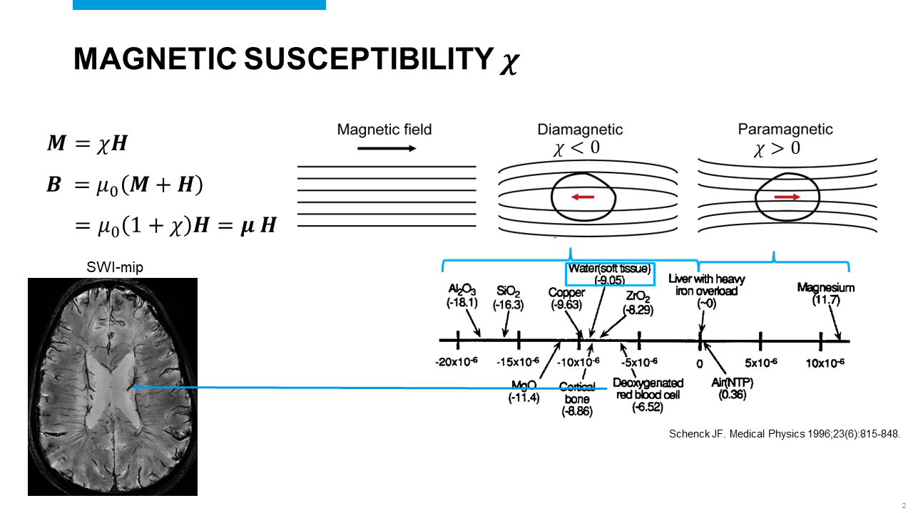 Slide #2
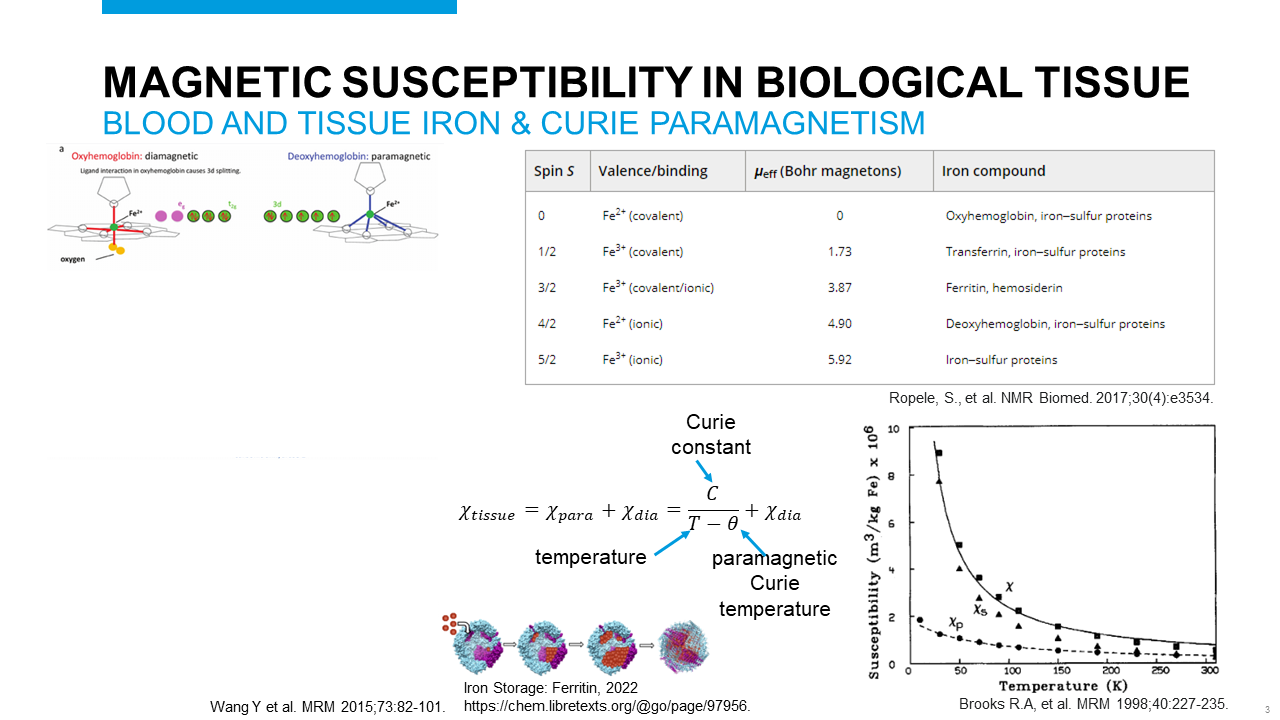 Slide #3
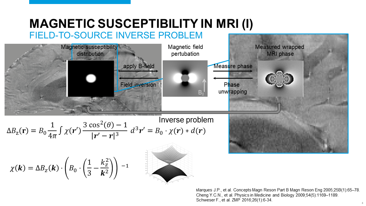 Slide #4
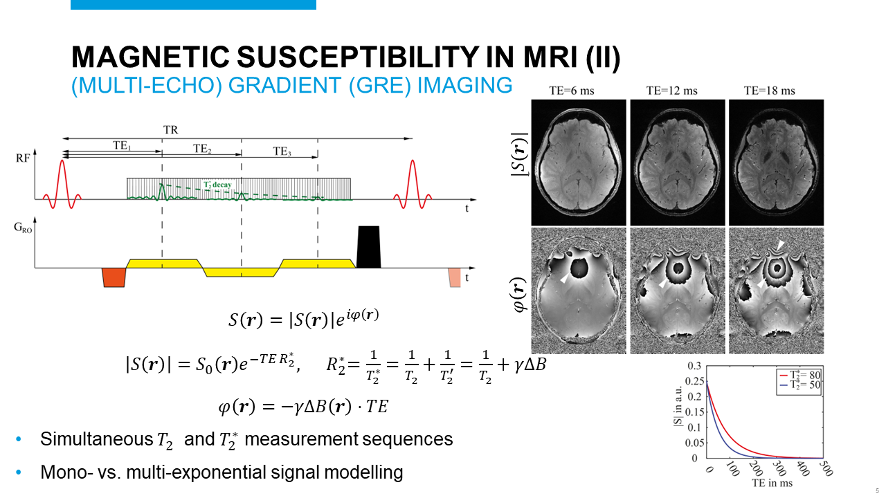 Slide #5
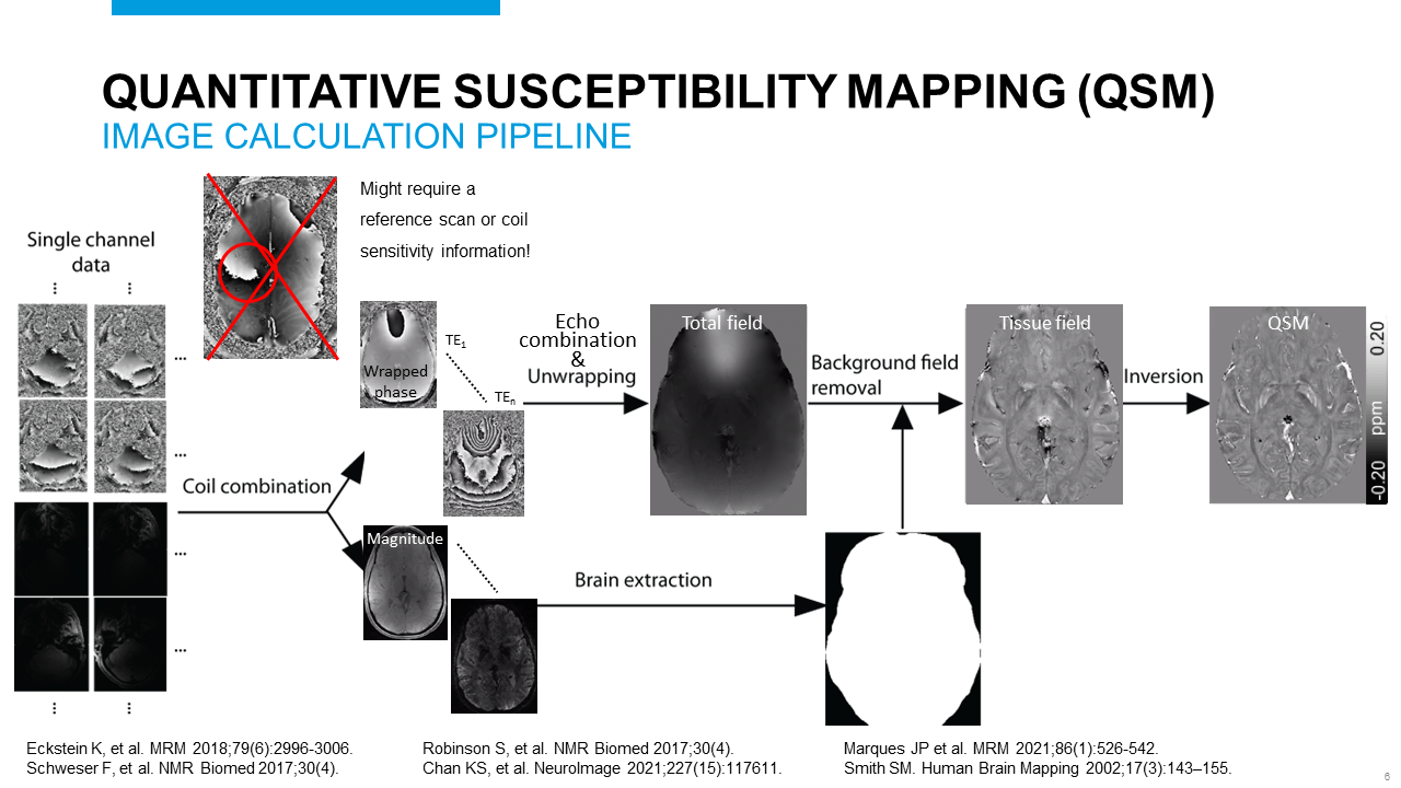 Slide #6
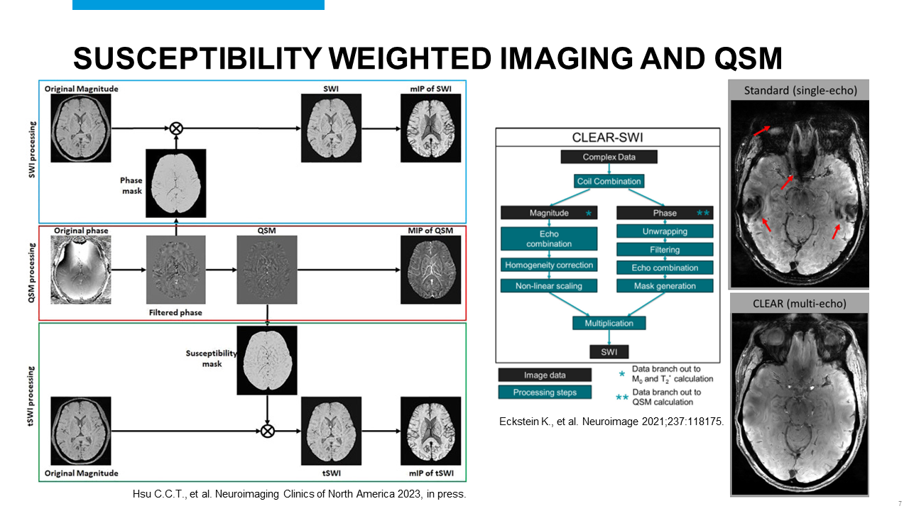 Slide #7
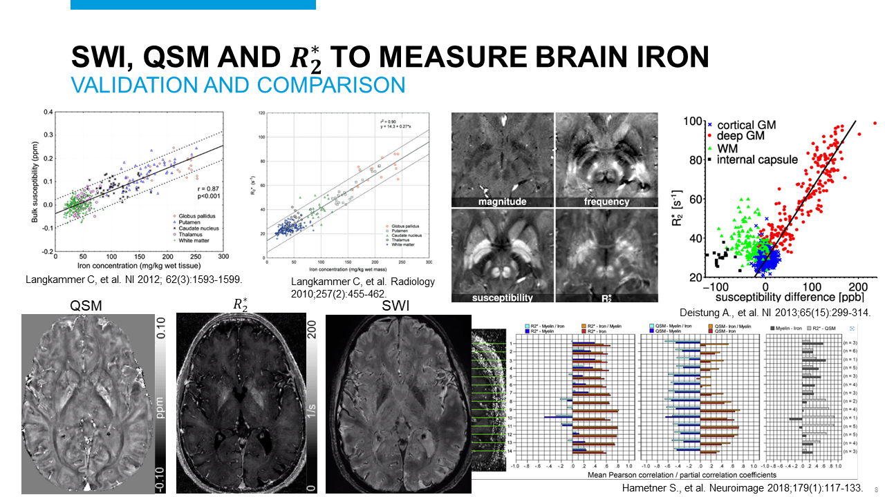 Slide #8
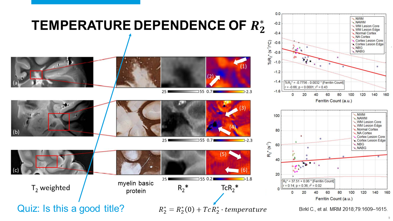 Slide #9
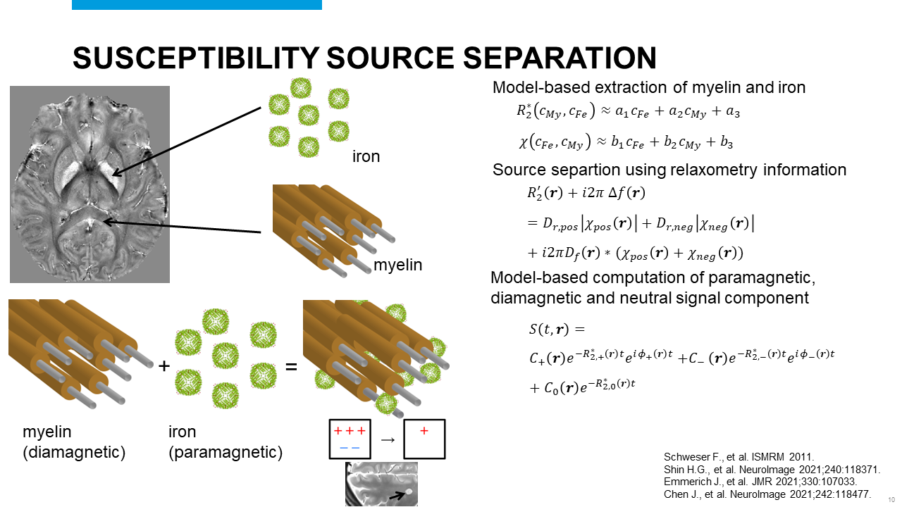 Slide #10
 Slide #11
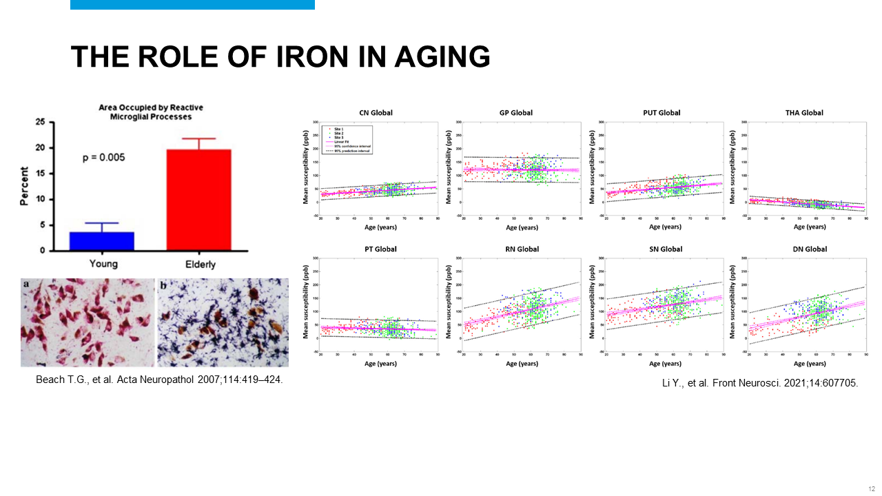 Slide #12
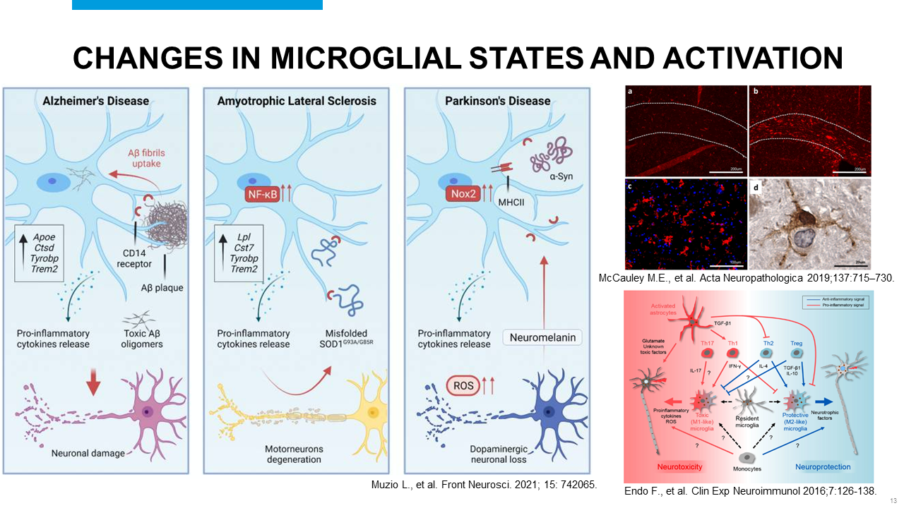 Slide #13
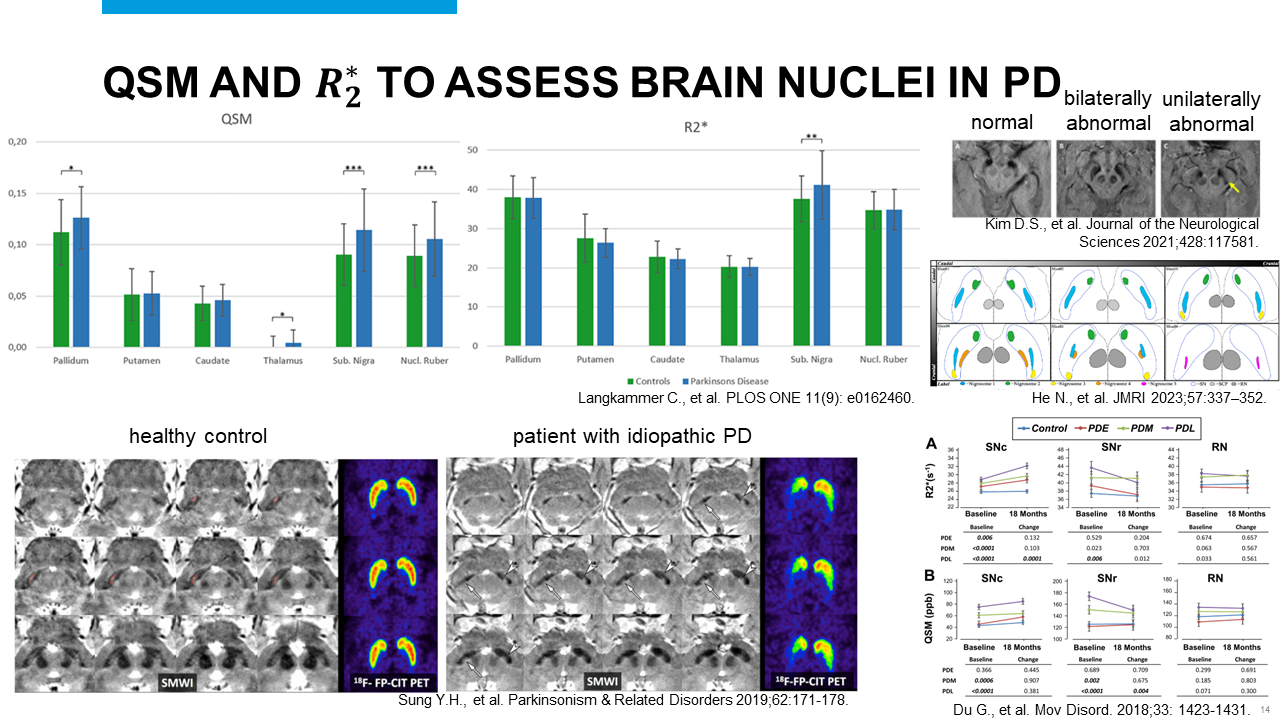 Slide #14
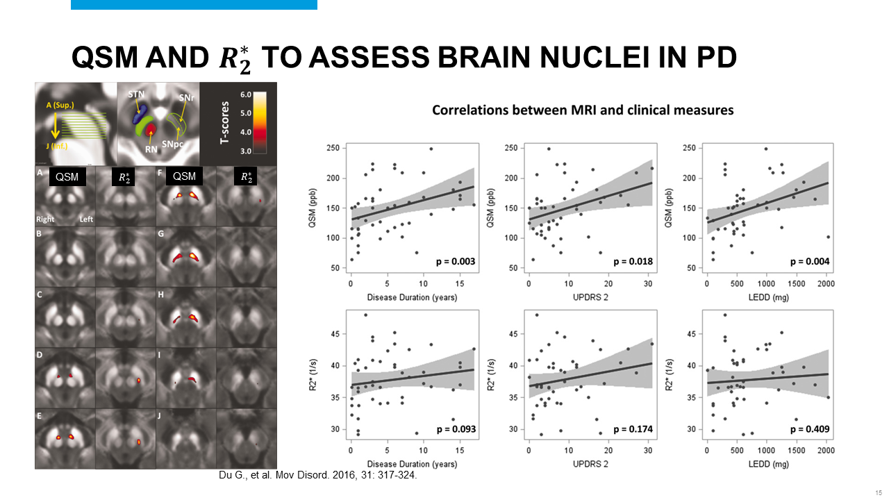 Slide #15
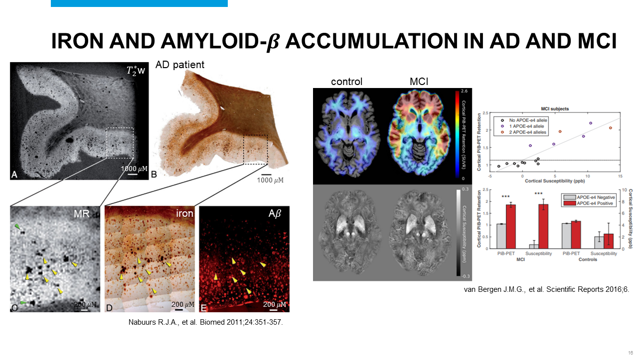 Slide #16
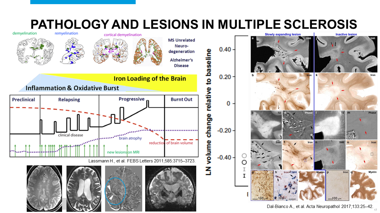 Slide #17
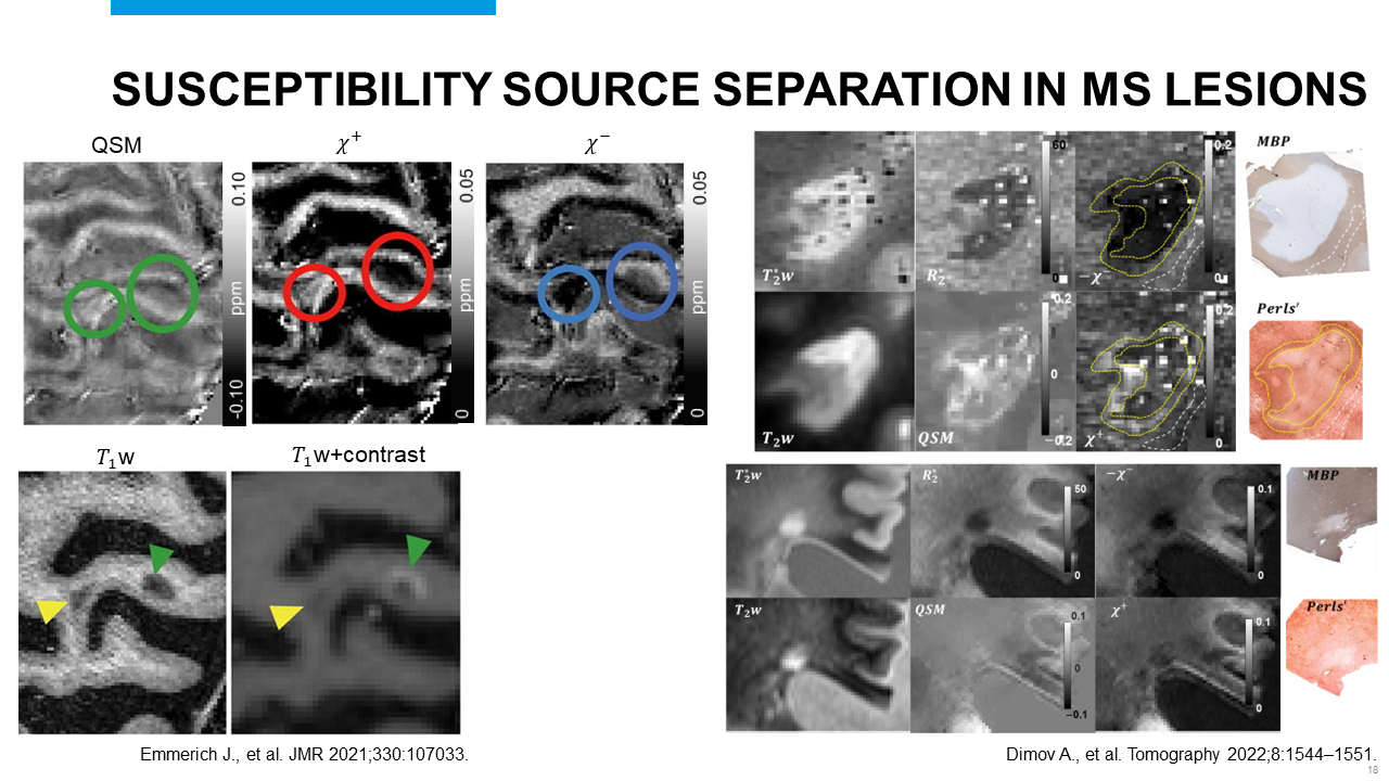 Slide #18
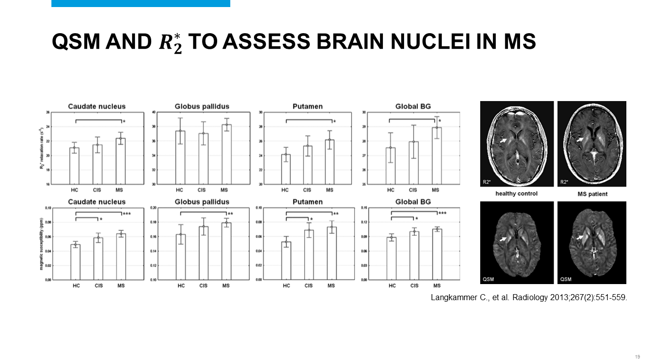 Slide #19
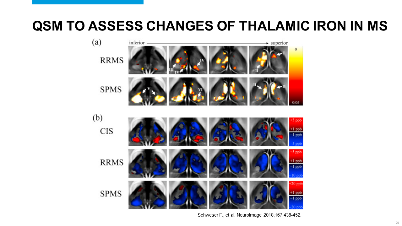 Slide #20
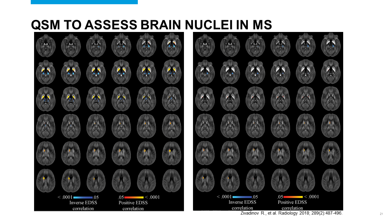 Slide #21
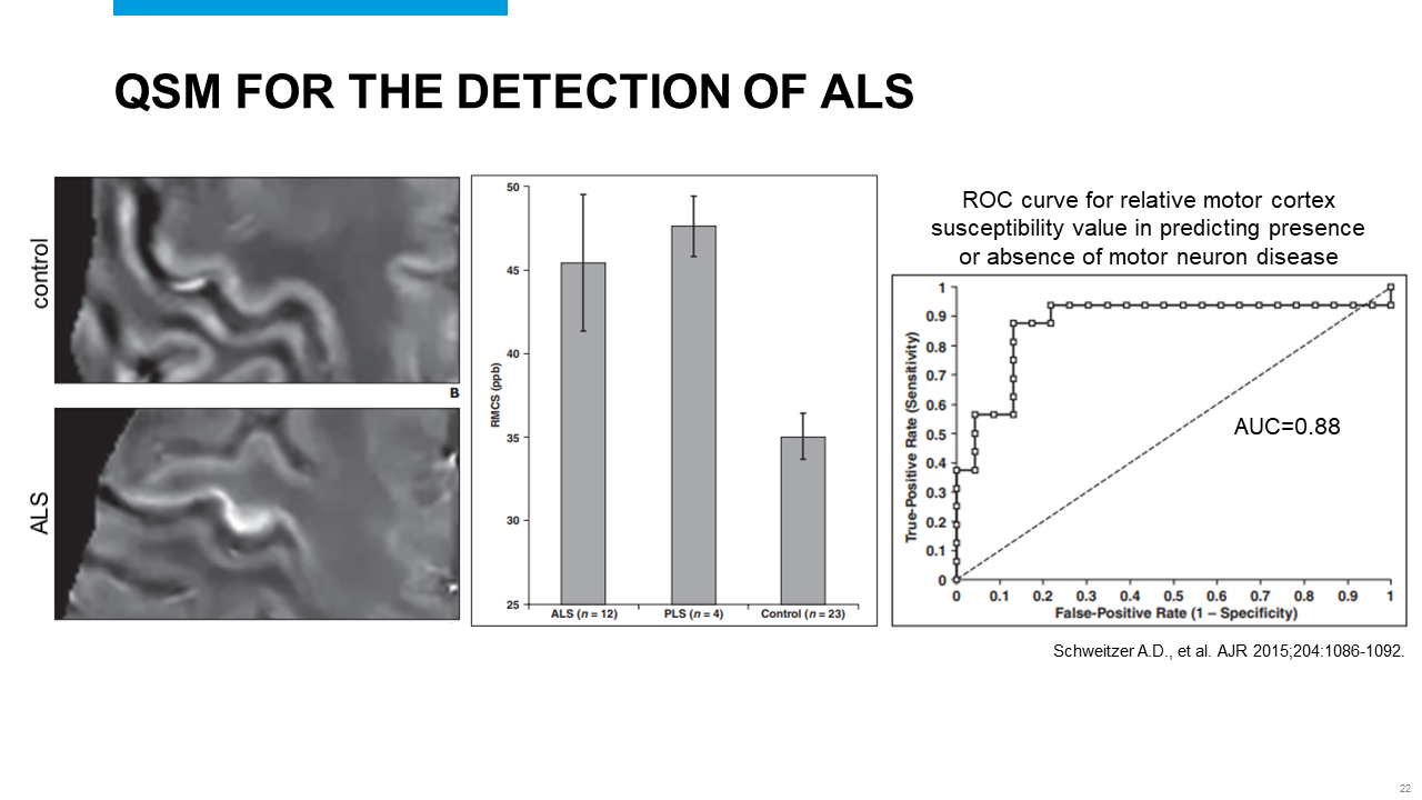 Slide #22
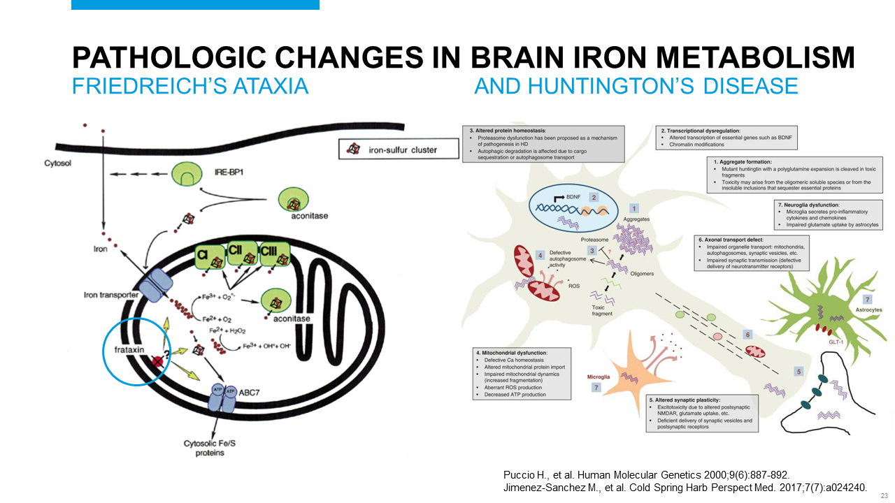 Slide #23
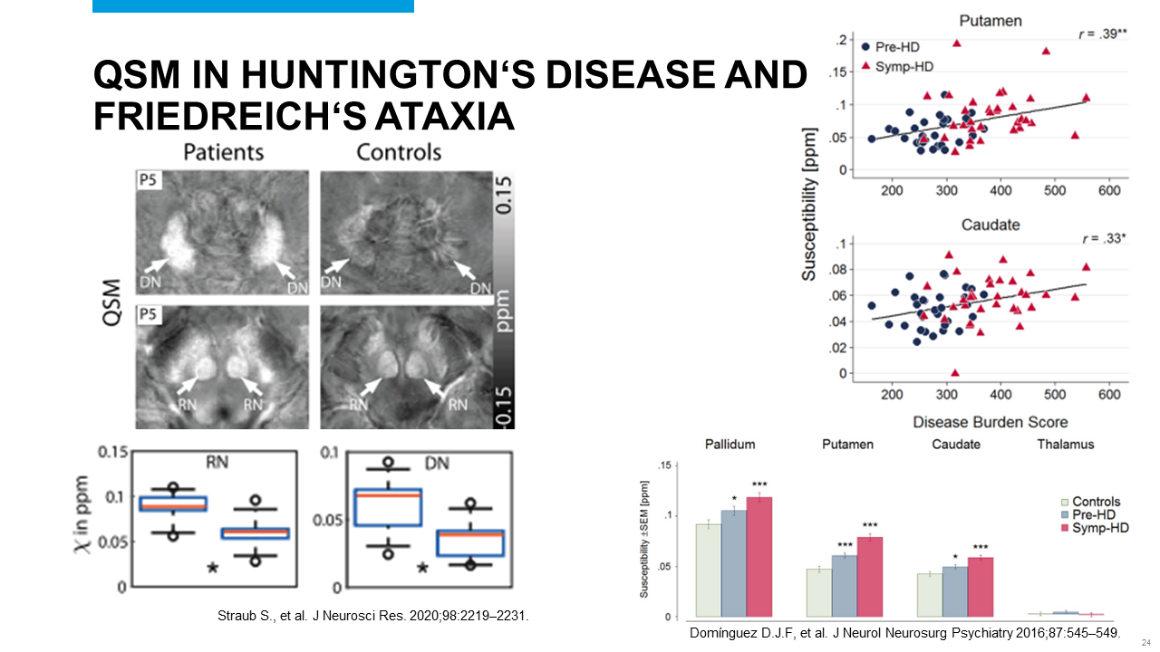 Slide #24
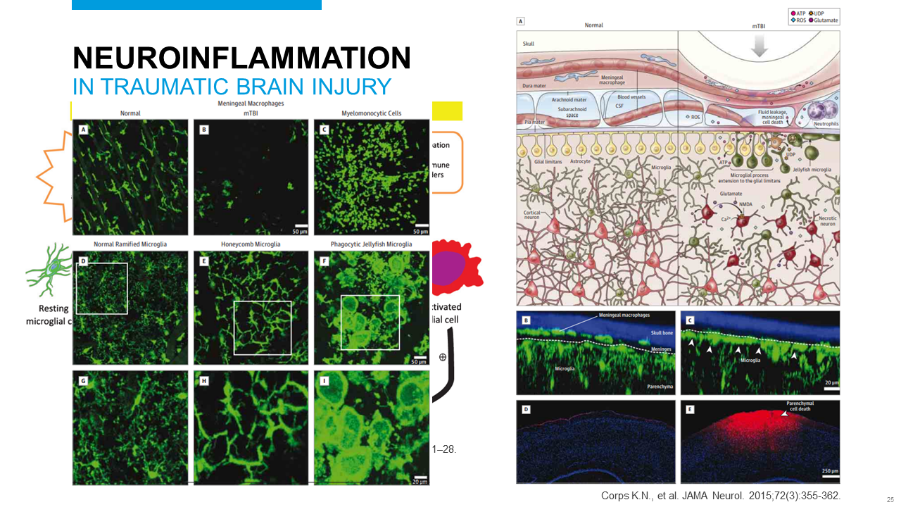 Slide #25
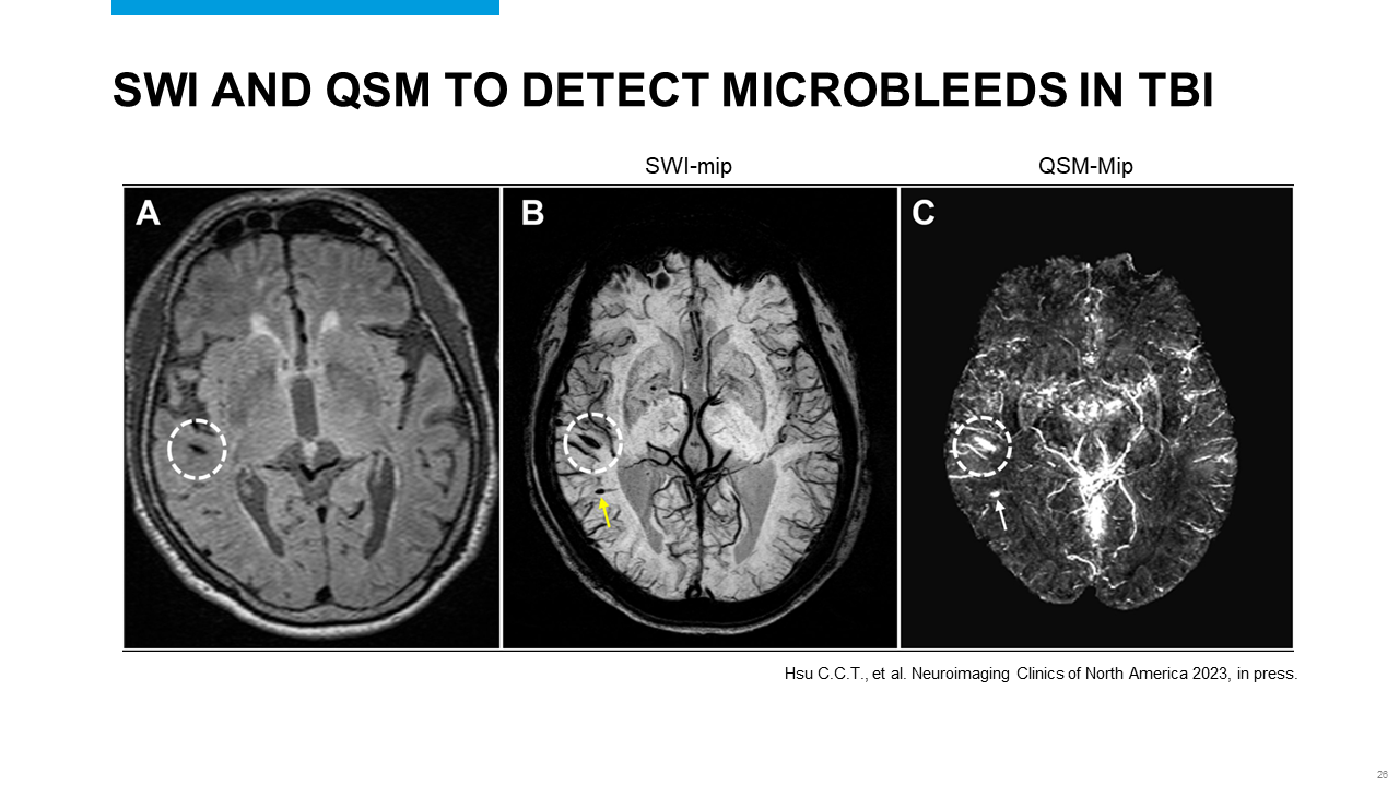 Slide #26
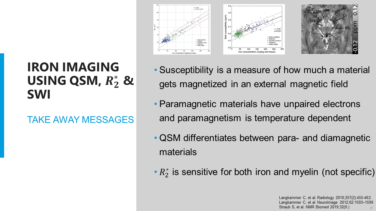 Slide #27
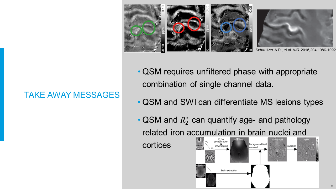 Slide #28