BOLD & Non-BOLD Contrasts in Human fMRI
Sriranga Kashyap1
1Krembil Brain Institute, University Health Network, Toronto, Canada
1Krembil Brain Institute, University Health Network, Toronto, Canada
Synopsis
Keywords: Contrast mechanisms: fMRI, Neuro: Brain
FMRI is a non-invasive method that allows scientists to study the brain function during task or at rest. The BOLD contrast is the workhorse of functional neuroimaging. A cascade of physiological events following neuronal activity (changes in blood oxygenation, flow and volume) culminates in the BOLD signal. The versatility of MRI enables imaging of blood flow and volume using techniques such as Arterial Spin Labelling (ASL) and Vascular Space Occupancy (VASO) respectively. In this talk, we will learn about BOLD and non-BOLD contrasts (CBF, CBV), discuss what they offer and how they differ in their application to human fMRI.Introduction
Functional magnetic resonance imaging (fMRI) is a non-invasive method that allows scientists to study the brain function during task or at rest typically carried out using the blood oxygenation level-dependent (BOLD) contrast1. The measured BOLD signal probes neuronal activity indirectly via a complex cascade of physiological events such as changes in blood flow, volume and oxygenation. The versatility of MRI allows to also perform fMRI using these intermediate stages using techniques such as Arterial Spin Labelling2 (ASL) and Vascular Space Occupancy3 (VASO) for blood flow and volumes respectively. Despite the fact that the earliest human fMRI studies4,5 published were carried out using CBV and CBF contrasts, BOLD fMRI has emerged the workhorse of modern neuroimaging. In this talk, we will look at BOLD and non-BOLD contrasts (CBF, CBV) and discuss what they offer, how they differ, and understand their strengths and limitations of these methods with respect to human fMRI.Overview and learning goals
1. BOLD fMRIBOLD fMRI is the most widely used method in neuroscience research today6. There are two methods by which BOLD signal can be measured using MRI. The first method uses gradient-echo (GE) to generate the T2*-weighting in the data which is highly sensitive to local changes in the magnetic field susceptibility but lacks spatial specificity i.e., GE-BOLD signals can occur as far as several millimetres from the actual site of neuronal activation depending on local vasculature (presence of intracortical and pial draining veins)7. The second method uses spin-echo (SE) which uses a 180° refocussing pulse to generate the T2-weighting and suppresses the signal in and around large veins resulting in a higher spatial specificity8. What are some of the limitations of BOLD fMRI? When and why would you consider non-BOLD alternatives for your study?
2. CBF fMRI
In the mechanism of BOLD signal generation, Cerebral Blood Flow (CBF) is a key physiological parameter. CBF is defined as the amount of blood delivered to grey matter per unit time and tissue mass (ml / 100 g / min). CBF is measured using the Arterial Spin Labelling (ASL) technique that uses RF pulses to label protons in inflowing arterial blood and the measured signal in the brain is composed of the signal of the grey matter attenuated by the T1 of the labelled blood. CBF-weighted data are less sensitive to detecting fMRI activation than BOLD data despite their increased spatial specificity. They are also more challenging to acquire as they require advanced pulse sequences and analysis methods9,10. What are different methods to acquire CBF fMRI? What are the benefits and limitations of this approach compared to BOLD fMRI?
3. CBV fMRI
Cerebral Blood Volume (CBV) weighted fMRI is typically carried out using the Vascular Space Occupancy (VASO) approach11. VASO is an inversion-recovery sequence that selectively nulls the blood signal while preserving the tissue signal in the surrounding. Unlike BOLD and CBF approaches, the VASO signal is a 'negative' contrast, that is, CBV increase following neuronal activation causes a decrease in the VASO signal. In other words, the measured VASO signal is porportional to 1 - CBV. What are the challenges to acquiring CBV fMRI? What are the benefits and limitations of this approach compared to BOLD fMRI?
Unlike standard BOLD fMRI, CBF and CBV fMRI signals can be quantified into physiological units to aid interpretation12. Non-BOLD approaches have shown promise for applications such as laminar fMRI at ultra-high field13 as well as clinical applications14.
Acknowledgements
I would like to thank Drs. Dimo Ivanov, Laurentius Huber, Benedikt Poser, Icaro Oliveira and Kamil Uludag for their suggestions, slides and advice.References
- Ogawa S, Lee TM, Kay AR, Tank DW. Brain magnetic resonance imaging with contrast dependent on blood oxygenation. Proc Natl Acad Sci U S A. 1990
- Williams, D. S., Detre, J. A., & Leigh, J. S. Magnetic resonance imaging of perfusion using spin inversion of arterial water. Proc Natl Acad Sci U S A. 1992
- Lu H, Golay X, Pekar JJ, Van Zijl PC. Functional magnetic resonance imaging based on changes in vascular space occupancy. Magn Reson Med. 2003
- Belliveau J, Kennedy D, McKinstry R, Buchbinder B, Weisskoff R, Cohen M, et al. Functional mapping of the human visual cortex by magnetic resonance imaging. Science. 1991
- Kwong KK, Belliveau JW, Chesler DA, Goldberg IE, Weisskoff RM, Poncelet BP, et al. Dynamic magnetic resonance imaging of human brain activity during primary sensory stimulation. Proc Natl Acad Sci. 1992
- Norris DG. Principles of magnetic resonance assessment of brain function. J Magn Reson Imaging. 2006
- Uludağ K, Müller-Bierl B, Uğurbil K. An integrative model for neuronal activity-induced signal changes for gradient and spin echo functional imaging. Neuroimage. 2009
- Norris DG. Spin-echo fMRI: The poor relation? Neuroimage. 2012
- Detre JA, Wang J. Technical aspects and utility of fMRI using BOLD and ASL. Clin Neurophysiol. 2002
- Liu TT, Wong EC. A signal processing model for arterial spin labeling functional MRI. Neuroimage. 2005
- Lu H, Hua J, van Zijl PC. Noninvasive functional imaging of cerebral blood volume with vascular-space-occupancy (VASO) MRI. NMR Biomed. 2013
- Pike GB. Quantitative functional MRI: concepts, issues and future challenges. Neuroimage. 2012
- Huber L, Uludağ K, Möller HE. Non-BOLD contrast for laminar fMRI in humans: CBF, CBV, and CMRO2. Neuroimage. 2019
- Tjandra T, Brooks JC, Figueiredo P, Wise R, Matthews PM, Tracey I. Quantitative assessment of the reproducibility of functional activation measured with BOLD and MR perfusion imaging: implications for clinical trial design. Neuroimage. 2005
Figures
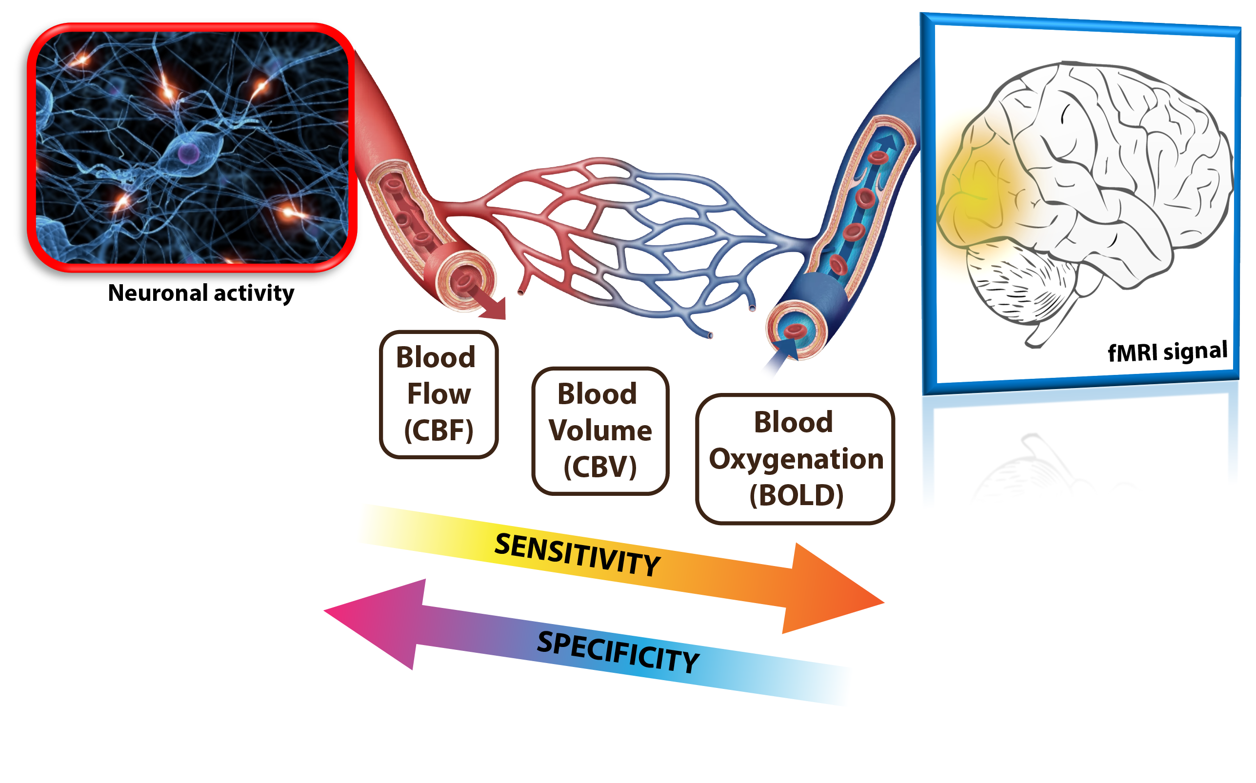
BOLD and non-BOLD contrasts in human fMRI