Gradient Coils
1Medical Physics Department of Radiology, University of Freiburg, Faculty of Medicine, Department of Radiology, Medical Physics, Freiburg, Germany
Synopsis
Keywords: Physics & Engineering: Hardware
The spatial encoding system, usually termed gradients, is crucial for magnetic resonance imaging (MRI). Gradient coils for MRI are usually designed using target field methods. The stream function method allows for approximating surface current densities by discrete wires. One of the main limiting factors of gradient coils in MRI is given by physiological constraints, namely peripheral nerve stimulation (PNS). As PNS properties of smaller coils are beneficial, head insert coils are experiencing a revival. Further localized, application-specific coils might be used to overcome today’s limitations. Different approaches have been used to implement complex wire geometries into realizable coil designs.Overview
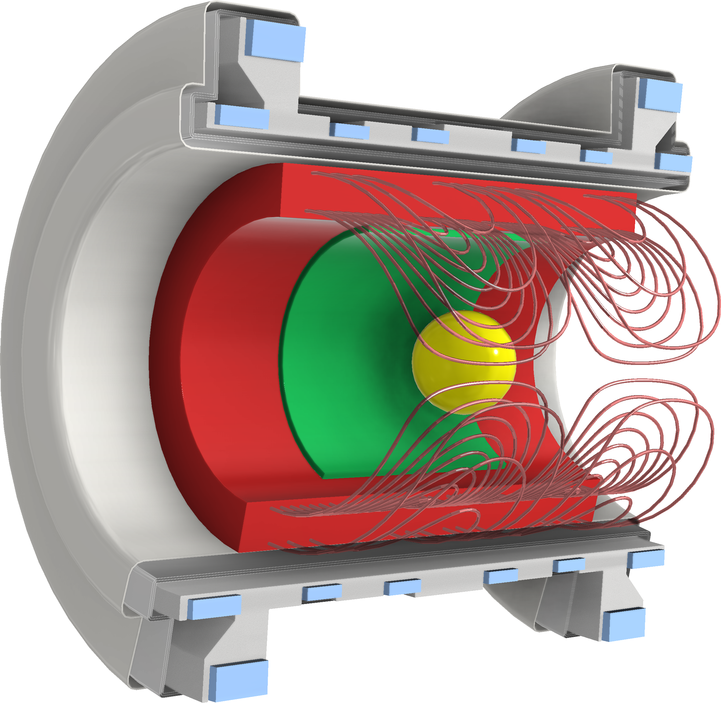
MR Imaging became possible by the introduction of switchable gradient fields that allowed imposing a spatially-dependent variation of the precession frequency on spins within the object under investigation. Gradient coils in MRI are usually designed to generate three orthogonal spatially-varying fields along the spatial dimensions x,y and z. Conventionally, the spatial variations in the z-component of these magnetic fields are designed to have a constant spatial derivative along the corresponding dimension. Linearly varying spatial encoding magnetic fields (spatial encoding magnetic fields (SEMs)) a.k.a. gradients result in a linear relation between k-space and image domain, allowing for image reconstruction via the computationally inexpensive fast Fourier transform (FFT).Whole-body gradient coils in clinical MR systems are usually able to generate gradient strengths in the range of 30-80mT/m with rise times of 75-200T/m/s. This strength and switching speed is achieved with dedicated power amplifiers which can deliver up to 1200A of current and multiple kV of voltage.
Fast switching of gradient coils may induce eddy currents within the cryostat. Due to cryogenic temperatures and associated low resistances these currents may decay over multiple seconds and generate fields which are detrimental to imaging. For avoiding eddy currents, gradient coils are magnetically screened with an additional outer shielding layer.
Oftentimes, gradient strengths and switching speeds that may technically be achieved are higher, than ones used for in vivo imaging. Whole-body imaging performance is often limited by physiological constraints set by the stimulation of peripheral nerves. This peripheral nerve stimulation (PNS) may be caused by voltages induced along the nerve fibers, strong enough to trigger discharges through ion channels. PNS manifests itself in muscle contractions or unpleasant sensations. Such unwanted stimulation which initially may appear as slight twitching, can reach unacceptable painful muscle contractions upon the increase of the gradient amplitude or switching rate. Due to the interaction between a time-dependent spatial variation of the induced voltages and individual subject anatomies, an accurate PNS prediction is complex and subject of active research.
In this talk the following topics are covered:
- Basic requirements of MRI gradient coils
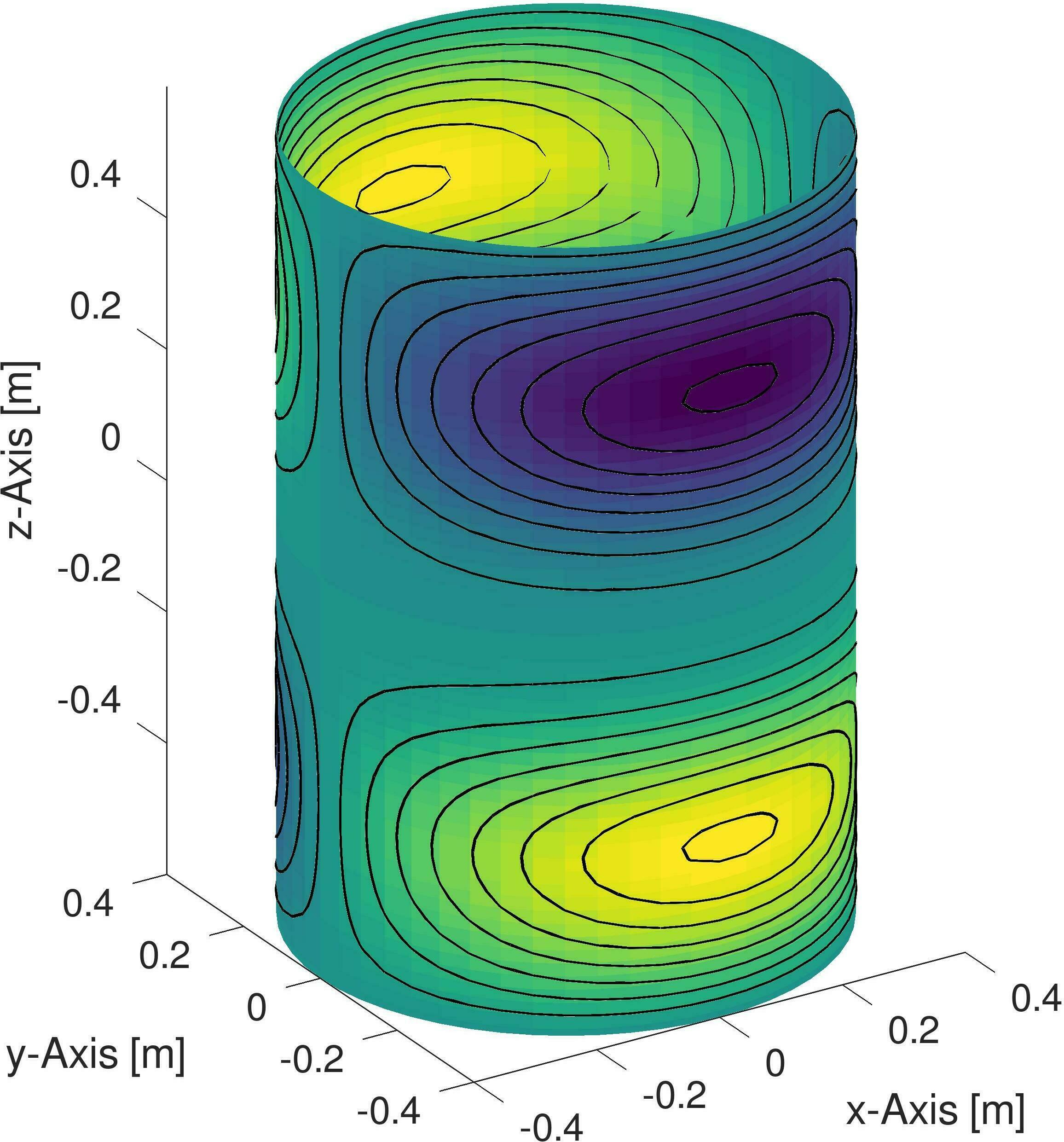
- Approaches to finding suitable wire patterns (target field method, concept of stream function method)
- Open source tools for coil design
- Additional obejectives during the optimization
- Coil implementation
- Novel gradient coil approaches
Acknowledgements
The author would like to acknowledge his colleagues Feng Jia, Philipp Amrein and Maxim Zaitsev for providing Materials for the presentation.
The following funding sources are gratefully acknowledged:
- German research council (DFG) project 468440804 "High-Power Diffusion Probe for Human Breast MRI – Phase 2"
- EU Horizon project 101078393 " MRITwins"
References
Further reading, more to follow in presentation slides:
Basic introduction to gradient coil design methods:
[1] Hidalgo-Tobon, S.S. (2010), Theory of gradient coil design methods for magnetic resonance imaging. Concepts Magn. Reson., 36A: 223-242. https://doi.org/10.1002/cmr.a.20163
Open source coil design
[2] Littin S, Jia F, Amrein P and Zaitsev M (2021) Methods: Of Stream Functions and Thin Wires: An Intuitive Approach to Gradient Coil Design. Front. Phys. 9:699468. https://doi.org/10.3389/fphy.2021.699468
[3] Amrein, P, Jia, F, Zaitsev, M, Littin, S. CoilGen: Open-source MR coil layout generator. Magn Reson Med. 2022; 88( 3): 1465- 1479. https://doi.org/10.1002/mrm.29294
Overview of recent developments of gradient coils:
[4] Gudino, N. and Littin, S. (2023), Advancements in Gradient System Performance for Clinical and Research MRI. J Magn Reson Imaging, 57: 57-70. https://doi.org/10.1002/jmri.28421
Figures