Beyond DCE: DWI & Emerging Techniques
1Radiology, Seoul National University Hospital and Seoul National University College of Medicine, Seoul, Korea, Republic of
Synopsis
Keywords: Body: Breast
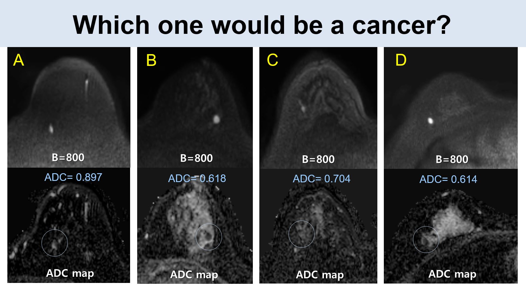
Diffusion MRI has emerged as an alternative and complementary technology for breast evaluation. It has already shown the clinical value of diffusion MRI for improving specificity leading to a decreased benign biopsy for suspicious lesions on contrast-enhanced breast MRI. In addition, studies are actively underway to evaluate its value as a stand-alone screening method. To help radiologists implement DWI in clinical practice and to inspire physicists to develop new technologies, this lecture will outline principles, standardized techniques, clinical applications, and research of diffusion MRI using ADC. Furthermore, advanced DWI techniques, including IVIM, DKI, and DTI, will be briefly reviewed.ADC quantifies mean bulk diffusion per pixel and is an established quantitative surrogate for tissue cellularity and structural features. Quantitative ADC can be used to downgrade suspicious lesions on contrast-enhanced MRI to reduce unnecessary biopsy. In the multicenter trials, 21% to 33% of unnecessary biopsy could be avoided based on DWI. In addition, DWI was found to be less sensitive to cancer detection than DCE-MRI, but DWI’s sensitivity was found to be superior to mammography or ultrasound. The cancer detection rate comparable to abbreviated MRI has been reported in one study. Thus, several prospective multicenter trials are underway to compare performance of DWI alone vs. DCE-MRI or other combinations of mammography or US for high to intermediate risk women. Stand-alone DWI screening is expected to be a valuable alternative for women of contraindications to contrast agents or intermediate-risk women.
Many studies have also demonstrated the ability of DWI using ADC, intravoxel incoherent motion (IVIM), non-Gaussian kurtosis imaging (DKI), and diffusion tensor imaging (DTI) in the characterization of breast tumors, prediction of molecular subtypes or responsiveness of neoadjuvant chemotherapy or as indicators of tumor aggressiveness or invasiveness of tumors. In the literature, significant correlations have been reported between various DWI parameters and breast cancer molecular subtype, invasive cancer (compared with in situ cancers), tumor grade, Ki-67 level, or lymphovascular invasion. In addition, the association between pretreatment ADC or changes in ADC and tumor response or pCR has also been extensively studied. However, various results have been reported depending on the MRI timing, imaging parameter measurement methods such as histogram analysis, advanced DWI modeling approaches, or definitions of responsiveness. With respect to advanced DWI techniques, IVIM, non-Gaussian DWI, and DTI metrics provide additional insights into breast pathophysiology, but they are still a field of research due to the lack of standardization of image acquisition and contradictory results.
In conclusion, the clinical value of DWI in breast is promising and has not yet been fully investigated.
Acknowledgements
No acknowledgement found.References
1. Le Bihan D, Iima M, Partridge SC. General principles and challenges of diffusion MRI. In: Iima M, Partridge SC, Le Bihan D, eds. Diffusion MRI of the breast. Elsevier; 2022:1-17.
2. Baltzer P, et al. Diffusion-weighted imaging of the breast—a consensus and mission statement from the EUSOBI International Breast Diffusion-Weighted Imaging working group. Eur Radiol 2020;30(3):1436-1450.
3. Baltzer PAT. Overview of breast DWI: diagnosis of suspicious lesions using DWI in combination with standard MRI. In: Iima M, Partridge SC, Le Bihan D, eds. Diffusion MRI of the breast. Elsevier; 2022:40-48.
4. Iima M, et al. Diffusion MRI of the breast: current status and future directions. J Magn Reson Imaging 2020;52:70-90.
5. Lee SH, Shin HJ, Moon WK. Diffusion-weighted magnetic resonance imaging of the breast: standardization of image acquisition and interpretation. Korean J Radiol 2021;22:9-22.
6. Shin HJ, Moon WK, Amornsiripanitch N, Partridge SC. Diffusion MRI as a stand-alone unenhanced approach for breast imaging and screening. In: Iima M, Partridge SC, Le Bihan D, eds. Diffusion MRI of the breast. Elsevier; 2022:86-107.
7. Zhu CR, Chen KY, Li P, Xia ZY, Wang B. Accuracy of multiparametric MRI in distinguishing the breast malignant lesions from benign lesions: a meta-analysis. Acta Radiologica 2021;62:1290-1297.
8. Rahbar H, et al. Utility of diffusion-weighted imaging to decrease unnecessary biopsies prompted by breast MRI: a trial of the ECOG-ACRIN cancer research group (A6702). Clin Cancer Res 2019;25:1756-1765.
9. Amornsiripanitch N, et al. Diffusion-weighted MRI for unenhanced breast cancer screening. Radiology 2019;293:504-520.
10. Ha SM, et al. Detection of contralateral breast cancer using DW MRI in women with newly diagnosed breast cancer: comparison with combined mammography and whole-breast ultrasound. Korean J Radiol. 2021;22:867–879.
11. Le Bihan D. What can we see with IVIM MRI? NeuroImage 2019;187:56-67.
12. Iima M, et al. IVIM and non-Gaussian DWI of the breast. In: Iima M, Partridge SC, Le Bihan D, eds. Diffusion MRI of the breast. Elsevier; 2022:116-143.
13. Kim JY, et al. Diffusion tensor magnetic resonance imaging of breast cancer: associations between diffusion metrics and histological prognostic factors. Eur Radiol 2018;28:3185-3193.
14. Partridge SC, Nissan N, Rahbar H, Kitsch AE, Sigmund EE. Diffusion-weighted breast MRI: clinical applications and emerging techniques. J Magn Reson Imaging 2017;45:337-355.
15. Sigmund EE, Furman-Haran E, Baltzer PAT, Partridge SC. Diffusion tensor imaging (DTI) of the breast. In: Iima M, Partridge SC, Le Bihan D, eds. Diffusion MRI of the breast. Elsevier; 2022:144-161.
Figures