5432
VitaLenz: A Convolutional Neural Network for the Detection of Magnetic Resonance Imaging Artifacts1Philips, Cleveland, OH, United States
Synopsis
Advances in MR acceleration techniques have produced a paradigm shift in MR productivity. In addition, the integration of artificial intelligence offers even more promise to integrate MR workflow and accelerate image acquisition. Recognizing the absence of operator assisted technologies we created VitaLenz, a convolutional neural network, to test the ability of artificial intelligence in detecting common MR imaging artifacts. VitaLenz was able to identify common MR image artifacts with high sensitivity, accuracy, and speed. Creation and use of this type of assistive technology can help ensure image quality and can also lead to faster clinical adoption of newer imaging techniques.
Purpose
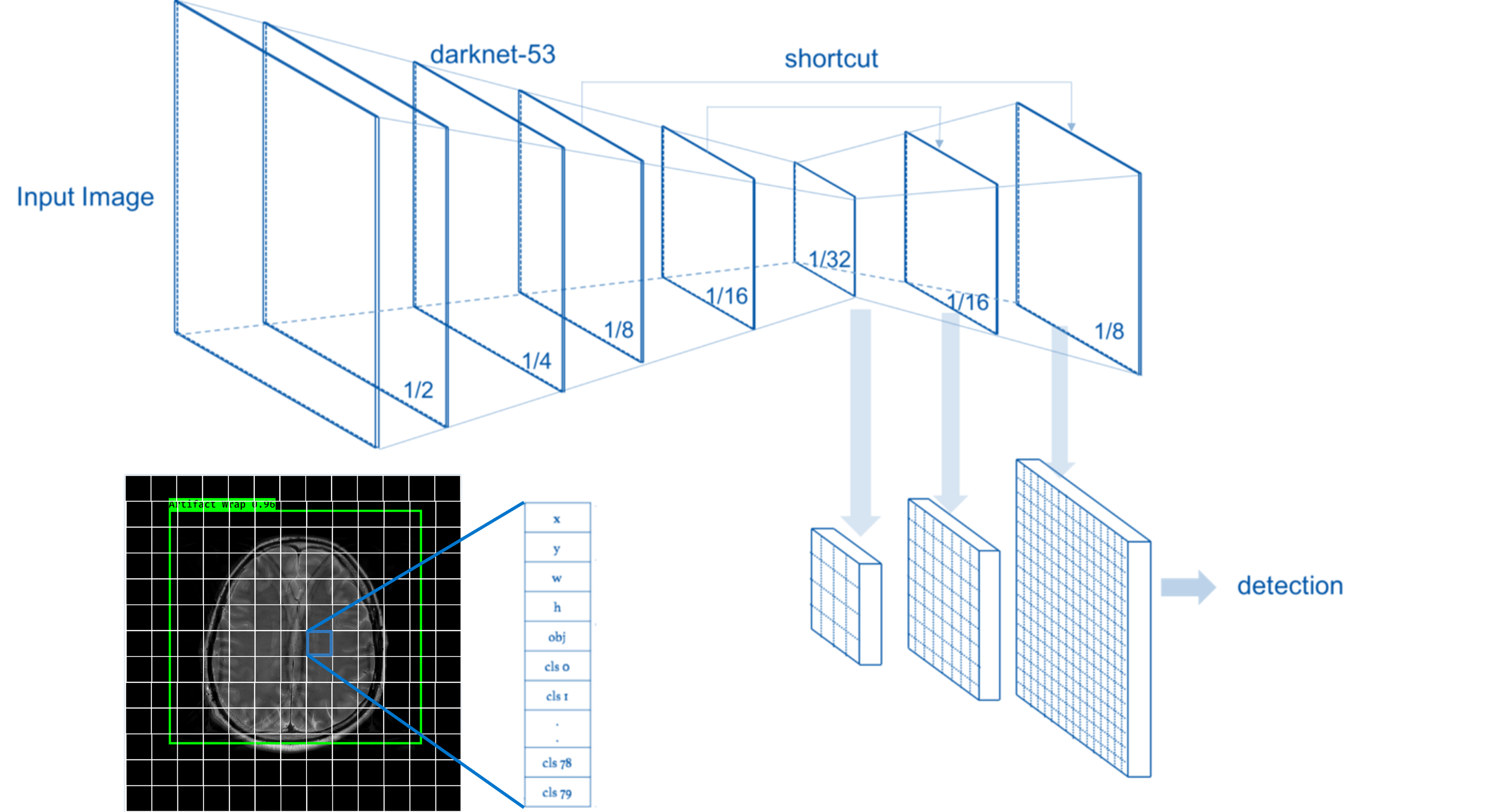
Advances in MR acceleration techniques like compressed sensing have produced a paradigm shift in MR productivity1. In addition, the integration of artificial intelligence (AI) offers even more promise to integrate MR workflow and accelerate image acquisition2. While the use of AI can drastically decrease scan times for patients and increase productivity, this presents a potential problem to ensure proper patient care. MR technologists in charge of running the scanner are faced with unprecedented challenges amid a Covid-19 pandemic that has led to staffing shortages, demand for higher productivity, increased MR safety concerns and patient documentation, all while ensuring image quality (IQ) is maintained. As we develop and introduce new technology to accelerate image acquisition and increase productivity it is imperative that we also create technology that assists the MR technologist by maintaining diagnostic IQ. Creation and use of this type of assistive technology will help ensure the patient receives a diagnostic scan and can also lead to faster clinical adoption of newer imaging techniques. Recognizing the absence of operator assisted technology we created VitaLenz, a convolutional neural network (CNN), to test the ability of AI in detecting common MR imaging artifacts in this proof-of-concept study (Figure 1).Methods
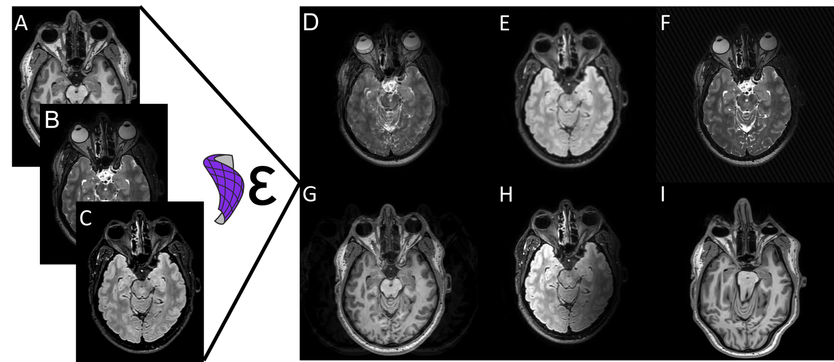
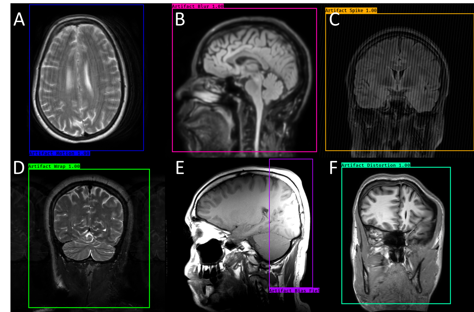
With the goal of assisting the MR technologist during scanning, a real-time object detection CNN was trained to identify MR image artifacts3. The training dataset consisted of 4606 brain images made up of three image contrasts (T1-weighted, T2-weighted, and FLAIR) and three imaging planes (axial, sagittal, and coronal). These images were augmented using TorchIO4, an open-source Python library, to produce six common image artifacts (motion, wrap, blurring, bias field, RF spike, and geometric distortion) (Figure 2). 570 test MR images5 were processed and evaluated for sensitivity, specificity, and accuracy. In addition, a subset of test images was evaluated by eight registered MR technologists with an average of 17.2 years of experience to compare VitaLenz versus a human reader.Results
Analysis of VitaLenz revealed a sensitivity of 81.25%, specificity of 66.67%, and accuracy of 74.19% (Figure 3). In comparison the sensitivity, specificity, and accuracy for the group of human readers was 85%, 86.96%, and 86.05% respectively. VitaLenz processing took on average 0.477 seconds per image to identify, label, and locate artifacts. This is almost eight times faster than the average time spent by readers to evaluate an image5.Conclusions
As advances in MR image acceleration continue to grow, it is important to also create tools that assist the technologist to ensure IQ is maintained. To this end we created and tested VitaLenz, a real-time object detection CNN. In this proof-of-concept study, VitaLenz was able to identify MR image artifacts with high sensitivity, accuracy, and speed. VitaLenz showed similar performance compared to the human reader group in sensitivity and accuracy. Although, VitaLenz showed lower specificity, its high sensitivity means that it still was able to identify images with artifacts in them that may need to be re-run or adjusted. Moreover, the speed at which the VitaLenz can evaluate images is significantly higher than that of the human readers and not subject to the fatigue of evaluating thousands of images that are acquired throughout a normal technologist’s shift. Deployment of an object detection AI solution to identify image artifacts on the scanner would allow for real-time automated assessment of IQ. This would serve to triage IQ as well as provide guidance for improving IQ when artifacts occur. AI is a powerful tool, that not only can help accelerate image acquisition, but also help maintain high IQ and produce diagnostic scans.Acknowledgements
No acknowledgement found.References
1. Mönch, Sebastian, et al. "Magnetic resonance imaging of the brain using compressed sensing–Quality assessment in daily clinical routine." Clinical Neuroradiology 30.2 (2020): 279-286.
2. Pezzotti, Nicola, et al. "Adaptive-CS-Net: FastMRI with adaptive intelligence." arXiv preprint arXiv:1912.12259 (2019).
3. Redmon, Joseph, and Ali Farhadi. "Yolov3: An incremental improvement." arXiv preprint arXiv:1804.02767 (2018).
4. Pérez-García, Fernando, Rachel Sparks, and Sébastien Ourselin. "TorchIO: a Python library for efficient loading, preprocessing, augmentation and patch-based sampling of medical images in deep learning." Computer Methods and Programs in Biomedicine 208 (2021): 106236.
5. McDonald, Robert J., et al. "The effects of changes in utilization and technological advancements of cross-sectional imaging on radiologist workload." Academic radiology 22.9 (2015): 1191-1198.
Figures