5428
The Quantitative Evaluation of Mild Traumatic Brain Injury with DSI and DTI MR Technology1Chongqing Emergency Medical Center, Chongqing, China
Synopsis
Compared with DTI (diffusion tensor imaging), DSI(diffusion spectrum imaging) has more accurate spatial resolution ability in mTBI(mild traumatic brain injury), and DSI-based fiber tracking technology has become an important tool for medium scale (mesocale) structural elucidation, building a bridge between microscopic and macroscopic scales, which provides the possibility for further exploration and integration of multi-scale analytical studies at the cellular level as well as at the subcellular level.
abstract
In this study, diffusion tensor imaging was compared with diffusion spectrum imaging to investigate the diagnostic value of this technique for microstructural changes in the brain tissue of mTBI patients.Methods
MRI Protocol:MRI exams were performed on a 3.0-T scanner (MAGNETOM Prisma, Siemens Healthcare, Erlangen, Germany) with a phasedarray 64-channel body coil. According to the inclusion and exclusion criteria, 6 patients with acute mild traumatic brain injury were collected and included in the mTBI group, and 6 healthy volunteers matched for age, gender, and handedness were recruited and included in the healthy control group during the same period.
Reconstruction:
The obtained DTI、DSI data were extracted from brain images after transforming the data format, head movement correction, removing the scalp and skull.Finally, diffusion tensor calculations were performed by the dtifit tool in the FDT toolbox.Based on the above preprocessed DTI data, we used the Tract-Based Spatial Statistics (TBSS) method provided by FSL software to perform voxel-wise statistical analysis of FA data from all study subjects, followed by MD, AD, and RD images that were also aligned to MNI space and projected onto FA framework for analysis.
Data Analysis:
Statistical analysis mainly used voxel-based independent sample t-test to test the difference between the two groups of data, and compared the difference between the whole brain FA, MD, AD, and RD values between the two groups of subjects, setting a statistical threshold of p < 0.05 and cluster size ≥100.
Results
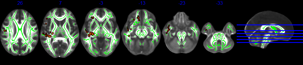
DTI showed that the MD value of the posterior limb of the right internal capsule in the case group was higher than that of the healthy control group (P<0.05). After TBSS treatment, both DSI and DTI had an effect on the posterior limb of the right internal capsule. Doctors subjectively reported that the image signal-to-noise ratio was high, the lesions showed clearly, the whole image distortion was small, and the mass area of the measureer was smaller than that of DTI.Discussion
In this study, TBSS analysis method was used, which did not require a preset region of interest (ROI), and overcame the shortcomings of voxel-basedanalysis (VBA) based analysis in the process of registration and smoothing, which could align and register the main white matter fiber tracts of different subjects, so as to achieve a more accurate intergroup comparison, so it was more accurate to locate the abnormal white matter regions and provide a reliable parameter for quantitative assessment of white matter lesions[1]. This study found that there were abnormalities in MD values in the posterior limb of the right internal capsule, and the study confirmed that [2] the pathological changes of MD values in the brain tissue after trauma were limited extracellular water diffusion caused by cytotoxic edema.In addition to fine display of cross fibers and better guidance of clinical surgery, DSI tracking technology can show the patterns of neural circuits between cerebellar cortex, deep cerebellar and brainstem nuclei, and thalamus, revealing the complex network connection of the cerebellum.Conclusion
The results showed that the proposed DSI scan protocol is feasible in clinical practice for 3.0T MRI. This modality allows precise diagnosis of mTBI in daily clinical practice, thereby improving patient outcomes.Acknowledgements
Meining Chen MR Scientific Marketing, SIEMENS Healthcare, Shanghai, Chinameining.chen@siemens-healthineers.com

References
[1] Yi Huiming, Jiangshan. Progress and application of spatial statistical methods based on tractography [J]. International Journal of Medical Radiology,2011,34(2):162-165.
[2] Marmarou Anthony et al. Predominance of cellular edema in traumatic brain swelling in patients with severe head injuries.[J]. Journal of neurosurgery, 2006, 104(5) : 720-30
Figures