5419
Comparing Dual Shimming with Average Single Shimming on Fat Suppression Techniques1Radiology and Biomedical Imaging, University of California San Francisco, San Francisco, CA, United States, 2GE Healthcare, Waukesha, WI, United States, 3Rad/PET/MRI Metabolic Service Center, Stanford University, Palo Alto, CA, United States
Synopsis
When acquiring fat suppression sequences, active shimming can be used to reduce magnetic field inhomogeneities which are inversely related to image quality and cause image artifact. Active shimming improves image quality by optimizing the homogeneity on an individual patient basis to optimize the final shimming and directly, the resulting image quality.
Background
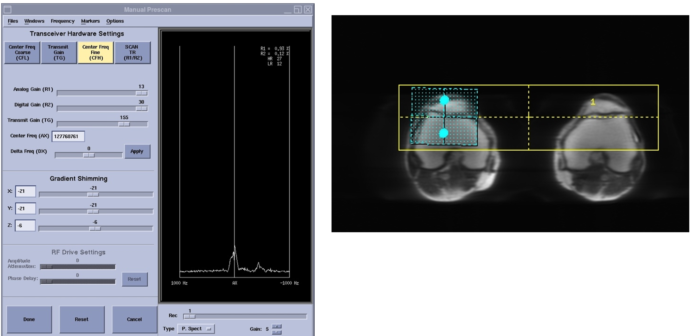
For simultaneous acquisition of bilateral knee [1a, 1c] on a PET/MR scanner (SIGNA, GE Healthcare, Waukesha, WI), shimming can be done in two ways: 1) an average of single shims, placed on each knee, can be calculated and used as manual input to increase the image quality. This is done by using the average X, Y, Z and center frequency manually for the right and left knee in fat suppression sequences[1c] ; 2) an automatic double shim on both knees during the ongoing PET/MR scans can be used to reduce timing and avoid any calculation error.[2c , 2d]Methods
In this study, a musculoskeletal radiologist with 14 years of experience, evaluated sequence performance in terms of spatial distortion, blurring, shading, and intensity loss for both shimming methods (using the average shimming of each knee and automatic shimming across both knees) on a PET/MR scanner. A Likert scale score (1-to-5) was assigned to each of the reviews.Results
We concluded that the two methods are similar, and no significant scoring difference was observed in terms of the image quality in the above mentioned metrics. Therefore, to reduce timing and avoid any subjective error during calculation, the automatic double shim method is preferred during PET/MR scans of the knees.Conclusions
To improve image quality, an average of single shimming is often used by optimizing the homogeneity of magnetic field. However, calculating averages on single shims can sometimes cause errors and is time consuming. Using automatic double shimming improves image quality, while reducing time and human error by avoiding manual calculations.Acknowledgements
We would like to thank Sharmila Majumdar, Ph.D. for her guidance and support on this project also, the assistance provided by Ms. Roya Habibi was greatly appreciated.References
1. F. Kogan, E. Levine, A, Chaudhary, et al., “Simultaneous Bilateral Knee MR Imaging,” Magn Reson Med. 2018 Aug; 80(2): 529–537.
2. https://archive.ismrm.org/2019/1310.html
3. Fillmer A, et al., Constrained image based B0 shimming accounting for “local minimum traps” in the optimization and field inhomogeneities outside the region of interest. MRM 2015; 73:1370-1380.
Figures
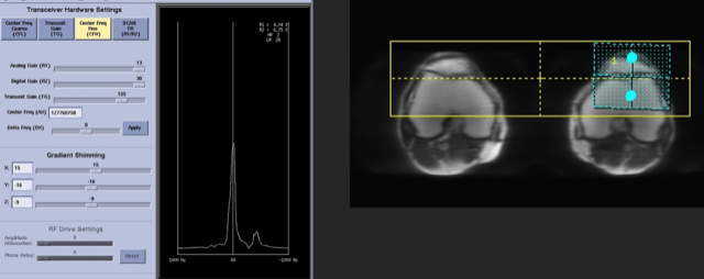
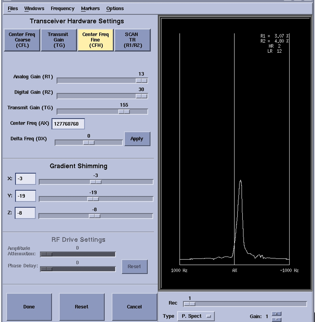
2a. Image of bilateral knees with dual shimming
2b. The gradient shim values and the center of frequency for automatic left knee shim (Volume 1)
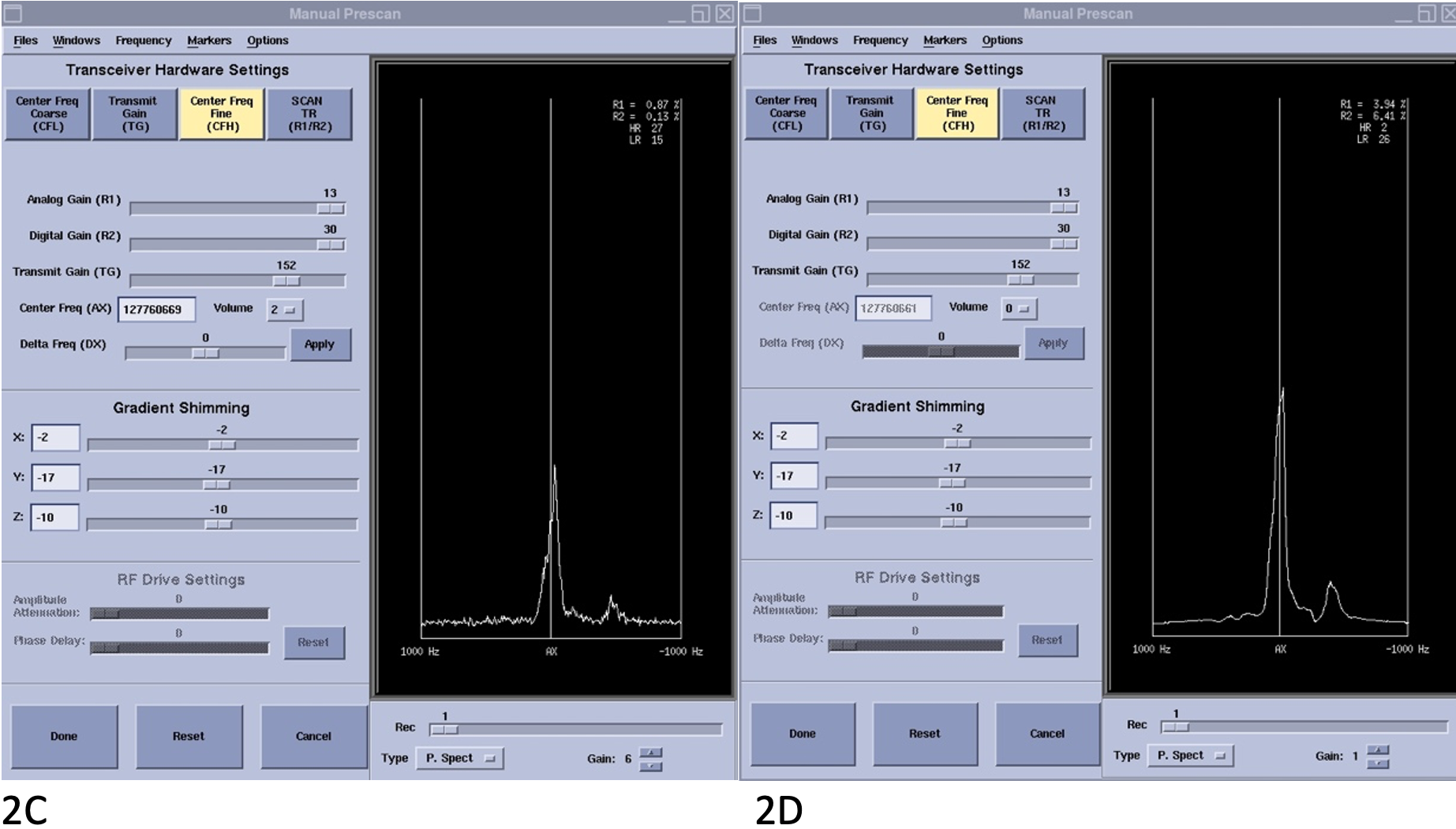
2c. The gradient shim values and the center of frequency for automatic right knee shim (Volume2)
2d. The gradient shim values and the center of frequency for automatic dual shimming (Volume 0)