5412
Dog breed size versus brain size and its inferences, and canine pathology case studies
Shiami Delina Luchow1
1MRI, Hunter Medical Research Institute/University of Newcastle, New Lambton Heights, Australia
1MRI, Hunter Medical Research Institute/University of Newcastle, New Lambton Heights, Australia
Synopsis
Colloquially intelligence is correlated to brain size. Although not accurate in humans, is this true in dogs? Dogs are diverse in shapes and sizes and are bred for their unique abilities and behavioural characteristics. However, during clinical MRI examinations, it was clear that although the body mass ranged from approximately 1kg to over 100kg in different breeds, the brain size did not vary drastically. This study will analyse the neurocephalic index of different breeds of dogs to evaluate if this correlates with the dog breeds’ unique abilities.
Introduction
Colloquially intelligence is correlated to brain size. Although not accurate in humans, is this true in dogs? Dogs are diverse in shapes and sizes and are bred for their unique abilities and behavioural characteristics. However, during clinical MRI examinations, it was clear that although the body mass ranged from approximately 1kg to over 100kg in different breeds, the brain size did not vary drastically. This study will analyse the neurocephalic index of different breeds of dogs to evaluate if this correlates with the dog breeds’ unique abilities.Method
The animal referral and emergency hospital in Newcastle, Australia refers small animals for clinical MRI scanning at the MRI suite at Hunter Medical Research Institute. Twenty of these dog MRI brain studies were chosen to analyse the neurocephalic index of each dog. The dogs were scanned on a Siemens, 3T Prisma scanner (Erlangen, Germany) and the T2 transverse fast spin echo with TR 3410ms, TE 56ms, and slice thickness of 2.5mm and no gap, was used for the measurements.Neurocephalic ratio is generally used to describe skull morphology and the slice with the largest area at the level of the parietal lobes was chosen for the measurement. The following formula was used for the calculation:
Neurocephalic index = (biparietal diameter/occipitofrontal diameter) x 100
Discussion
Dogs have long been man’s best friend and MRI is increasingly used for their clinical management. This gives a unique opportunity to understand the normal dog brain morphology of different breeds as well as evaluating pathology. Dogs are often chosen by their owners for the breed’s unique characteristics and do these characteristics correlate with brain morphology? The neurocephalic index was calculated for each dog and although head size differed remarkably between different breeds, the brain size was fairly consistent. This is similar to the findings by Hecht et al, 2019, where brain size did not proportionate to the body size. While the brain remained a similar size to a smaller dog, the temporalis muscle was much larger in bigger breeds resulting in the larger head size. The percentage difference from the largest to the smallest neurocephalic index was 31%. However this difference was within the small to medium breeds and not small to large breed size. Does this mean smaller dogs are more intelligent? Table 1 lists most common characteristic for each breed and they are varied and serves different purposes. Is this due to the selective breeding by humans and change of neuroanatomical characteristics as discussed in Hecht et al. Hence, brain size alone may not be a great indicator of the level of cognition and behaviour traits. Detailed analysis of the brain morphology with segmentation of different brain regions will be useful, which is the next step for this project.Case studies
MRI is a valuable tool in the clinical management of pet animals and it is the gold standard for assessing the central nervous system and musculoskeletal system in small animals. Below are several case studies that illustrate the usefulness of MRI in a diagnosis of cranial pathology.Case study 1:
- Three-year-old English bulldog presented with seizures and queried space occupying lesion.
- Slight increase in T2 signal in the right basal ganglia region seen on the T2 FSE and FLAIR sequences. Contrast enhancement seen in this region and the adjacent meninges. No space occupying lesion.
- Diagnosis – Meningoencephalitis
- Outcome – the dog successfully treated with immunosuppressants and remains well. Without MRI the dog may have been euthanised for suspected brain tumour.
Case study 2:
- Nine-year-old bulldog presented with decerebellate posture.
- Inflammation and fluid seen in the inner ear and optic nerves in T2 and FLAIR images as well as enhancement post contrast.
- Diagnosis – Otitis interna, optic neuritis.
- Outcome – the dog successfully treated.
Case study 3:
- Nine-year-old bulldog presented with epilepticus for 36 hours.
- T2 hyperintense space occupying lesion involving right temporal and parietal lobes causing midline shift. Diffusion restriction seen in this region with corresponding decrease in ADC.
- Diagnosis – neoplasia/lymphoma with non-haemorrhagic infarct.
- Outcome – owner considering chemo and radiation therapy.
Case study 4:
- Five-year-old golden retriever presented with seizures.
- Heterogeneous lesion in the left fronto-parietal region with high and low T2 signal with surrounding oedema and high signal in T1, indicating recent haemorrhage with corresponding susceptibility in SWI. Contrast enhancement of the lesion post contrast.
- Outcome – owner considering euthanasia.
Soft tissue contrast, resolution and techniques available in MRI in unparalleled when imaging the central nervous system and this can offer valuable diagnostic data for veterinary imaging for clinical management and owner peace of mind. At times it can lead to great outcomes such as the example in the first case study.
Acknowledgements
Colleagues at HMRI Imaging Centre
Staff at AREC particularly vet surgeon Julian Lunn
References
Hecht et al, Significant neuroanatomical variation in domestic dog breeds, The Journal of neuroscience, September 25, 2019: 39(39) 7748-7758Figures
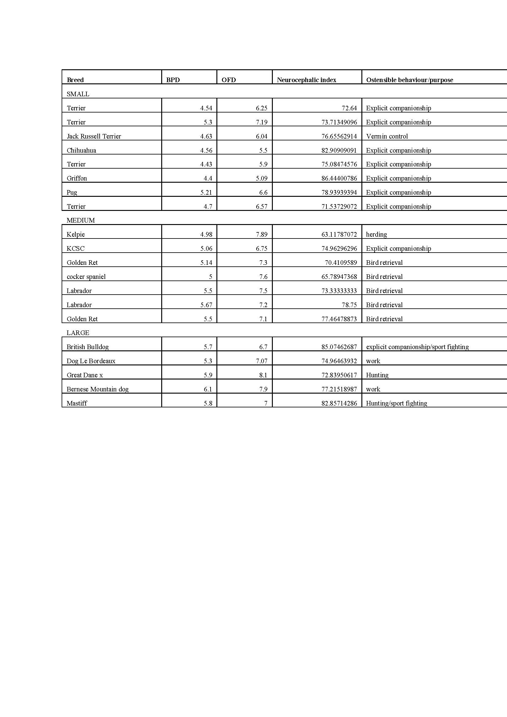
table 1: Results - Breed type, Neurocephalic index and breed characteristics.
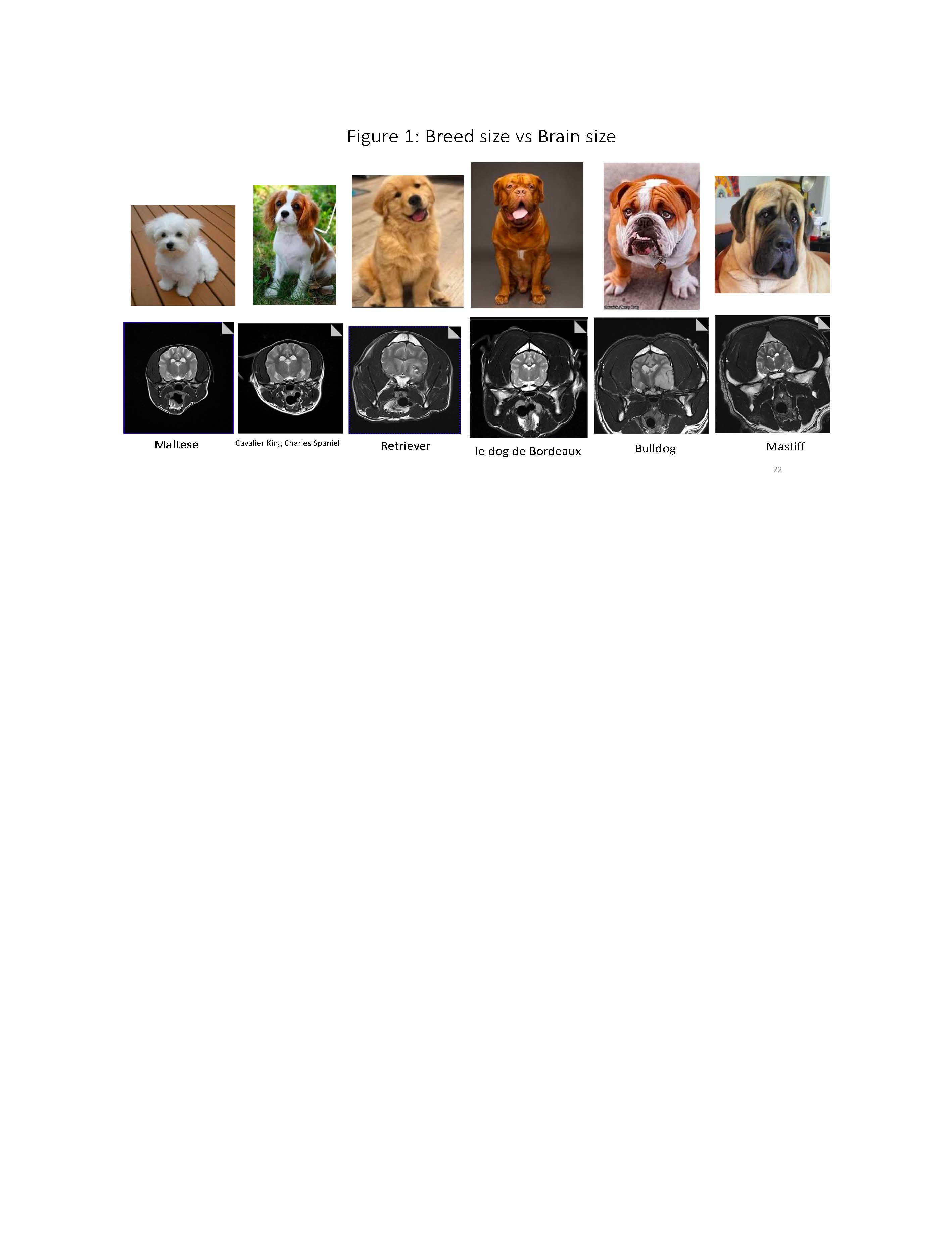
Figure 2: Dog breed vs brain size
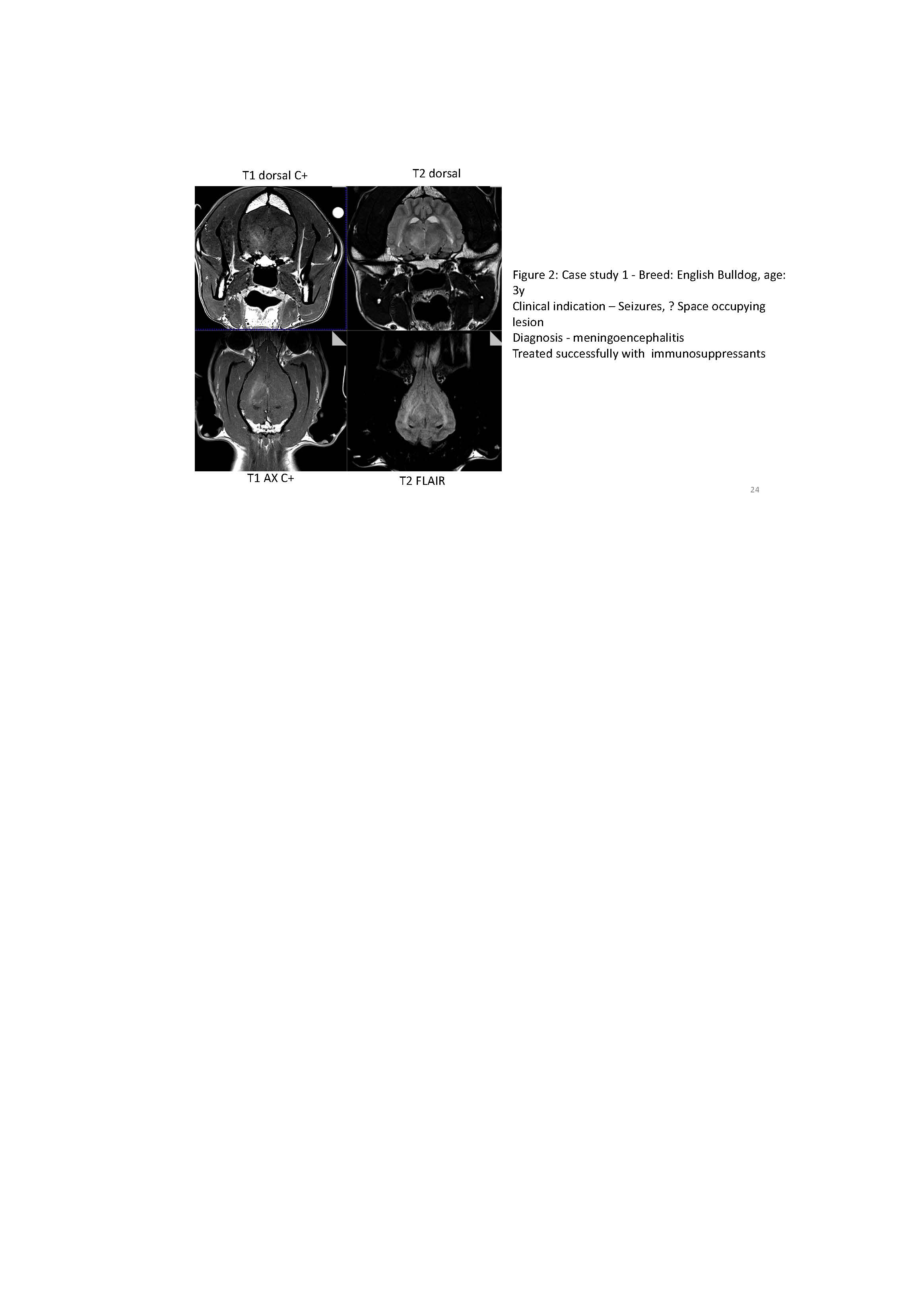
Figure 3: case study 1
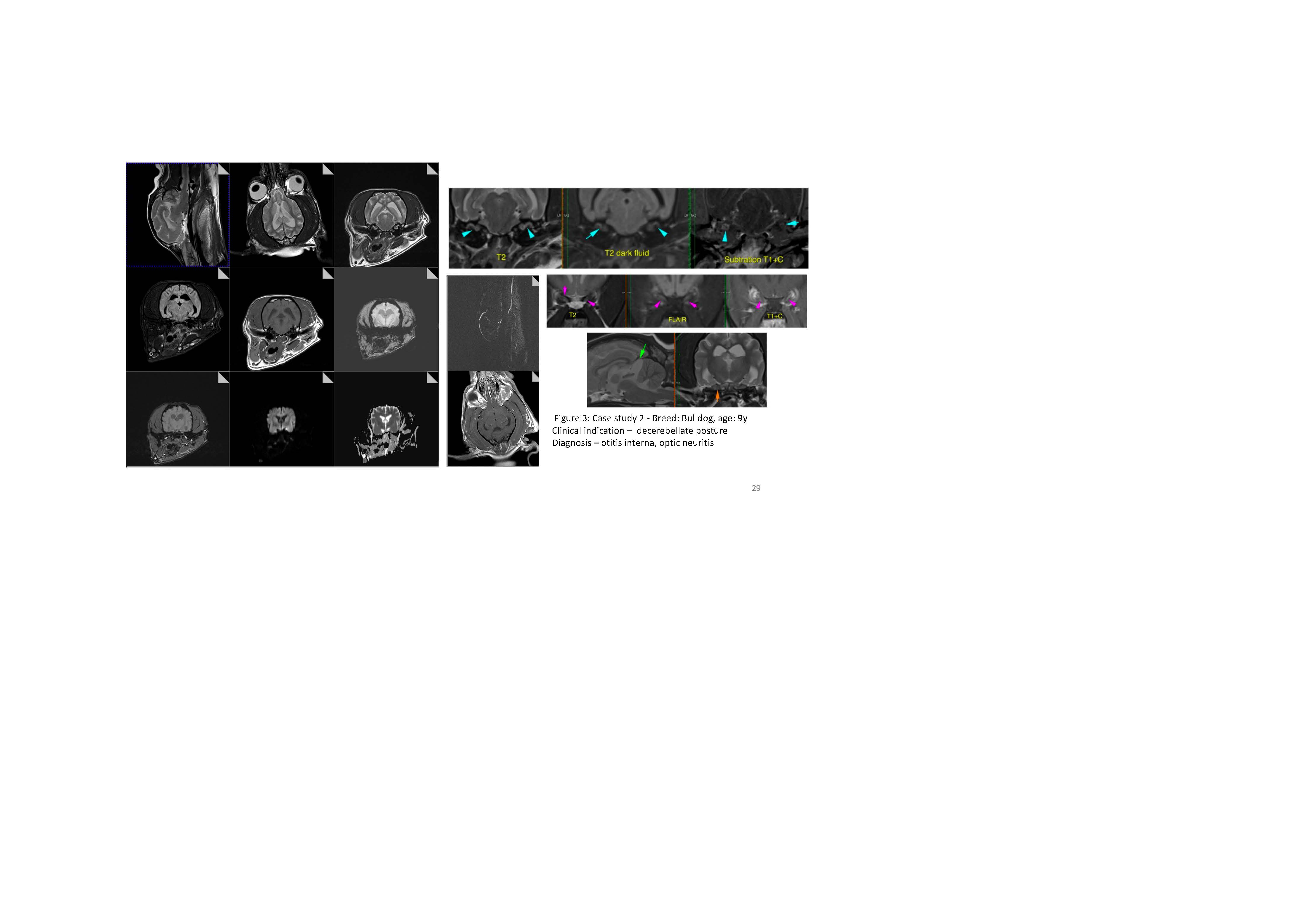
Figure 4: case study 2

Figure 5: case study 3 and 4
DOI: https://doi.org/10.58530/2023/5412