5405
Is Veterinary MRI feasible in a human facility?1MRI, Hunter Medical Research Institute/University of Newcastle, New Lambton Heights, Australia, 2HMRI Imaging Centre, Hunter Medical Research Institute/University of Newcastle, New Lambton Heights, Australia
Synopsis
Magnetic resonance imaging is continuing to grow for clinical diagnosis in veterinary practice. Although MRI is the gold standard for imaging the central nervous system and musculoskeletal pathology in animals, the use of MRI is limited due to the difficulty in accessing and the higher running cost. It is a limited resource disadvantaging many vet animals and their owners of care and cost. Is it feasible for a human MRI facility to image animals? This paper discusses how this was achieved at the Hunter Medical Research Institute Imaging Centre, Newcastle, Australia, and the safety procedures that were necessary for approval.
Introduction
The local Animal Referral & Emergency Centre (AREC) approached the HMRI-IC for possible MRI services for their small animals, particularly cats and dogs. This is favourable for AREC as the animals were generally transported 2 hours each-way to the nearest major city for diagnostic MR services which was inconvenient and costly. This would also support the running cost of MR facility, and provide a unique and needed service for domesticated animals and their carers. These initial discussions lead to a year-long approval process as the facility provided high-end human research imaging for the HMRI and the university of Newcastle (UoN). The major concerns were safety for human participants from the animals and disease/allergens and safety for staff. Additionally, the presence of small animals in a research institute would raise ethical questions about the work being conducted.Method and Discussion
A standard operating procedure (SOP) document was drafted taking the following considerations into account:- Companion animals may represent a vector for certain infectious disease as well as a trigger for individuals with specific allergic conditions. With appropriate hygiene practices this risk would be minimal to staff and users of the MRI facility. To date, there is no evidence to support the role of companion animals in the transmission of multi-resistant bacterial infections in Australia. However, appropriate protocols should be adhered to minimise the risk of accidental transmission. Jutzeit 2019 reported that MRI scanner used for both dogs and humans was routinely cleaned after animal scanning, there was substantially lower bacterial load compared with scanners used exclusively for humans (Jutzeit, 2019).
- A question regarding allergy to animals were included in the human MRI safety questionnaire.
- All veterinary staff attending the MRI exam must complete MRI safety training prior to attending and can only enter zones 3 and 4 accompanied by a radiographer.
- All MRI personnel potentially exposed to animals complete the Laboratory Animal Allergy Workers Questionnaire and consult the Animal Hazards Procedure. Appropriate Personal Protective Equipment (PPE) will also be worn to minimise exposure to animal allergens.
- Animal scanning only occur after hours or on a designated weekday after human scanning is conducted. A period of 8 hours elapse between the last animal scan and next human scan.
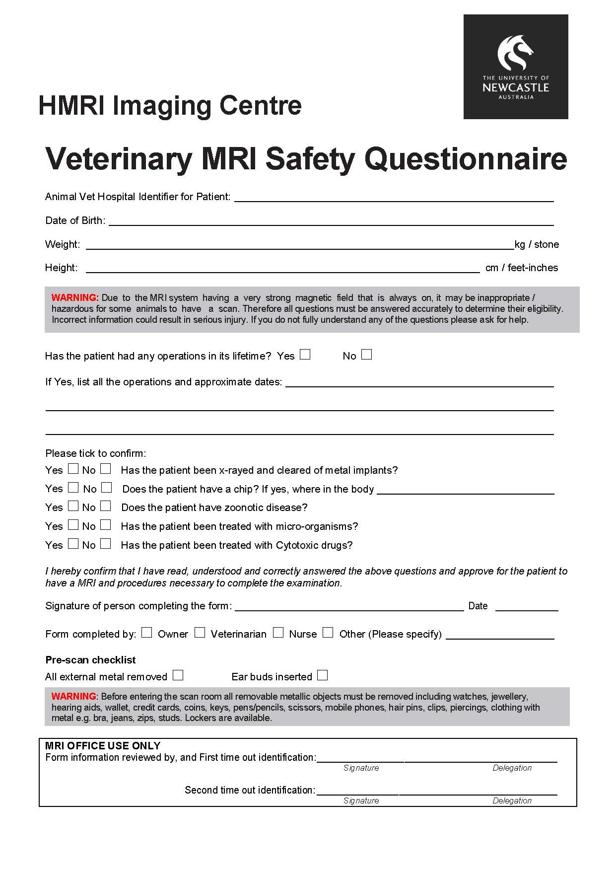
- Animals confirmed as metal free by a full body x-ray conducted by veterinary staff prior to being booked into the MRI.
- Animals are under the direct care and supervision of a licensed veterinarian at all times, and they are confirmed free of known infectious diseases, ectoparasites, and other external contaminants (e.g. soil, urine, faeces).
- Vet MRI safety questionnaire completed and signed prior to the examination.
- All animal preparation (including sedation but excluding anaesthesia), clipping for cannula access and ECG placement are to be performed at the veterinary surgery prior to transportation to the MRI suite.
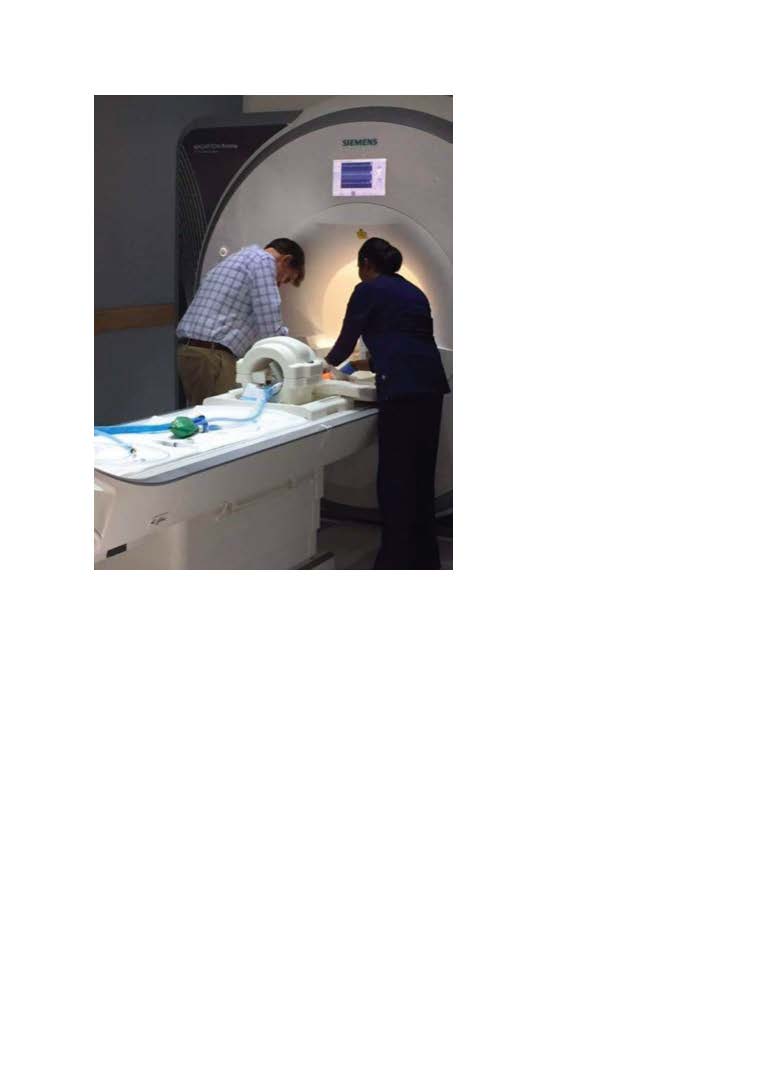
- Animal enters via a separate entrance at the back of the facility to minimise exposure and moved to the MRI-safe trolley for anaesthesia.
- The anaesthetic equipment remains outside the magnet room with MRI compatible tubing passed through the waveguide connecting to the mask which is applied to the animal.
- Taped-on earbuds applied to the animal to protect against MRI generated noise.
- MRI-safe trolley and the scanner table are covered with an impermeable drape topped with an absorbent pad for the collection of body fluids. The animal restrained with straps.
- The animal monitored whilst in the scanner using pulse oximetry, heart rate and ECG. A separate pulse sensor used for the animals.
- All areas touched by animals or animal equipment wiped down with hospital grade sanitiser in the MRI Suite. Hygiene and Infection Control
- Disposable absorbent pads used to minimise direct contact of animals with surfaces which come into contact with human patients.- Animals known to have common zoonotic diseases not enter the suite unless all personnel involved have been properly trained.
- Personnel must wear PPE when handling animals and during the imaging session. Gloves removed, and hands washed before touching control panels, video equipment, telephones, doorknobs, elevator buttons, or other objects in shared spaces, including the control room.
- All drapes removed and disposed of in the clinical waste bins, and the scanner bed and coils cleaned with standard hospital disinfectant.
- No human patients/participants to enter the MR Suite until it is cleaned and sufficient time has elapsed for room air change to have been completed. The facility air conditioning system was fitted with high-efficiency particulate absorbing filters which capture pollen, dirt, dust, moisture, bacteria (0.2-2.0 μm), virus (0.02-0.3 μm), and submicron liquid aerosol (0.02-0.5 μm) and maintained according to industry standards.
- waste generated is disposed of as clinical waste immediately after the conclusion of the animal scan and cleaned. All animal procedure waste is bagged and removed with the animal.
The SOP was cleared by the the UoN and HMRI health and safety committees and, inspection and risk assessment were performed, granting approval for scanning animals. The facility has been successfully scanning animals for number of years without incident or negative outcomes for human participants, providing valuable diagnostic information to the veterinary colleagues and pet owners.
Acknowledgements
Colleagues at HMRI Imaging Centre and UON health and safety staff.
Staff at AREC, particularly vet surgeon Dr Julian Lunn.
References
1. Berns G et al, Clinical findings in dogs trained for awake MRI, Frontiers in Veterinary Science, August 2018: Vol 52.
2. Gutzeit G et al, Would it be safe to have a dog in the MRI scanner before your own examination? A multicentre study to establish hygiene facts related to dogs and men, European Radiology, 2019 Feb;29(2):527-534
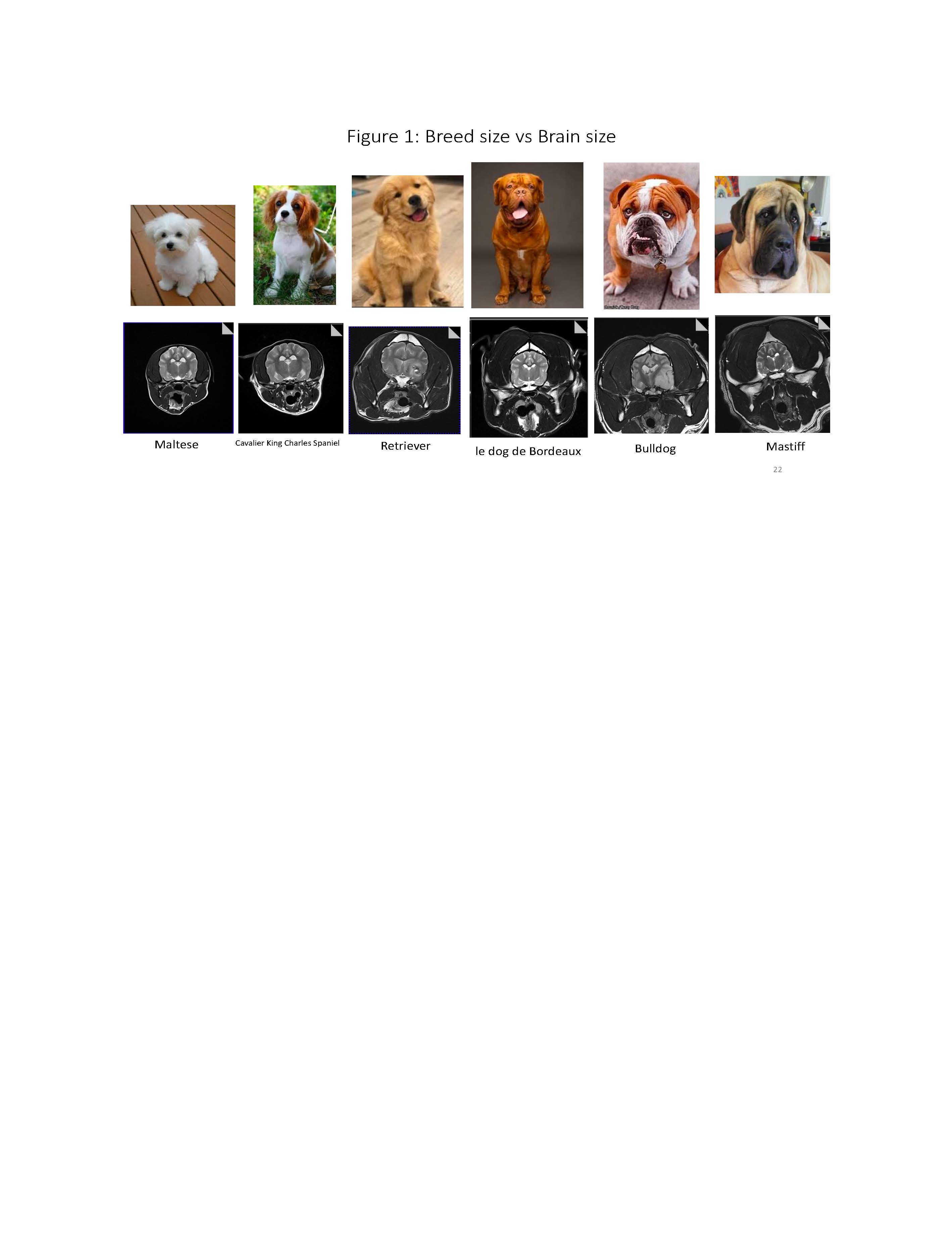
Figures