5400
Longitudinal Clinical Study of Patients with Iron Rim Lesions in Multiple Sclerosis1Clinical Neurology, University of Nottingham, Nottingham, United Kingdom, 2Princess Nourah bint abdulrahman University, Riyadh, Saudi Arabia, 3University of Nottingham, Nottingham, United Kingdom, 4School of Medicine, Faculty of Health and Medical Sciences, Taylor’s University, Taylor's, Malaysia, 5Department of Neurology, Cooper Neurological Institute, Cooper Neurological Institut, Camden, PA, United States, 6Queen's Medical Centre, Nottingham, United Kingdom, 7Clinical Neurology, University of nottingham, Nottingham, United Kingdom
Synopsis
Iron rim lesions in Multiple Sclerosis disability
Background
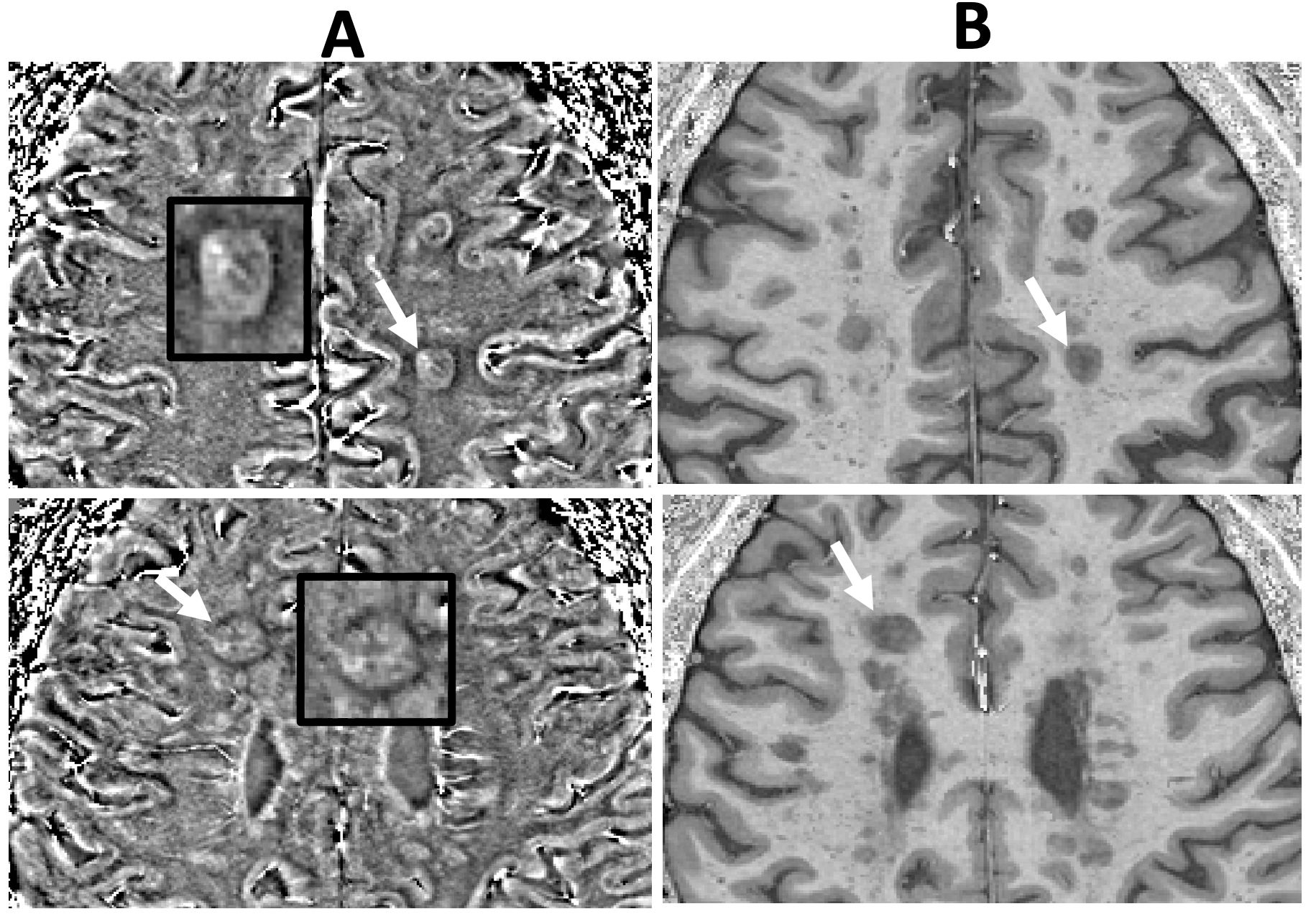
Iron rims (IR) surrounding white matter lesions (WML) are suggested to predict a more severe disease course. Only small longitudinal cohorts of patients with and without IRL have been reported so far.Aim
To assess whether the presence and number of iron rim lesions (IRLs) in patients with clinically isolated syndrome (CIS) and multiple sclerosis (MS) are associated with long-term disability or progressive disease.Material and Method
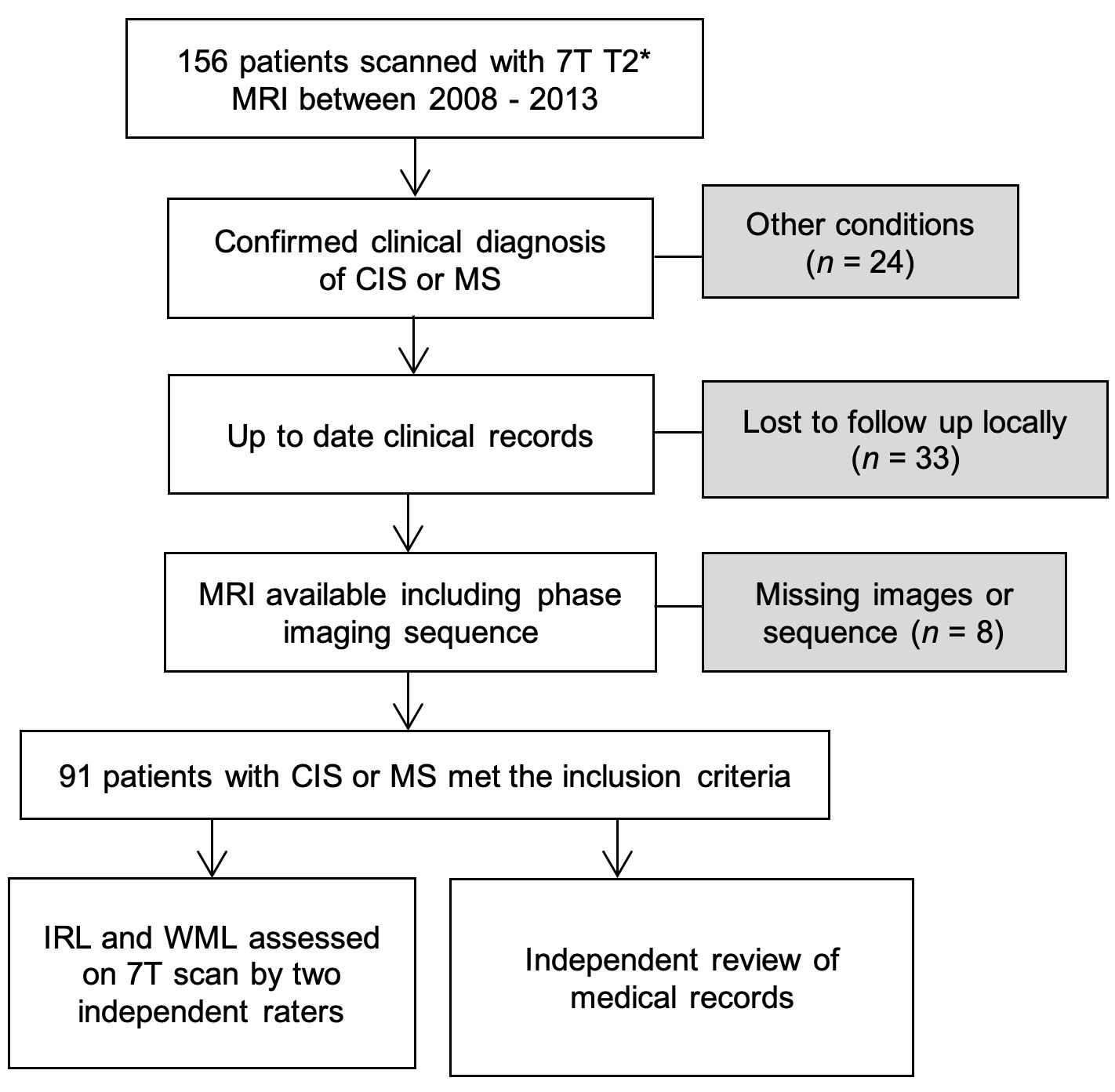
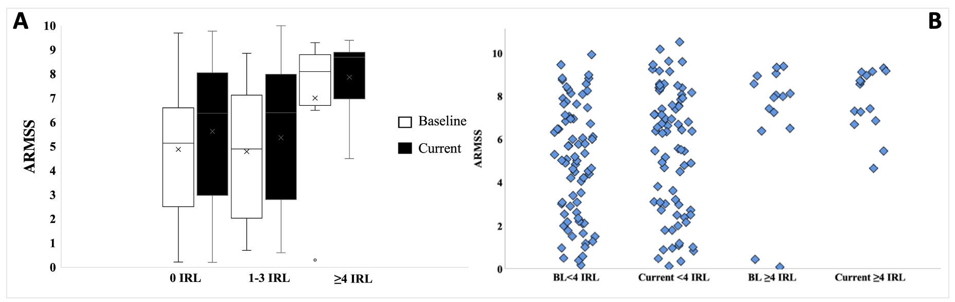
Ninety-one CIS/MS patients were recruited between 2008 and 2013 and scanned with 7T MRI. Age-Related Multiple Sclerosis Score (ARMSS), was calculated at the time of scan and at the latest clinical follow-up after 9 years. WMLs were assessed for the presence of IRL using SWI-filtered phase images.Results
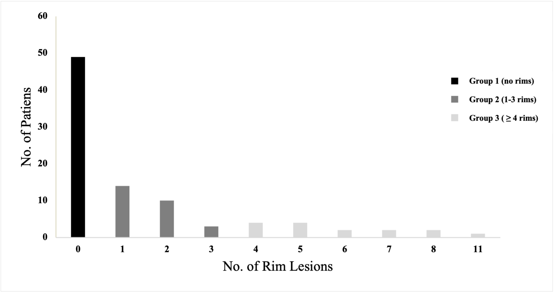
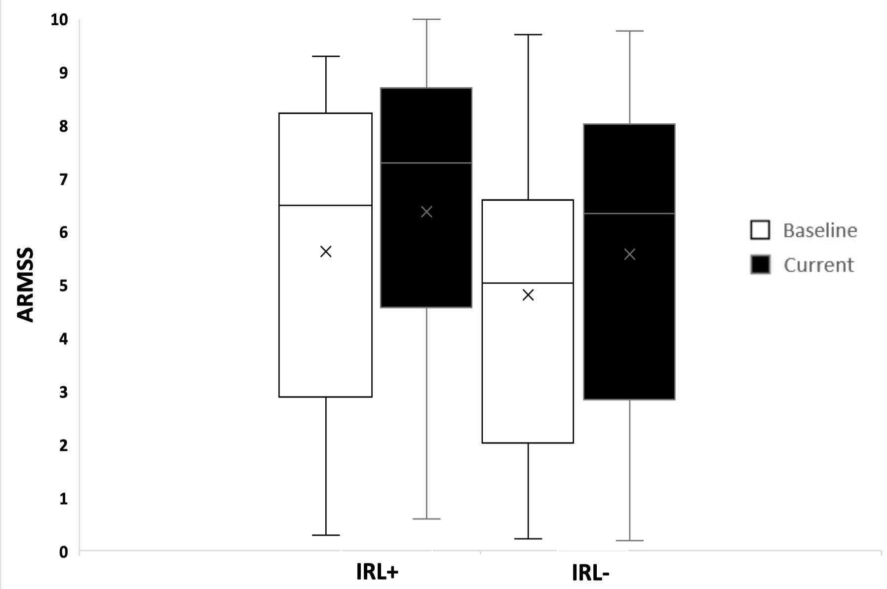
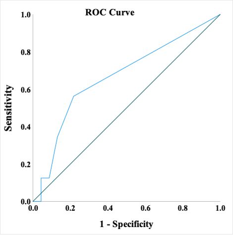
132 IRLs were detected in 42 patients (46%). 9% of WMLs had IR. 54% of the cohort had no rims, 30% had 1-3 rims and 16% had ≥4. Patients with IRL at baseline had a higher EDSS and ARMSS. Presence of IRL was also a predictor of long-term disability, especially in patients with ≥4 IRL. IRLs have a greater impact on disability compared to the WML number and volume.Conclusions
The presence and number of perilesional IR at a baseline scan hold prognostic value for long-term clinical disability in MS.Acknowledgements
Potential Conflict of Interests The author(s) declared the following potential conflicts of interest with respect to the research, authorship and/or publication of this article: A.I.A., A.H, A.A., SY.L., and O.M. nothing to declare. C.A. has received speaker honorarium from the Multiple Sclerosis Academy. R.F. has participated on advisory boards, received travel grants and attended educational programmes sponsored by Roche and Novartis. C.S.C. has received grants, personal fees, and nonfinancial support from Biogen; personal fees and nonfinancial support from Novartis, Teva, Merck, and Sanofi Genzyme. N.E. is a member of the advisory board for Biogen, Merck, Novartis and Roche; he has received grant income from the MS Society, MRC, PCORI and NIHR.References
1 Dal-Bianco, A, Kolbrink, S, Pusswald, G, Grabner, G, Kronnerwetter, C, Reiter, et al. Do 7T observed iron rim lesions in patients with multiple sclerosis serve as a marker for neuropsychological deficits? Mult. Scler. J. 2019, 25, 732.
2 Dal-Bianco A, Grabner G, Kronnerwetter C, Weber M, Höftberger R, Berger T, et al. Slow expansion of multiple sclerosis iron rim lesions: pathology and 7 T magnetic resonance imaging. Acta Neuropathol. 2017 Jan;133(1):25-42.
3 Altokhis AI, Alotaibi AM, Felmban GA, Constantinescu CS, Evangelou N. diagnostics Iron Rims as an Imaging Biomarker in MS: A Systematic Mapping Review. Diagnostics. 2020; Available from: www.mdpi.com/journal/diagnostics
4 Dal-Bianco A, Grabner G, Kronnerwetter C, Weber M, Kornek B, Kasprian G, et al. Long-term evolution of multiple sclerosis iron rim lesions in 7 T MRI. Brain. 2021 Apr 12;144(3):833-847.
5 Dal-Bianco, A, Grabner, G, Kronnerwetter, C, Weber, M, Berger, T, Leutmezer, F, et al. Iron rim lesions in multiple sclerosis at 7 Tesla magnetic resonance imaging: A 7 year prospective longitudinal study. Mult Scler. J. 2019, 25, 50–5.
6 Mehta V, Pei W, Yang G, Li S, Swamy E, et al. (2013) Iron Is a Sensitive Biomarker for Inflammation in Multiple Sclerosis Lesions. PLoS ONE 8(3): e57573. doi:10.1371/journal.pone.0057573. / CC BY 2.5
Figures