5393
RA synovitis segmentation based on unsupervised learning and TIC signal data on DCE-MRI
YiJun Mao1,2, Wanxuan Fang2, Yujie An2, Hiroyuki Sugimori1, Shinji Kiuch3, and Tamotsu Kamishima1
1Faculty of Health Sciences, Hokkaido University, Sapporo, Japan, Sapporo, Japan, 2Graduate School of Health Sciences, Hokkaido University, Sapporo, Japan, Sapporo, Japan, 3AIC Yaesu Clinic, Tokyo, Japan, Tokyo, Japan
1Faculty of Health Sciences, Hokkaido University, Sapporo, Japan, Sapporo, Japan, 2Graduate School of Health Sciences, Hokkaido University, Sapporo, Japan, Sapporo, Japan, 3AIC Yaesu Clinic, Tokyo, Japan, Tokyo, Japan
Synopsis
Keywords: Rheumatoid Arthritis, DSC & DCE Perfusion The volume of synovitis change is one of the most important pathological features of rheumatoid arthritis. By quantitative analysis of the enhancement of synovitis, we can define the degree of the disease, and determine the treatment and diagnosis. Considering the time-consuming of manual outlining and visual assessment, this study uses machine learning methods to conduct quantitative analysis of TIC, and proposes an unsupervised learning method with excellent results, which is expected to be an alternative for the gold-standard manual synovitis contour outlining.
Background or Purpose
Dynamic contrast-enhanced magnetic resonance imaging (DCE-MRI) provides a lot of important clinical information for the inflammatory degree of rheumatoid arthritis (RA)[1]. At the same time, research shows that the volume of synovitis has a strong relationship with the degree of RA, and correct quantification of the amount of pannus can help to evaluate the disease and treatment status of RA patients[2]. Early diagnosis and disease control based on quantitative results can effectively prevent the possibility of further joint injury and disability[3]. Time – intensity curve (TIC) is one of the most obvious and important features in DCE-MRI. In recent years, experiments on the shape analysis of TIC to quantify synovitis of RA have been carried out. Among them, the type 4 TIC shape has been confirmed as synovitis’ features, which obtain the obvious signal enhancement in the early stage, followed by washout phase[4]. Therefore, inflammatory activity can be estimated by quantifying the increase of synovitis signal intensity on DCE-MRI. To automatically quantify synovitis in patients with rheumatoid arthritis on DCE-MRI, TIC shape classification based on Resnet-50, TIC data classification based on one-dimensional convolutional neural network, and TIC signal data clustering based on unsupervised learning, three automatic measurement methods were compared. This study mainly focuses on the synovitis segmentation based on unsupervised learning, which is evaluated as an alternative for the gold-standard manual synovitis contour outlining.Methods
In this study, the TIC data was used to train a classifier. Our TIC data was manually done by an experienced MSK radiologist on 3T DCE-MRI of 26 cases with different phases. The obtained TIC data can be ground in bones, muscles, and synovitis. In all three methods, the method of TIC shape classification based on Resnet-50 is very time-consuming, and due to the large amount of noise in DCE-MRI, the curve shape is difficult to be well classified. The method of TIC data classification based on one-dimensional convolutional neural network is easy to over fit and requires a lot of manual labeling, but the results are not accurate enough. Therefore, a lightweight unsupervised learning method is proposed to solve the above problems. In pre-processing, a median filter is used to remove isolated noise and preserve tissue edges. Phase only correlation (POC) is used to correct the position difference between phases. In view of the various sampling phase number and sampling time interval of DCE-MRI data, linear interpolation is used to upsample TIC data with the same standard of time interval, and redundant time series data is trimmed. To increase the characteristics of high signal synovitis data, a power function mapping is used to increase the variance between the target category and other categories. The fuzzy c-means (FCM) clustering method is used to train a classifier with the data after dimension reduction based on the principal component analysis (PCA), which can improve the classification accuracy. Compared with k-means method, the membership grade is considered in the classification, which can improve the accuracy. Comparing with the gold-standard manual synovitis contour outlining, cross-validation was used to evaluating the segmentation performance of three models.Results
Quantitative and qualitative evaluations on 112 joint regions of interest show that our unsupervised learning method improves the segmentation accuracy and speed significantly. (Dice: 0.772±0.083, Accuracy:0.992±0.008, Sensitivity:0.751±0.094, Specificity:0.998±0.001, Training and prediction time: less than 1 second) In contrast, the other two methods are difficult to evaluate due to the unclear boundary segmentation of the results after visual evaluation and the large difference between the quantitative and manual annotation of synovitis.Conclusion
Compared with the previous classification methods of TIC shape analysis and pharmacokinetic modeling on DCE-MRI, this study, as a prospective study, focuses on the TIC data itself and suggests that unsupervised learning is a fast and accurate classification method for RA synovitis segmentation, which is expected to replace manual synovitis contour outlining for quantitative analysis and to be optimized for clinical application.Acknowledgements
No acknowledgement foundReferences
1 Cimmino MA, Innocenti S, Livrone F, Magnaguagno F, Silvestri E, Garlaschi G. Dynamic gadolinium-enhanced magnetic resonance imaging of the wrist in patients with rheumatoid arthritis can discriminate active from inactive disease. Arthritis Rheum 2003; 48: 1207–13. 2Carotti M, Galeazzi V, Catucci F, Zappia M, Arrigoni F, Barile A, et al. Clinical utility of eco-color-power Doppler ultrasonography and contrast enhanced magnetic resonance imaging for interpretation and quantification of joint synovitis: a review. Acta Biomed. 2018;89(1-S):48-77. 3Nam, Jackie L, Maria Antonietta D'Agostino. Role of ultrasound imaging in individuals at risk of RA. Best Practice & Research Clinical Rheumatology, 2017, 31(1): 71-79. 4. Kobayashi Y, Kamishima T, Sugimori H, Ichikawa S, Noguchi A, Kono M, et al. Quantification of hand synovitis in rheumatoid arthritis: Arterial mask subtraction reinforced with mutual information can improve accuracy of pixel-by-pixel time-intensity curve shape analysis in dynamic MRI. J Magn Reson Imaging. 2018.Figures
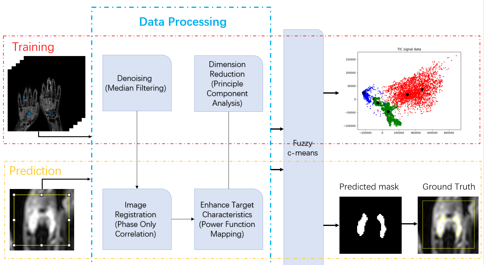
Fig.1 The flow of the algorithm
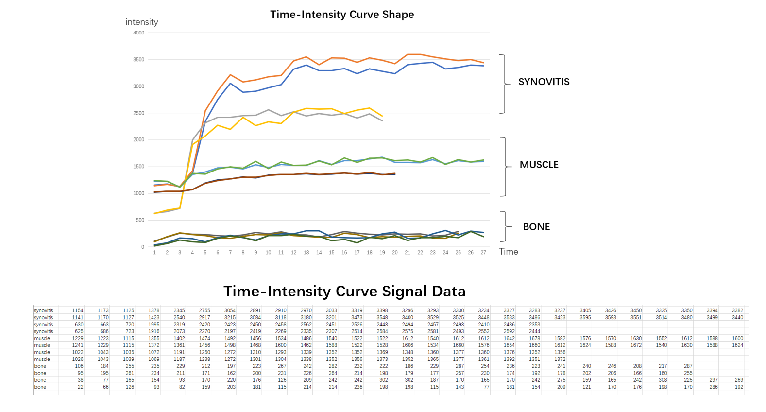
Fig.2 TIC Shape. Specific performance of different tissues on TIC shape under different phases. TIC Signal Data. Specific TIC signal values derived from DCE-MRI.
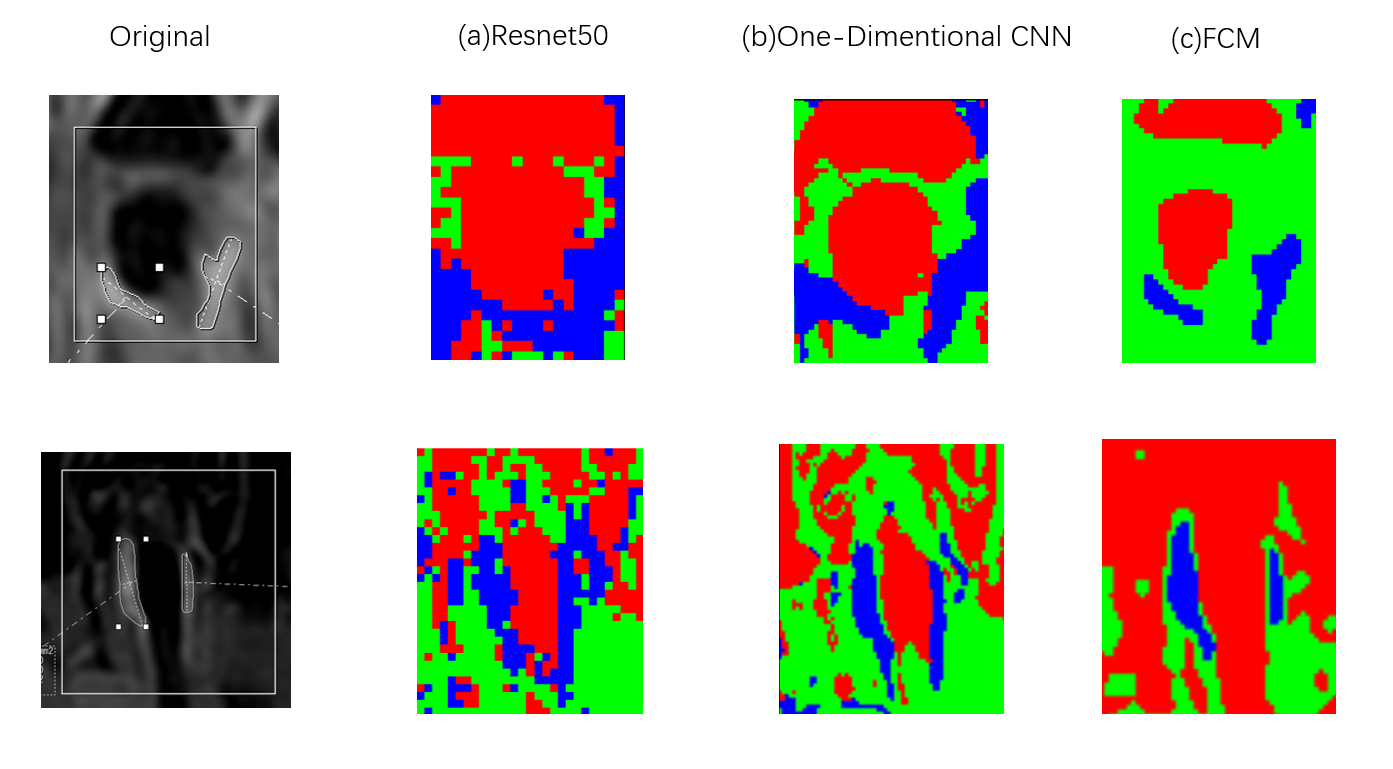
Fig.2 Examples of segmentation of synovitis based on different methods. (a) TIC shape classification based on Resnet-50, (b) TIC data classification based on one-dimensional
convolutional neural network, (c) TIC signal data clustering based on unsupervised
learning. Blue: synovitis, Green: muscle, Red: bone.
DOI: https://doi.org/10.58530/2023/5393