5390
The benefits of ZTE to standard MRI practice1MRI, CUH Addenbrookes UK, Newmarket, United Kingdom
Synopsis
In theory a ZTE or Zero Echo Time could visualise and benefit MRI imaging of any joint. It can be used in a wide spectrum of developmental, traumatic, inflammatory, rheumatologic and oncologic conditions. It may remove the need for CT with detailed depiction of bone anatomy. It opens the doors for more MRI based research into many musculoskeletal conditions and morphometric analysis. One MRI examination with a ZTE sequence allows cross referencing of sequences aiding diagnosis, prognostication and surgical guidance in soft tissue and bone with precise measurements that involve bony landmarks.
Background
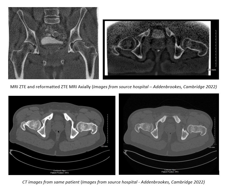
What is ZTE?ZTE or Zero Echo Time is an MRI sequence that visualises tissues such as bone with the shortest T2 values (Aydingöz Ü, 2022) giving images with a CT like appearance (Lu A., Sep 2019). Signal is acquired immediately after applying the radiofrequency pulse resulting in a near zero echo time with the next radiofrequency pulse following in a very short repetition time (Glick, May 2021). This rapid switch from transmit to receive mode enables acquisition of quickly decaying signal starting at a near zero echo time, capturing what little signal is present - especially in cortical bone (Aydingöz Ü, 2022). Absence of on and off gradient switching gives a virtually silent acquisition (Aydingöz Ü, 2022). ZTE sequences are resilient to artefacts caused by motion and magnetic field homogeneities with great signal to noise ratio and scan time efficiencies (Breighner RE., Nov 2017) (Lu A., Sep 2019). They have advantages in depicting small bone fragments in a trauma setting and subtle cortical erosion (Breighner RE., Nov 2017).
Benefits and where it is showing success
ZTE may remove the need for CT with detailed depiction of bone anatomy (Glick, May 2021) making an MRI examination a 1 stop imaging examination for conditions such as femoroacetabular impingement giving both soft tissue and bone imaging within the same examination (Carl M, Autumn 2021). Substantial agreement is being found in morphometric measurements, with reproducibility of many lytic or sclerotic lesions - although spatial resolution is still inferior to CT or radiography (Aydingöz Ü, 2022). It can be used in a wide spectrum of developmental, traumatic, inflammatory, rheumatologic and oncologic conditions. It has shown value in bone morphology, assessment of fractures, displaced bone fragments, shoulder instability, calcification (ossification of ligaments, calcific tendinitis) spinal foramina stenosis, skull assessment (suture closure, trauma) and assessment of bone erosions in bone tumours (Carl M, Autumn 2021). It may demonstrate a wide range of structural abnormalities and disease or healing processes (Aydingöz Ü, 2022). Drawbacks of ZTE are readily offset by cross checking with other MRI sequences. Correlation with other standard MR images is essential for correct characterisation of misleading signal intensity on ZTE such as hemosiderin deposition, gas and microscopic fragments within joints or tissue mimicking calcification or ossification) (Aydingöz Ü, 2022).
Teaching Points
- Benefits of ZTE imaging
- Which body parts and disease processes is ZTE showing most value in
- The physics behind the sequence and why the sequence is so quiet
- Pitfalls of ZTE imaging
Summary/Conclusions
In theory ZTE could visualise and benefit MRI imaging of any joint. It is having most success at 3T because although feasible at 1.5T and 3T, hardware requirements including fast transmit receive switching, precise RF wave form transmission, high gradient performance and gradient calibration are better at 3T. The quest for shorter imaging times favour 3T for better signal to noise ratio (Aydingöz Ü, 2022). Sequences must be isotropic to allow multiplanar and radial formatting as currently undertaken with CT (Aydingöz Ü, 2022) & (Lu A., Sep 2019).ZTE can be used to visualise a wide spectrum of developmental, traumatic, inflammatory, rheumatological and oncological conditions. It opens the doors for more MRI based research into many musculoskeletal conditions and morphometric analysis (Aydingöz Ü, 2022) & (Weiger M B. D., 2013) including new possibilities for research in healing and non-healing fractures. One MRI examination with a ZTE sequence allows cross referencing of sequences aiding diagnosis, prognostication and surgical guidance in soft tissue and bone with precise measurements that involve bony landmarks. It brings promising possibilities in routine patient services and research (Aydingöz Ü, 2022).
Acknowledgements
Thank you for the support and knowledge of...
Dr Andrew Grainger, MSK Radiologist Consultant, CUH Addenbrookes Hospital UK
Dr Emma Gerety, MSK Radiologist Consultant, CUH Addenbrookes Hospital UK
Bruno Carmo, MRI Manager, CUH Addenbrookes Hospital UK
References
Aydingöz Ü, E. Y. (2022). Zero Echo Time Musculoskeletal MRI: Technique, Optimization, Applications and Pitfalls. Radiographics 42, 1398-1414.
Breighner RE., E. Y. (Nov 2017). Technical Developments: Zero Echo Time Imaging of the Shoulder, Enhanced Osseous Detail by Using MR Imaging . Radiology 286 (3) [online] https://pubs.rsna.org/doi/10.1148/radi.
Breighner, R. (Spring 2018). GE: Utility of ZTE MR for bone imaging. [online] https://www.gesignapulse.com/signapulse/spring_2018/MobilePagedArticle.action?articleId=1396201#articleId1396201.
Carl M, P. R. (Autumn 2021). oZTEo: enabling MR as a one-stop shop for soft tissue and bone imaging Tech Trends. gesignapulse.com.
Glick, Y. (May 2021). Zero Echo Time. [online] https://radiopaedia.org/articles/zero-echo-time-imaging?lang=gb (Accessed: 23/10/2022).
Lee, C. J. (2020). Research Article: CT-like MRI using the zero-TE technique for osseous changes of the TMJ Dentomaxillofacial . Radiology 49 [online] https://www.birpublications.org/doi/pdf/10.1259/dmfr.
Lu A., G. K. (Sep 2019). Zero TE MRI for Craniofacial Bone Imaging. American Journal of Neuroradiology 40 (9) [online] http://www.ajnr.org/content/40/9/1562, 1562-1566 .
Weiger M, B. D. (2013). ZTE Imaging in Humans. Magnetic Resonance in Medicine 70 [online] https://onlinelibrary.wiley.com/doi/pdf/10.1002/mrm.24816, 328-332 .
Weiger M, B. D. (2014). ZTE Imaging with T1 Contrast. Proceedings of the International Society for Magnetic Resonance in Medicine 22, 4262.
Figures