5380
DW-MRI B-tensor encoding acquisition and processing on a pre-clinical Bruker scanner.1Universidad Nacional Autonoma de Mexico, Queretaro, Mexico, 2Centro de Investigacion en Matematicas, A.C., Guanajuato, Mexico
Synopsis
Keywords: Diffusion/other diffusion imaging techniques, Diffusion/other diffusion imaging techniques, Pipeline
We present how we implemented diffusion weighted B-tensor encoding acquisitions on a preclinical 7 T Bruker scanner using free software shared by the DW-MRI community. We also present an auxiliary free repository that contains helpful tools that facilitate the implementation. This educational poster is intended to aid other users interested in acquiring B-tensor encoded DW-MRI with Bruker scanners.Introduction
Diffusion-weighted MRI (DW-MRI) is a technique capable of characterizing microstructural properties in tissue1. While the technique has shown high sensitivity, standard approaches lack specificity to certain microstructural properties that can be important in neurodegeneration events2.Advanced DW-MRI acquisition schemes are able to provide additional information about tissue microenvironment3. Diffusion gradient waveforms with complex shapes4 measure q-space trajectories, rather than a single point in q-space, and are summarized by a B-tensor5 (a generalization of the b-value and b-vector). Different measurements using different B-tensors (size, shape, and orientation) enable DW-MRI to assess microstructural details that are otherwise inaccessible to the classic pulse-shaped diffusion gradient acquisition6.
Because B-tensor encoding schemes are an active topic of investigation, these sequences are not readily available in MRI systems by default. Still, the DW-MRI community has developed a series of resources and tools (see, for instance, https://github.com/filip-szczepankiewicz/fwf_seq_resources) that enable researchers to implement B-tensor encoding in different MRI systems. In this work, we present the complete acquisition and processing pipeline that we implemented for our preclinical Bruker system, highlighting the different open source tools used. We hope this information will be useful for other researchers interested in implementing these advanced DW-MRI acquisitions on their sites.
Methods
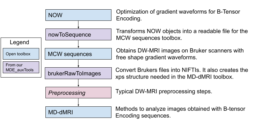
Figure 1 shows a flowchart for an acquisition and analysis pipeline for b-tensor encoding images on a preclinical Bruker system. We briefly describe each step in the following subsections.Waveform design for B-tensor encoding
The NOW toolbox (Numerical Optimization of gradient Waveforms)7, available in https://github.com/jsjol/NOW is a MATLAB package that optimizes gradient waveforms to acquire B-tensor encoding images. This toolbox optimizes the waveform to a specific B-tensor shape but limits it to specified valid hardware parameters of the MRI scanner to be used. The most recent version also features compensation for concomitant gradients8 and motion9.
Free-gradient waveform sequence in a Bruker scanner
The “MCW sequences” available on the Preclinical Neuro MRI repository https://osf.io/ngu4a/ can be used to obtain B-tensor encoding images. These sequences can acquire DW-MRI using free diffusion gradient waveforms (such as the ones obtained through NOW) on Bruker systems with Paravision 6.0.1 software. Installation and use of these sequences are documented in the corresponding manual; familiarity with Paravision is required.
Processing of B-tensor images
Processing of B-tensor encoded images is an active field of investigation. The freely-available Multidimensional diffusion MRI Matlab toolbox (https://github.com/markus-nilsson/md-dmri)10 is able to perform various forms of analysis on B-tensor DWI.
The MDE_auxTools repository
We provide useful functions that aid in the implementation of B-tensor encoding sequences on a Bruker scanner (https://github.com/RicardoRios46/MDE_auxTools). These functions are intended to be used as connectors between tools developed by the DW-MRI scientific community such that complete acquisition pipelines can be built (Figure 1).
Results
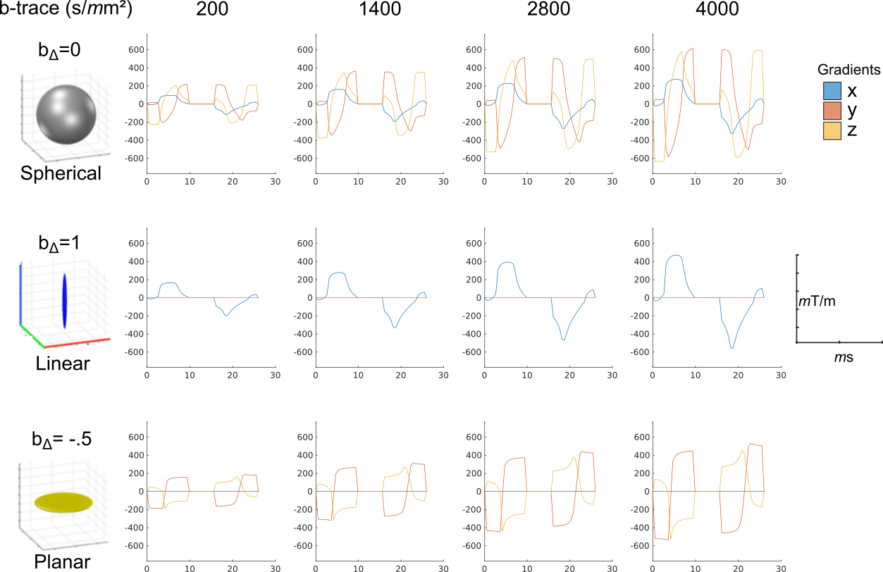
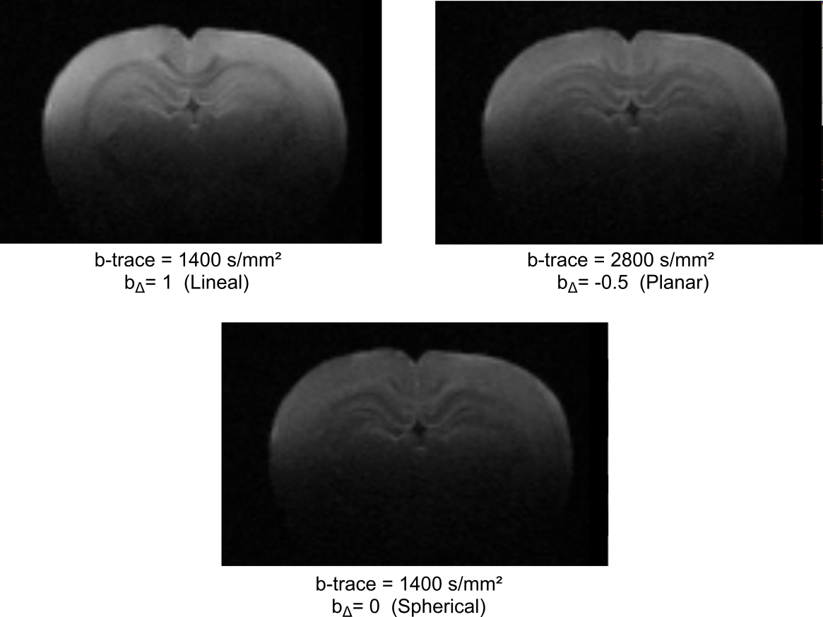
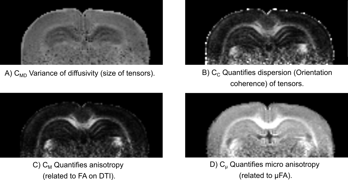
We implemented the pipeline in Figure 1 on a 7 T Bruker system to obtain B-tensor encoded DWI of ex vivo rat brains using a Cryoprobe. Figure 2 shows an example of diffusion gradient waveforms optimized with NOW for our Bruker system. Spherical and linear waveforms are frequency-tuned. Figure 3 shows example images with different B-tensors (B-values and B-shapes) acquired with the waveforms illustrated in Figure 2. Figure 4 shows q-space trajectory Imaging (QTI)5 metrics computed with the MD-MRI toolbox.Conclusion
In this work, we show an implementation of B-tensor encoded DW-MRI acquisition on a pre-clinical Bruker scanner and their subsequent analysis. We highlight the different software and steps needed for the implementation and a complementary toolbox to facilitate this process. Other institutions interested can do a similar approach with the presented pipeline.Acknowledgements
Imaging was performed at the National Laboratory for Magnetic Resonance Imaging (Conacyt, UNAM, CIMAT, UAQ). Funding by CONACYT (FC 1782) and UNAM-DGAPA (IG200117, IN204720).References
1. Novikov, D. S., Fieremans, E., Jespersen, S. N. & Kiselev, V. G. Quantifying brain microstructure with diffusion MRI: Theory and parameter estimation. NMR Biomed. 32, e3998 (2019).
2. Lampinen, B. et al. Towards unconstrained compartment modeling in white matter using diffusion‐relaxation MRI with tensor‐valued diffusion encoding. Magn. Reson. Med. 84, 1605–1623 (2020).
3. Topgaard, D. Multidimensional diffusion MRI. J. Magn. Reson. 275, 98–113 (2017).
4. Szczepankiewicz, F., Westin, C.-F. & Nilsson, M. Gradient waveform design for tensor-valued encoding in diffusion MRI. J. Neurosci. Methods 348, 109007 (2021).
5. Westin, C.-F. et al. Q-space trajectory imaging for multidimensional diffusion MRI of the human brain. NeuroImage 135, 345–362 (2016).
6. Narvaez, O., Svenningsson, L., Yon, M., Sierra, A. & Topgaard, D. Massively Multidimensional Diffusion-Relaxation Correlation MRI. Front. Phys. 9, 793966 (2022).
7. Sjölund, J. et al. Constrained optimization of gradient waveforms for generalized diffusion encoding. J. Magn. Reson. 261, 157–168 (2015).
8. Szczepankiewicz, F., Westin, C.-F. & Nilsson, M. Maxwell-compensated design of asymmetric gradient waveforms for tensor-valued diffusion encoding. Magn. Reson. Med. 82, 1424–1437 (2019).
9. Szczepankiewicz, F. et al. Motion-compensated gradient waveforms for tensor-valued diffusion encoding by constrained numerical optimization. Magn. Reson. Med. 85, 2117–2126 (2021).
10. Nilsson, M. et al. An open-source framework for analysis of multidimensional diffusion MRI data implemented in MATLAB. (2018).
Figures