5373
Effects of Diffusion Time and Echo Time Changes of Diffusion MRI on Clinical Brain Neoplasm Diagnosis
Masaaki Hori1,2, Tomoko Maekawa2, Kouhei Kamiya1,2, Akifumi Hagiwara2, Koji Kamagata2, and Shigeki Aoki2,3
1Toho University Omori Medical Center, Tokyo, Japan, 2Radiology, Faculty of Medicine, Juntendo University, Tokyo, Japan, 3Faculty of Health Data Science, Juntendo University, Chiba, Japan
1Toho University Omori Medical Center, Tokyo, Japan, 2Radiology, Faculty of Medicine, Juntendo University, Tokyo, Japan, 3Faculty of Health Data Science, Juntendo University, Chiba, Japan
Synopsis
Keywords: Tumors, Tumor
In this exhibit, we outline the effect of TE shortening on the diffusion-weighted imaging (DWI) in clinical practice and the associated shortening of diffusion time on recent clinical MRI scanner for brain neoplasms diagnosis. In general, shortening the diffusion time reduces the contrast of lesions that show an abnormally high signal on DWI, and the apparent diffusion coefficient values also changes toward a larger value, which may lead the radiologist to err in differential diagnosis or grading of the neoplastic lesions. Therefore, it is important for radiologists to be aware of these effects when diagnosing brain neoplasms.Purpose:
With advances in hardware and software on clinical MRI scanners, especially 3Tesla, the echo time (TE) of diffusion-weighted (DW) imaging using single-shot echo-planar imaging are being set up with shorter than before. Of course, in principle this is true in terms of improved image quality, but at the same time diffusion times are often automatically shortened. Also, clinicians are often unaware that changes in TE and diffusion time can change the contrast of the DW imaging and its quantitative value. The purpose of this exhibit is to present the effects of diffusion time and TE changes of diffusion MR imaging on clinical brain neoplasms diagnosis.Outline of contents:
1. First, the representative case images are shown when the TE time is set shorter on a 3T MRI system in clinical settings. Usually, the diffusion time is shortened along with the TE shortening. As a result, both the contrast of the brain parenchyma and the contrast of neoplastic lesions in the brain change and the corresponding apparent diffusion coefficient (ADC) values also change (Figure 1). In the past, 1.5T MRI systems had a TE of approximately 100-120 ms for DW imaging, whereas the current state-of-the-art 3T MRI systems have a TE = 60 ms is set as a standard value in the latest clinical 3T MRI systems. Therefore, when referring to past images, it is necessary to consider not only differences in static magnetic field strength but also differences in TE and diffusion time.2. In DW images, if the diffusion time is fixed and TE is varied, the contrast of the b=0 image is defined by the value of TE, and the contrast of the trace image, which is used for clinical diagnosis, also changes (Figure 2). In general, diffusion-weighted images are known to be TE-dependent1. Therefore, the radiologist should be aware of the TE value and its effect on the DW images, or the differential diagnosis may be incorrect.
3. Next, we show what can happen in DW images when TE is fixed and diffusion time is varied. The radiologist must first recognize that with newer MRI systems, the diffusion time is shorter than before as the TE is shortened. Shorter diffusion times reduce the contrast of brain neoplasms that show abnormally high signal on DW images. In some cases, there is a risk of unrecognizability2, 3 (Figure 3). In addition, changes in image contrast, i.e., various brain tumors known to show abnormally high signal on DW images and reduced ADC (e.g., germinoma, CNS lymphoma, glioblastoma, etc.) do not show typical imaging findings, which may lead the radiologist to err in differential diagnosis or grading of the neoplastic lesions4 (Figure 4 and 5).
Summary:
In this exhibit, we outline the effect of TE shortening on the DW imaging in clinical practice and the associated shortening of diffusion time on recent clinical MRI scanner for brain neoplasms diagnosis. In general, shortening the diffusion time reduces the contrast of lesions that show an abnormally high signal on DWI, and the ADC value also changes toward a larger value. It is important for radiologists to be aware of these effects when diagnosing brain neoplasms.Acknowledgements
This work was supported by JSPS KAKENHI Grant Number 22K15850.References
- Veraart J, et al. Diffusional kurtosis imaging: the quantification of non-Gaussian water diffusion by means of magnetic resonance imaging. Magn Reson Med. 2005;53(6):1432–1440. TE dependent Diffusion Imaging (TEdDI) distinguishes between compartmental T2 relaxation times. Neuroimage.2018 Nov 15;182:360-369.
- Boonrod A, et al.Reduced visualization of cerebral infarction on diffusion-weighted images with short diffusion times. Neuroradiology. 2018 Sep;60(9):979-982.
- Hori M, et al. Teaching Neuroimages: Obscured Cerebral Infarction on MRI. Clin Neuroradiol. 2017 Dec;27(4):519-520.
- Maekawa T, et al.Differentiation of high-grade and low-grade intra-axial brain tumors by time-dependent diffusion MRI. Magn Reson Imaging. 2020 Oct;72:34-41.
Figures
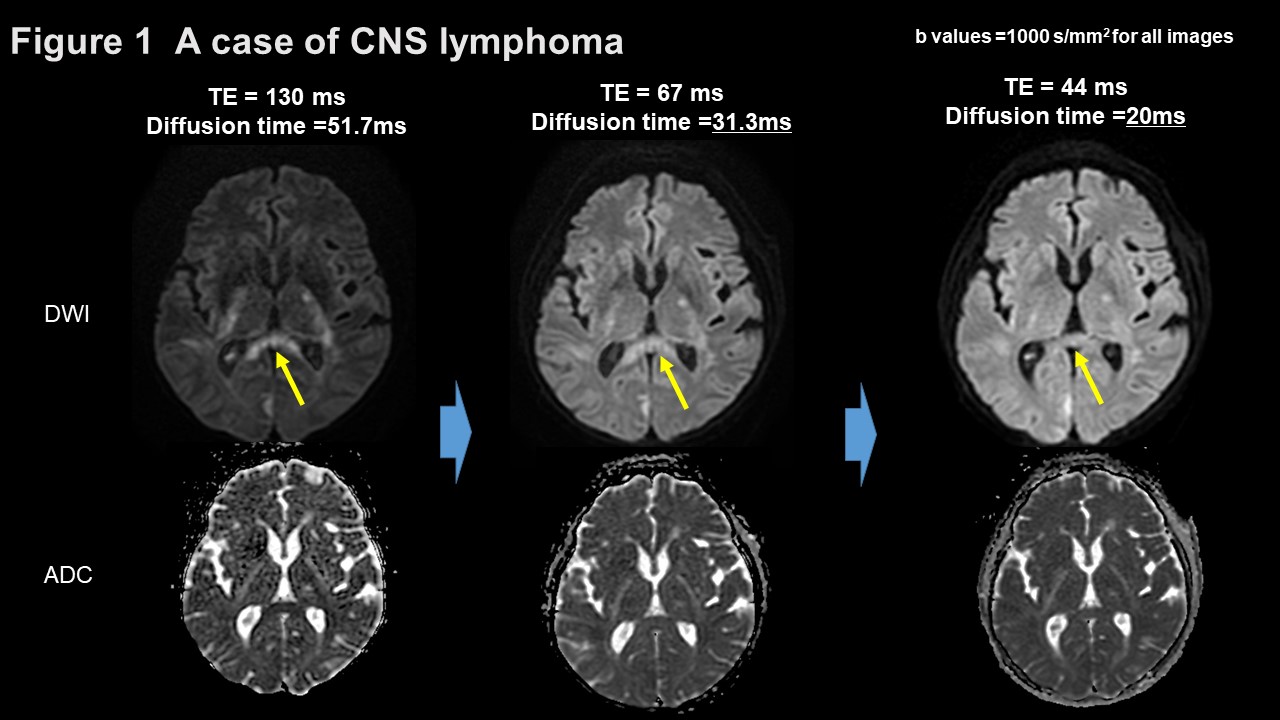
Figure 1
This is a case of CNS lymphoma. All diffusion-weighted images were acquired on
the same 3T MRI; as the echo time is set shorter, the diffusion time decreases.
The high intensity lesions, such as in the corpus callosum, are obscured
(arrows) as the diffusion time becomes shorter. Corresponding apparent
diffusion coefficient (ADC) maps show different ADC values in the lesions. Therefore,
ADC values are diffusion time dependent and TE dependent.

Figure 2
A case of meningioma. T2WI shows the lower intensity mass indicating meningioma
(arrow). As echo time shortens, the tumor is seen at progressively higher
signals in both b0 images and diffusion-weighted images.
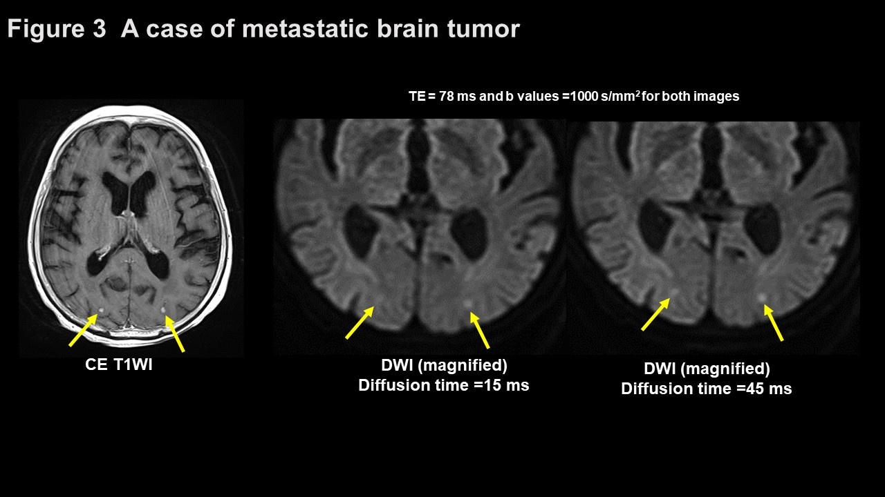
Figure 3
Diffusion-weighted images(DWI) and contrast enhanced T1WI images show the
abnormal hyper intensity and abnormal enhancements in bilateral occipital
lobes, indicating metastatic brain tumors(arrows). Lesion are more clearly
demonstrated on DWI of diffusion time 45ms.
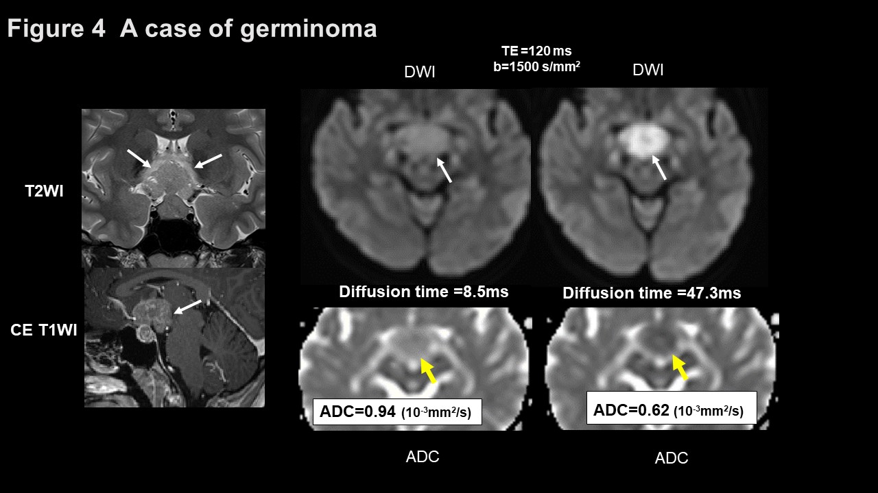
Figure 4
Contrast-enhanced T1WI and T2WI show the enhanced suprasellar tumor, indicating
germinoma (arrows). Diffusion-weighted image (DWI) of short diffusion time
shows the lower intensity and higher apparent diffusion coefficient value,
compared with DWI of long diffusion time image (arrows). DWIs were obtained
with oscillating gradient spin–echo sequence.
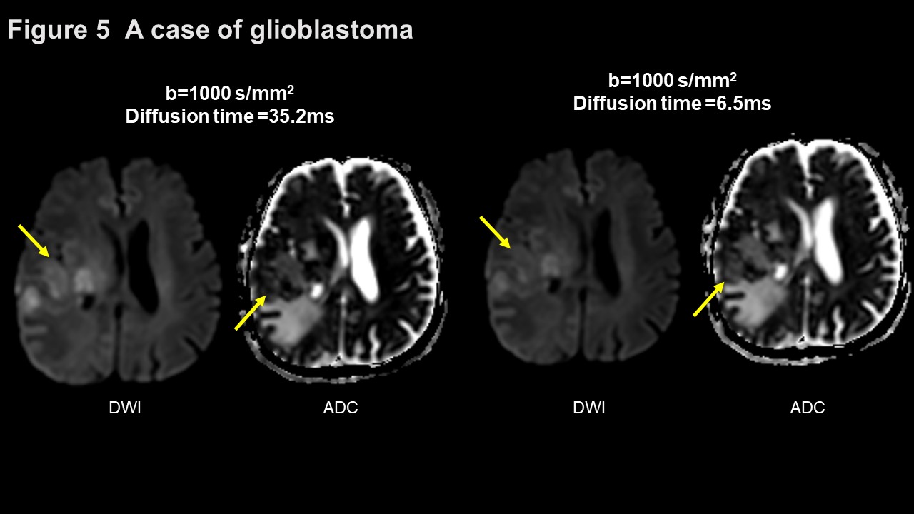
Figure 5
Diffusion-weighted image (DWI) of short diffusion time shows the lower intensities
and higher apparent diffusion coefficient values, compared with DWI of long
diffusion time image (arrows) in the glioblastoma. DWIs were obtained with oscillating
gradient spin–echo sequence.
DOI: https://doi.org/10.58530/2023/5373