5362
Automatic detection method for the stationary period of the coronary arteries for whole-heart coronary MR angiography using deep learning1Department of Medical Radiological Technology, Faculty of Health Sciences, Kyorin University, Mitaka-shi, Tokyo, Japan, 2Radiology Department, Tokyo Teishin Hospital, Chiyoda-ku, Tokyo, Japan
Synopsis
Keywords: Vessels, Cardiovascular, Coronary MRA
We developed a new method using a convolutional neural network to obtain the stationary periods of the coronary arteries for whole-heart coronary magnetic resonance angiography. A time-domain segmentation method using U-net was proposed. Two motion curves, which were obtained from the motion of the coronary arteries between cine frames, were vertically arranged, converted to motion images, and used to extract the stationary periods. The results demonstrate that the proposed method can accurately determine the stationary period of the coronary arteries in humans, and it is expected to be a fully automatic determination method for the stationary period.Introduction
Whole-heart coronary magnetic resonance angiography (MRA)1 requires data acquisition to be performed during the stationary period of the coronary arteries. Therefore, it is important to determine the stationary period accurately. However, the detection of the stationary period is currently time-consuming and operator-dependent because it is visually determined by the operator from cine images. Several methods have been developed2-5. However, fully automatic methods have not yet been realized.The typical automatic method consists of two steps. Step 1 consists of detecting the position of the coronary artery in cine images and obtaining the motion curve by tracking the position in all cine phases. Step 2 consists of extracting the coronary stationary period from the magnitude of the motion curve. For example, a method using a template method in Step 1 and a threshold method in Step 2 has been reported4. However, for example, the shape of coronary arteries changes with the cardiac phase; therefore, it is difficult to detect the positions in all cardiac cycles with the fixed template. A problem with the threshold method in Step 2 is that the coronary arteries move slightly at the beginning, end and middle of the stationary period, and the threshold method dose not handle properly this movement. Therefore, it is difficult to obtain the stationary period from the motion curve, like the operator extracts.
To mitigate these problems, we developed a method that uses a single-shot multi-box detector (SSD)6 to detect the position of the coronary arteries in a previous study, which demonstrated an improvement in the detection rate7. However, the threshold method was still used for Step 2. In this study, we developed a new method that uses a CNN to obtain the stationary period of the coronary arteries from motion curves.
Methods
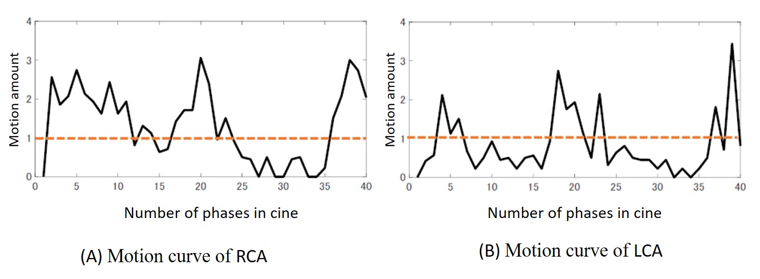
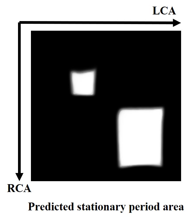
Steady-state free precession cine images (4 chamber, 31–63 phases) of 17 healthy volunteers were obtained using 3T MRI. For Step 1, an SSD, which is used for the detection of objects, was used to detect the position of the coronary arteries (Figure 1)7. Using this position, the movement of the coronary arteries in the temporally adjacent cine images was calculated, and two motion curves were obtained, one for the left coronary artery (LCA) and one for the right coronary artery (RCA) (Figure 2).For Step 2, we proposed a time-domain segmentation method as a new method to obtain stationary periods from motion curves. Two motion curves were arranged vertically and horizontally in an image, and the image intensity was obtained by multiplying them, which was used as the magnitude of motion in the image (Figure 3A).
Next, we set the regions of the stationary period determined by humans as the training images (labels) (Figure 3B) and input them into a U-net8 for learning, which is often used in image segmentation. For the test, the motion images obtained from the residual cine images were input into the U-net, and the stationary period of the LCA and that of the RCA were obtained from the 2D areas of the stationary period extracted by the U-net. Of the images, data from 13 volunteers were used for the training and data from 4 volunteers were used for the testing.
In the evaluation, the root mean square error (RMSE) was calculated between the frame numbers correspond to each start and end of the stationary period determined by the proposed method and the corresponding one determined by the human operator. The RMSE was calculated for each RCA and LCA in all cine phases, and the total RMSE with RCA + LCA was also calculated and used as a measure of the accuracy of the stationary period.
Results and Discussion
Figure 4 shows the extracted area of the coronary stationary period using the proposed method. The area of the stationary period on the motion image appeared as an area on the diagonal line of the motion image. This suggests that there is some degree of correlation between the stationary period of the LCA and that of the RCA. This means that the proposed method is expected to be more robust against noise, etc., than the conventional method, in which RCA and LCA are analyzed independently.Concerning the accuracy of extracted stationary periods, in the case of the threshold method, the RMSE was 1.9 phase for the RCA, 3.7 phase for the LCA, and 2.9 phase for all cine phases. In the case of the proposed method, the RMSE was 1.3 phase for the RCA, 2.2 phase for the LCA, and 1.8 phase for all cine phases. The proposed method exhibited fewer error phases than the conventional method. This is thought to be because the judgment of minute movements at the beginning, end and middle of the stationary period is learned from human data, so the result is closer to that of a human. Further investigation is needed because this study was conducted using a relatively small amount of data.
Conclusion
With the proposed method, the stationary period of the coronary arteries can be determined more closely to that of humans, and it has been shown to be useful as a fully automatic determination method for the stationary period of coronary arteries.Acknowledgements
No acknowledgement found.References
[1] Weber OM, Martin AJ, Higgins CB. Whole-heart steady-state free precession coronary artery magnetic resonance angiography. Magn Reson Med. 2003;50:1223–1228.
[2] Bi X. Magn Reson. Automatic data acquisition window determination for coronary MRA. J Cardiovasc Magn Reson. 2007;9(2):234–235.
[3] Jahnke C, Paetsch I, Nehrke K, et al. A new approach for rapid assessment of the cardiac rest period for coronary MRA. J Cardiovasc Magn Reson. 2005;7(2):395–399.
[4] Sato T, Maruyama K, Okada T, Kuhara S, Togashi K, Minato K. Automatic ROI placing for selecting optimal data acquisition window for magnetic resonance coronary angiography. Society for Cardiovascular Magnetic Resonance (SCMR). 2008.
[5] Ninomiya A, Kuhara S, Okada T, et al. Feasibility study of Global-to-Local Tandem Method for detecting the coronary stationary period in whole-heart magnetic resonance coronary angiography (WH MRCA). J Cardiovasc Magn Reson. 2010;(Suppl 1):34.
[6] Liu W, Anguelov D, Erhan D, et al. SSD: single shot multibox detector. Proceedings of the European Conference on Computer Vision (ECCV), 2016, arXiv:1512.02325:[cs.CV].
[7] Kasai R, Endo Y, Shibo H, Amanuma M, Kobayashi K, Kuhara S. A feasibility study of an automatic detection method of the stationary period of the coronary artery using the convolutional neural network (CNN). ESMRMB Congress. 2020;33(Suppl 1):S198 P01.22.
[8] Ronneberger O, Fischer P, Brox T. U-Net: convolutional networks for biomedical image segmentation. 2015;arXiv:1505.04597v1 [cs.CV].
Figures
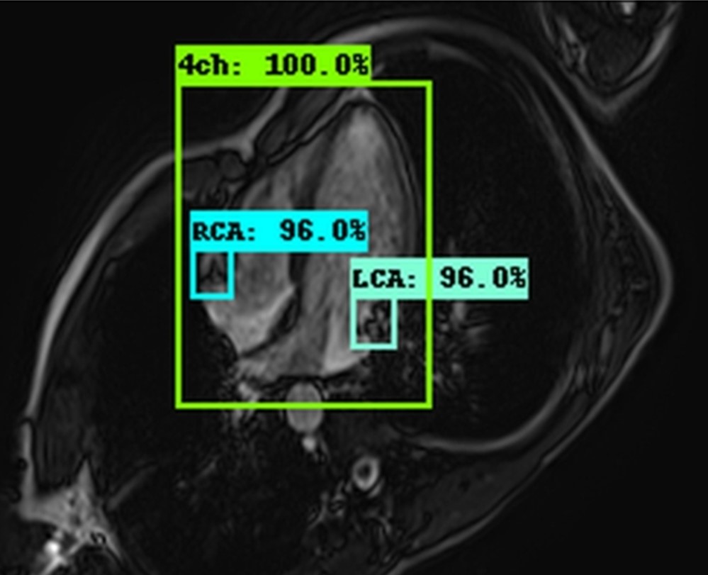
Detected positions of the right and left coronary arteries using SSD.
ROIs represent the detected positions of the coronary arteries, which are called the bounding boxes. The number of each bounding box represents the likelihood probability7.